The Future of Orthopedics: How 3D Printing and Additive Manufacturing Are Revolutionizing Hip Prostheses
This comprehensive article examines the transformative impact of 3D printing and additive manufacturing (AM) on the development, production, and application of hip prostheses.
The Future of Orthopedics: How 3D Printing and Additive Manufacturing Are Revolutionizing Hip Prostheses
Abstract
This comprehensive article examines the transformative impact of 3D printing and additive manufacturing (AM) on the development, production, and application of hip prostheses. Tailored for researchers, scientists, and biomedical engineers, it provides a multi-faceted analysis covering foundational principles, advanced manufacturing methodologies, process troubleshooting, and clinical validation. The article explores material innovations like titanium alloys and bioceramics, key AM techniques including Selective Laser Melting (SLM) and Electron Beam Melting (EBM), and the critical challenges of quality assurance and regulatory pathways. It concludes by evaluating clinical outcomes, comparing AM implants to traditional counterparts, and outlining future research directions in personalized medicine and bio-integrated implants.
From CAD to Implant: Understanding the Core Principles of 3D Printed Hip Prostheses
1.0 Introduction and Application Notes
Within the paradigm of orthopedic implant manufacturing, particularly for cementless hip prostheses, a fundamental shift is occurring from subtractive to additive methodologies. This transition is driven by the pursuit of enhanced osseointegration through controlled surface and structural porosity, patient-specific anatomical matching, and material efficiency.
Subtractive Manufacturing (SM): The conventional paradigm. Involves machining (milling, turning) a solid block of material (e.g., Ti-6Al-4V, Co-Cr alloy) to obtain the final implant geometry. This process is material-wasteful and inherently limits design complexity, typically producing only solid or superficially textured implants.
Additive Manufacturing (AM): The emergent paradigm. Constructs implants layer-by-layer from digital models, primarily using Powder Bed Fusion (PBF) techniques like Selective Laser Melting (SLM) or Electron Beam Melting (EBM). This enables the fabrication of complex, three-dimensional porous lattice structures (meta-biomaterials) that mimic the mechanical properties of bone and facilitate bone ingrowth.
Table 1: Quantitative Comparison of Key Manufacturing Paradigms for Ti-6Al-4V Hip Stems
| Parameter | Subtractive Manufacturing (CNC Machining) | Additive Manufacturing (Laser PBF/SLM) |
|---|---|---|
| Material Utilization | 10-20% (High waste) | 95-98% (Unfused powder recyclable) |
| Achievable Porosity | Surface-only (via grit-blasting, HA coating) | Volumetric, controlled (30-80% porous lattice) |
| Pore Size Range | Non-structural (µm-scale texture) | Structural (200-800 µm, designed for bone ingrowth) |
| Elastic Modulus | ~110 GPa (Solid Ti-6Al-4V) | 1-20 GPa (Tunable via lattice design) |
| Design Lead Time | Long (Fixture & toolpath programming) | Short (Direct from CAD to build) |
| Unit Cost (High Volume) | Lower | Higher |
| Unit Cost (Low Volume/Custom) | Very High | Competitive to Lower |
| Key Limitation | Design constraints, waste | Post-processing needs, powder handling |
2.0 Experimental Protocols
Protocol 2.1: In Vitro Assessment of Osseointegration Potential for AM vs. SM Surfaces
Objective: To compare the early-stage osteogenic cell response on AM-fabricated porous lattices versus SM-produced smooth and grit-blasted surfaces.
Materials: Human Osteoblast-like Cells (SaOS-2 or MG-63), Ti-6Al-4V discs (Ø15mm x 2mm): (a) SM-machined polished, (b) SM-grit-blasted, (c) AM-porous lattice (500µm pore size). Cell culture medium (α-MEM + 10% FBS), AlamarBlue assay kit, RNA extraction kit, qPCR reagents.
Methodology:
- Sample Preparation & Sterilization: Clean all samples ultrasonically. Sterilize by autoclaving (SM samples) or gamma irradiation (AM porous samples to avoid powder trapping).
- Cell Seeding: Seed cells at a density of 25,000 cells/cm² onto sample surfaces placed in 24-well plates. Allow 2 hours for initial adhesion before adding medium.
- Metabolic Activity (Day 1, 3, 7): At each time point, incubate with 10% AlamarBlue reagent for 3 hours. Measure fluorescence (Ex 560nm/Em 590nm).
- Gene Expression Analysis (Day 7): Extract total RNA from cells on samples. Perform reverse transcription and qPCR for osteogenic markers (Runx2, ALPL, COL1A1). Normalize to GAPDH.
- Statistical Analysis: Perform one-way ANOVA with post-hoc Tukey test (n=6, p<0.05).
Protocol 2.2: Mechanical Characterization of Manufactured Lattice Structures
Objective: To determine the compressive mechanical properties of AM-generated lattices versus solid AM and SM materials.
Materials: ASTM F2924-compliant Ti-6Al-4V powder, SLM machine, CNC machine. Samples: (a) SM-solid cube (10mm side), (b) AM-solid cube (10mm side), (c) AM-porous cubes with varying unit cells (e.g., Diamond, Gyroid) and strut thicknesses (10mm side).
Methodology:
- Fabrication: Fabricate all AM samples on a single build plate using optimized SLM parameters (Laser power 200W, scan speed 800 mm/s, layer thickness 30µm). Stress-relieve per ASTM F3001.
- Geometric Verification: Perform micro-CT scanning to measure actual strut thickness, pore size, and porosity relative to CAD model.
- Compression Testing: Perform quasi-static uniaxial compression test per ISO 13314. Use a 100kN load cell, displacement rate of 1 mm/min. Record load-displacement data until 50% strain.
- Data Analysis: Calculate effective Elastic Modulus (from linear elastic region), Yield Strength (0.2% offset), and plateau stress. Correlate with measured porosity.
3.0 Visualization of Workflows and Relationships
Title: Additive vs Subtractive Manufacturing Process Chain
Title: Integrated Research Workflow for Implant Evaluation
4.0 The Scientist's Toolkit: Key Research Reagent Solutions
Table 2: Essential Materials for Orthopedic AM Research
| Item / Reagent | Function / Application | Key Consideration |
|---|---|---|
| Gas-Atomized Ti-6Al-4V ELI Powder | Raw material for Laser PBF. Spherical morphology ensures consistent powder flow and fusion. | Must meet ASTM F2924 (Grade 23). Particle size distribution (15-45µm) is critical. |
| Osteoblast Cell Line (e.g., MG-63) | In vitro model for assessing biocompatibility and osteogenic response. | Choose human-derived line for clinical relevance. Monitor mycoplasma contamination. |
| AlamarBlue Cell Viability Reagent | Fluorometric assay for quantifying metabolic activity of cells on sample surfaces. | Non-destructive, allows longitudinal tracking on the same sample. |
| TRIzol Reagent | For simultaneous lysis and stabilization of RNA from cells grown on metallic implants. | Effective for difficult-to-lyse cells adhering to rough/porous surfaces. |
| Micro-CT Scanner (e.g., SkyScan) | Non-destructive 3D imaging for quantifying bone ingrowth into pores and verifying lattice geometry. | Requires high resolution (<10µm voxel size) for trabecular-level analysis. |
| Bone Morphogenetic Protein-2 (BMP-2) | Positive control for in vitro osteogenic differentiation assays and potential coating for implants. | High cost; use at optimized concentrations to avoid adverse effects. |
| Simulated Body Fluid (SBF) | In vitro bioactivity test to assess apatite-forming ability of surface-modified implants. | Solution ion concentrations must closely match human blood plasma. |
| Polymethylmethacrylate (PMMA) Embedding Kit | For histology preparation. Infiltrates and supports bone-implant interface for sectioning. | Requires careful vacuum cycling to fully infiltrate deep porous structures. |
Application Notes
In the additive manufacturing (AM) of hip prostheses, material selection dictates biomechanical performance, osseointegration, and long-term implant survivability. Ti-6Al-4V remains the dominant alloy due to its excellent specific strength and biocompatibility, commonly processed via Laser Powder Bed Fusion (L-PBF). Recent trends focus on lattice structure design to lower elastic modulus, reducing stress shielding. Cobalt-chrome (CoCr) alloys are pivotal for articulating surfaces (e.g., femoral heads) due to superior wear resistance and hardness, often fabricated via L-PBF or Electron Beam Melting (EBM). Porous tantalum, produced via Laser Powder Bed Fusion or chemical vapor infiltration, offers exceptional biocompatibility and a high degree of porosity (75-85%), promoting rapid bone ingrowth. Bioceramics, including hydroxyapatite (HA) and tricalcium phosphate (TCP), are used as coatings or composite materials to impart bioactivity, typically deposited via binder jetting or post-AM surface modification techniques like plasma spraying.
Table 1: Key Mechanical and Physical Properties of AM Materials for Hip Prostheses
| Material | Typical AM Process | Yield Strength (MPa) | Elastic Modulus (GPa) | Porosity (%) | Key Application in Hip Prosthesis |
|---|---|---|---|---|---|
| Ti-6Al-4V (ELI) | L-PBF | 895 - 1100 | 110 - 120 | 0-3 (solid); 50-70 (lattice) | Femoral stem, acetabular cup |
| Cobalt-Chrome (Co-28Cr-6Mo) | L-PBF / EBM | 900 - 1200 | 220 - 230 | <1 | Femoral head, articulating surfaces |
| Porous Tantalum | L-PBF of polymer template + CVD | N/A (scaffold) | 2.5 - 4.0 | 75 - 85 | Acetabular augments, porous coatings |
| Bioceramic (HA) | Binder Jetting | 40 - 100 (compressive) | 30 - 100 | 20 - 50 | Bioactive coatings, bone graft substitutes |
Table 2: In-Vivo Biological Performance Metrics
| Material | Osseointegration Rate (Relative) | Bone Ingrowth Depth at 12 weeks | Typical Coating Thickness (µm) | Reference Study Model |
|---|---|---|---|---|
| Ti-6Al-4V (grit-blasted) | Baseline | ~1.0 mm | N/A | Canine femoral implant |
| Ti-6Al-4V with HA coating | 1.5x - 2x | ~1.8 mm | 50 - 150 | Ovine model |
| Porous Tantalum | 2x - 3x | 2.0 - 3.5 mm | N/A (bulk porous) | Human retrieval studies |
| CoCr (as-polished) | Low | Minimal | N/A | Simulator wear studies |
Experimental Protocols
Protocol 1: L-PBF Fabrication and Post-Processing of Ti-6Al-4V Lattice Femoral Stem
- Powder Preparation: Use gas-atomized Ti-6Al-4V (Grade 23, ELI) powder, particle size 15-45 µm. Dry powder in vacuum oven at 120°C for 4 hours.
- Machine Setup: Calibrate L-PBF system (e.g., EOS M 290) with argon atmosphere (<0.1% O2). Preheat build platform to 200°C.
- Print Parameters: Laser power: 250-300 W, scan speed: 1200 mm/s, hatch spacing: 0.11 mm, layer thickness: 30 µm. Use a stripe or chessboard scan strategy.
- Stress Relief: Perform heat treatment at 800°C for 2 hours in argon, furnace cool.
- Hot Isostatic Pressing (HIP): 920°C, 100 MPa argon, for 2 hours.
- Surface Finishing: Use grit blasting with Al2O3 (250 µm) followed by ultrasonic cleaning in acetone, ethanol, and deionized water.
Protocol 2: Osteoblast Cell Seeding & Proliferation Assay on Porous Tantalum
- Sample Sterilization: Autoclave porous tantalum discs (10mm dia. x 5mm height, 80% porosity) at 121°C for 30 minutes.
- Cell Culture: Use human osteoblast-like cells (MG-63 or hFOB 1.19). Culture in DMEM/F-12 with 10% FBS and 1% penicillin/streptomycin at 37°C, 5% CO2.
- Seeding: Place sterilized sample in 24-well plate. Seed cells at density of 2x10^4 cells/cm² in 50 µL medium, allow 2 hours for attachment, then add 1 mL medium.
- Proliferation Assay (MTT): At days 1, 3, and 7, add MTT reagent (0.5 mg/mL) and incubate for 4 hours. Remove medium, add DMSO to solubilize formazan crystals. Measure absorbance at 570 nm using a plate reader.
- Analysis: Normalize absorbance values to day 1 control (tissue culture plastic). Perform statistical analysis (n=6) using one-way ANOVA.
Protocol 3: Binder Jetting of Hydroxyapatite for Acetabular Cup Coating
- Powder Preparation: Use hydroxyapatite powder (Ca/P ratio 1.67), particle size <50 µm. Dry at 150°C overnight.
- Printing: Load powder into binder jetting system (e.g., ExOne Innovent+). Use layer thickness of 100 µm. Jet colloidal silica binder.
- Depowdering: Carefully remove printed green part from powder bed using compressed air.
- Sintering: Sinter in high-temperature furnace with air atmosphere. Ramp at 2°C/min to 600°C (binder burnout), hold 1 hour. Ramp at 5°C/min to 1250°C, hold 2 hours. Cool at 3°C/min to room temperature.
- Characterization: Measure density via Archimedes' principle. Characterize phase purity via X-ray diffraction (XRD).
Visualizations
L-PBF Workflow for Metallic Implants
Osseointegration Signaling Cascade
The Scientist's Toolkit: Research Reagent Solutions
Table 3: Essential Reagents for AM Hip Prosthesis Research
| Item | Function in Research | Example Product/Catalog |
|---|---|---|
| Gas-Atomized Ti-6Al-4V (ELI) Powder | Raw material for L-PBF fabrication of load-bearing components. | AP&C, 15-45 µm, Grade 23 |
| Colloidal Silica Binder | Binds ceramic powder particles in binder jetting process. | ExOne Binder 1020 |
| Simulated Body Fluid (SBF) | In-vitro bioactivity test for apatite formation on bioceramics. | Kokubo Recipe, 1.5x SBF |
| AlamarBlue / MTT Reagent | Cell viability and proliferation assay on material surfaces. | Thermo Fisher Scientific, DAL1100 |
| Osteogenic Media Supplement | Induces osteogenic differentiation in cell culture studies. | Sigma-Aldrich, Dexamethasone, β-glycerophosphate, Ascorbate |
| Micro-CT Contrast Agent (e.g., Hexabrix) | Enhances soft tissue/bone contrast for ex-vivo implant integration analysis. | Guerbet, Oxilan-350 |
| Ringer's Solution | Electrolyte solution for in-vitro corrosion testing (ASTM F2129). | Baxter, 2B2324Q |
| Alpha-MEM, no nucleosides | Cell culture medium for osteoblast precursor cells. | Gibco, 12561-056 |
This document details the integrated digital workflow from medical imaging to additive manufacturing (AM) for patient-specific hip prostheses. Within the broader thesis on 3D printed hip implants, this pipeline is foundational for creating implants that address anatomical variability, improve bone integration, and optimize biomechanical performance. The protocols below enable the translation of patient anatomy into a functional, manufacturable design.
Application Notes & Protocols
Protocol: Medical Image Acquisition & Preprocessing
Objective: To obtain high-fidelity DICOM data of the hip joint suitable for 3D reconstruction.
Detailed Methodology:
- Imaging Parameters: For quantitative anatomical modeling, specific CT protocols are required.
- Scanner: Use a multi-slice CT scanner (≥64 detector rows).
- Voltage: 120 kVp.
- Current: Auto-mA or fixed ≥200 mAs to reduce noise.
- Slice Thickness: ≤1.0 mm (preferably 0.625 mm).
- Reconstruction Kernel: Use a bone or sharp kernel to enhance edge definition.
- Field of View (FOV): Adjust to encompass the entire acetabulum and proximal femur. Matrix size: 512 x 512.
- Patient Positioning: Supine, feet-first, with legs in neutral rotation. Use a positioning aid to minimize motion.
- DICOM Export: Export full series in DICOM format, ensuring all metadata is preserved.
- Preprocessing in ImageJ/FIJI:
- Import DICOM series.
- Apply a 3D median filter (radius 1 voxel) to reduce noise while preserving edges.
- Use the "Threshold" tool (Hounsfield Unit range: 150–3000 for cortical bone; 100–400 for trabecular bone) to create an initial segmentation mask.
- Save processed stack for segmentation.
Table 1: Optimized CT Imaging Parameters for Hip 3D Modeling
| Parameter | Recommended Value | Rationale |
|---|---|---|
| Slice Thickness | 0.625 - 1.0 mm | Balances detail with manageable file size. |
| Voltage (kVp) | 120 | Standard for adult pelvic imaging. |
| Current (mAs) | 200-300 (or Auto-mA) | Ensures high signal-to-noise ratio. |
| Pitch | ≤1.0 | Minimizes helical artifacts. |
| Reconstruction Kernel | Bone (Sharp) | Enhances bone-tissue interface clarity. |
| In-Plane Resolution | ≤0.5 mm | Captures fine anatomical features. |
Protocol: 3D Anatomical Model Segmentation & Processing
Objective: To generate a watertight, anatomically accurate 3D model from segmented medical images.
Detailed Methodology:
- Software: Utilize dedicated software (e.g., 3D Slicer, Mimics).
- Segmentation:
- Import preprocessed DICOM stack.
- Perform semi-automatic region-growing segmentation using the threshold range defined in Protocol 2.1.
- Manually correct errors using brush and erase tools, particularly at the acetabular rim and femoral head-neck junction.
- Create separate masks for the pelvis and the femur.
- 3D Model Generation:
- Calculate 3D models from masks using the "Model Maker" module.
- Apply surface smoothing (Laplacian smoothing, 10 iterations, relaxation factor 0.5) to reduce stair-step artifacts without significant shape loss.
- Model Validation:
- Measure critical anatomical dimensions (e.g., femoral head diameter, neck-shaft angle) on the 3D model and compare to manual measurements on 2D slices. Acceptable error: <1.0 mm.
- Export model as an STL file.
Protocol: Design for Additive Manufacturing (DfAM) of a Cementless Acetabular Cup
Objective: To apply DfAM principles to design a hip acetabular cup with a porous lattice structure for enhanced osseointegration.
Detailed Methodology:
- Anatomical Fit:
- Import the pelvic STL into CAD software (e.g., SolidWorks, FreeCAD).
- Design a solid cup backside that is a negative impression of the patient's reamed acetabulum, maintaining a uniform 2 mm interference fit for press-fit stability.
- Lattice Structure Integration:
- Define a region on the cup's outer surface (the bone-interface zone) for porous lattice.
- Generate a unit cell lattice (e.g., Diamond, Gyroid, or TPMS) with a pore size of 600 ± 200 μm and porosity of 70 ± 5%—parameters proven to facilitate bone ingrowth.
- Apply the lattice to the defined zone, ensuring a 1-2 mm solid transition zone at the implant's rim and screw holes.
- DfAM Optimization:
- Orient the cup in the build chamber with the dome facing the build platform to minimize support structures in the critical bone-contact zone.
- Apply support structures only on the smooth inner hemisphere (bearing surface side).
- Perform a virtual build simulation (in software like Netfabb) to check for thermal warping or support failure.
- Output: Export final design as an AMF or 3MF file to preserve lattice metadata.
Table 2: Key DfAM Parameters for Titanium Acetabular Cup
| Parameter | Target Value | Functional Rationale |
|---|---|---|
| Pore Size | 600 μm (range: 400-800 μm) | Optimizes osteoblast migration and bone ingrowth. |
| Porosity | 70% (range: 65-75%) | Balances mechanical strength with biological fixation. |
| Stiffness Gradient | Lattice vs. Solid Core | Reduces stress shielding by matching bone modulus. |
| Wall Thickness (Lattice) | 100-150 μm | Ensures printability via Laser Powder Bed Fusion (PBF-LB). |
| Surface Roughness (As-built) | Ra 20-40 μm | Enhances initial mechanical interlock. |
Visualization: Digital Workflow Diagram
Diagram 1: Digital workflow from scan to implant.
The Scientist's Toolkit: Research Reagent & Material Solutions
Table 3: Essential Resources for Digital Workflow Research
| Item/Category | Function in Workflow | Example/Note |
|---|---|---|
| 3D Slicer | Open-source software platform for medical image segmentation and 3D model generation. | Critical for protocol standardization in academic research. |
| Materialise Mimics | Industry-standard software for advanced medical image processing and 3D design. | Offers robust tools for lattice integration and DfAM. |
| Netfabb (Autodesk) | Specialized software for preparing, analyzing, and simulating additive manufacturing builds. | Performs essential lattice validation and support generation. |
| Ti-6Al-4V ELI Powder | Titanium alloy powder for PBF-LB printing of final implants. | Grade 23, spherical, 15-45 μm particle size. ASTM F3001 standard. |
| Geomagic Control X | 3D metrology software for validating printed implant geometry against CAD. | Uses structured-light scanning for deviation analysis. |
| Synopsys Simpleware | Software for image-based meshing and FE model generation from scan data. | Bridges anatomy to biomechanical simulation. |
| ASTM F2924 | Standard specification for additive manufacturing of Ti-6Al-4V via PBF. | Defines material, mechanical property, and quality requirements. |
Historical Evolution and Regulatory Milestones for 3D Printed Implants
Application Notes: Evolution of Regulatory Frameworks
The regulatory pathway for 3D-printed implants, particularly hip prostheses, has evolved from a "one-size-fits-all" model to a patient-specific, data-driven paradigm. The following table summarizes key quantitative milestones.
Table 1: Global Regulatory Milestones for 3D-Printed Orthopedic Implants
| Year | Regulatory Body/Region | Milestone/Standard/Approval | Key Quantitative Impact |
|---|---|---|---|
| 2009 | ASTM International | ASTM F2924: Standard Specification for Additive Manufacturing Titanium-6 Aluminum-4 Vanadium with Powder Bed Fusion | First consensus-based material standard for Ti-6Al-4V in AM. |
| 2012 | US FDA | First 510(k) clearance for a 3D-printed cranial implant | Opened pathway for Class II PACS devices. |
| 2016 | US FDA | First 510(k) clearance for a 3D-printed spinal interbody fusion cage | Established "substantial equivalence" precedent for load-bearing porous structures. |
| 2017 | US FDA | Finalized "Technical Considerations for Additive Manufactured Medical Devices" Guidance | Provided pre-submission checklist for design, software workflow, material, build, post-processing, and testing. |
| 2019 | US FDA | De Novo classification for a patient-specific total temporomandibular joint (TMJ) implant | Set a precedent for Class II regulatory pathway for truly patient-specific (PASS) devices, requiring unique verification/validation. |
| 2020 | China NMPA | Approval of 3D-printed acetabular cup for total hip arthroplasty (based on GB/T 39146-2020) | Over 10,000 units implanted domestically within first two years. |
| 2021 | EU MDR (2017/745) | Fully Applicable | Mandated stricter clinical evidence, post-market surveillance (PMS), and Unique Device Identification (UDI) for patient-specific implants. |
| 2022 | Health Canada | Updated guidance on "Custom-Made 3D-Printed Medical Devices" | Clarified distinction between custom-made and patient-matched devices, with specific requirements for each. |
| 2023 | US FDA | Cleared first 3D-printed total ankle implant system | Expanded anatomical sites for major joint replacement. |
| 2024 | ISO/ASTM | ISO/ASTM 52930:2024 Additive manufacturing — Qualification principles — Requirements for industrial additive manufacturing processes | Latest harmonized standard for qualifying AM production sites, critical for implant manufacturing. |
Table 2: Key Performance Metrics for AM vs. Traditional Hip Implants (Representative Research)
| Metric | Traditional (Cemented/Standard Porosity) | Additive Manufactured (Lattice/Trabecular) | Measurement Method/Evidence |
|---|---|---|---|
| Porosity Range | 30-50% (sintered beads) | 55-80% (designed lattice) | Micro-CT analysis |
| Elastic Modulus (GPa) | ~110 (solid Ti alloy) | 1.5 - 3.5 (lattice structures) | Mechanical compression testing (ISO 13314) |
| Bone Ingrowth Depth | 1-2 mm at 12 months | Up to 4+ mm at 6 months | Histomorphometry in ovine models |
| Pull-Out Strength | Baseline | 150-220% of baseline at 12 weeks | Biomechanical testing in synthetic bone models (ASTM F543) |
| Surface Roughness (Ra, µm) | 3-8 | 20-60 (as-printed, non-blasted) | Confocal microscopy / White light interferometry |
Experimental Protocols for Critical Validations
Protocol 2.1:In VitroFatigue Testing of AM Acetabular Cup Lattice Structures
Objective: To evaluate the fatigue performance of a Ti-6Al-4V ELI acetabular cup with a porous ingrowth region under physiologically relevant cyclic loading. Materials:
- AM-fabricated acetabular cup (as-built, post-processed: stress-relieved, HIP, etched).
- Servo-hydraulic biaxial testing machine (e.g., Instron 8874).
- Polymer bone substitute block (rigid polyurethane foam, 0.64 g/cm³, per ASTM F1839).
- Cobalt-chrome femoral head (mating component).
- Phosphate-buffered saline (PBS) bath at 37°C ± 2°C.
Procedure:
- Fixture Assembly: Mount the acetabular cup into a rigid fixture that simulates pelvic bone fixation, ensuring only the porous region is unsupported. Press-fit the CoCr femoral head into the cup.
- Environmental Control: Submerge the assembly in a temperature-controlled PBS bath.
- Load Application: Apply a sinusoidal cyclic load. The load profile should approximate the in vivo hip joint contact force during walking (peak load: 2.5 - 3.0 times body weight, e.g., 2300 N for a 75kg patient, at 2 Hz).
- Cycling & Monitoring: Run for a minimum of 10 million cycles (equivalent to ~10 years of service). Continuously monitor load and displacement. Inspect visually and with microscopy at 1M cycle intervals for crack initiation.
- Failure Analysis: Upon completion or failure, perform micro-CT scanning and scanning electron microscopy (SEM) on the lattice to assess for micro-cracking, strut failure, or deformation.
Protocol 2.2: PreclinicalIn VivoOsseointegration Assessment in Ovine Model
Objective: To quantify and compare the rate and quality of bone ingrowth into a novel AM porous titanium acetabular component against a conventional sintered bead control. Materials:
- Test article: AM Ti-6Al-4V acetabular plug with 700µm pore size lattice.
- Control article: Ti-6Al-4V plug with 300-500µm sintered bead coating.
- Mature skeletally healthy sheep (n=8 per group per time point).
- Surgical instruments for implantation in femoral condyle or iliac crest.
- Fluorochrome labels (e.g., Calcein Green, Alizarin Red, Tetracycline).
Procedure:
- Implantation: Under general anesthesia and aseptic conditions, create a critical-size defect in the weight-bearing region of the femoral condyle. Press-fit the test and control implants into contralateral limbs in a randomized design.
- Fluorochrome Labeling: Administer sequential fluorochrome labels intravenously at predefined intervals (e.g., 2, 4, and 6 weeks pre-euthanasia) to dynamically label new bone formation.
- Termination & Harvest: Euthanize animals at 4, 12, and 26 weeks. Excise the implant-bone complex with a margin of native bone.
- Micro-CT Analysis: Scan specimens at high resolution (e.g., 10µm isotropic voxel). Calculate bone volume/total volume (BV/TV) within a region of interest 1mm from the implant surface, bone-implant contact (BIC%), and bone ingrowth depth.
- Histomorphometry: Process undecalcified sections using methylmethacrylate embedding. Perform thin sectioning and staining (e.g., Toluidine Blue, Stevenel's Blue/Van Gieson Picrofuchsin). Under fluorescent microscopy, measure mineral apposition rate (MAR) from fluorochrome labels. Quantify BIC% and tissue composition (bone, fibrous tissue, marrow) within the porous structure.
Mandatory Visualizations
Title: Patient-Specific Implant (PSI) Development & Regulatory Workflow
Title: Biological Pathway for Osseointegration of AM Lattice
The Scientist's Toolkit: Research Reagent Solutions
Table 3: Essential Materials for In Vitro Biomimetic Testing of AM Implants
| Item | Function & Rationale | Example Product/Catalog |
|---|---|---|
| Alpha-Modified Eagle's Medium (α-MEM) | Standard cell culture medium for osteoblast precursors and mesenchymal stem cells (MSCs), providing essential nutrients and vitamins. | Gibco 12571063 |
| Fetal Bovine Serum (FBS) | Provides growth factors, hormones, and attachment factors necessary for cell proliferation and differentiation on test surfaces. | HyClone SH30071.03 |
| Ascorbic Acid, β-Glycerophosphate, Dexamethasone | Critical components of osteogenic differentiation media, stimulating collagen matrix production, mineralization, and osteoblast maturation. | Sigma-Aldrich A8960, G9422, D4902 |
| AlamarBlue or PrestoBlue | Resazurin-based cell viability and proliferation assays. Used to quantify metabolic activity of cells seeded on AM porous scaffolds over time. | Invitrogen DAL1025 / A13261 |
| Phalloidin (e.g., Alexa Fluor 488 conjugate) | High-affinity actin filament stain. Used in fluorescence microscopy to visualize cell cytoskeleton and adhesion morphology on complex AM topographies. | Invitrogen A12379 |
| Quant-iT PicoGreen dsDNA Assay | Ultrasensitive fluorescent nucleic acid stain. Quantifies cell number/DNA content within 3D porous scaffolds after lysing, assessing cell infiltration and growth. | Invitrogen P11496 |
| Simulated Body Fluid (SBF) | Ion concentration similar to human blood plasma. Used in bioactivity studies to assess the apatite-forming ability (osteoconductivity) of AM surface treatments. | Prepared per Kokubo protocol or Biorelevant.com SBF-2 |
| Human Mesenchymal Stem Cells (hMSCs) | Primary cells capable of osteogenic differentiation. The gold-standard cell type for in vitro evaluation of an implant material's biocompatibility and osteoinductive potential. | Lonza PT-2501 / ATCC PCS-500-012 |
Application Notes: Advancing Hip Prosthesis Design via Additive Manufacturing
Customization: Patient-Specific Implants
Additive manufacturing (AM) enables the production of hip prostheses tailored to individual patient anatomy. This is achieved through segmentation of patient CT/MRI data to create a 3D model of the acetabulum and proximal femur, followed by topological optimization of the implant design to match bone morphology and density. This personalized approach significantly improves biomechanical compatibility, reduces stress shielding, and enhances surgical planning accuracy.
Complex Geometries: Lattice and Trabecular Structures
The principal advantage of AM, particularly laser powder bed fusion (LPBF) and electron beam melting (EBM), is the fabrication of complex, porous lattice structures that mimic the natural trabecular bone. These structures are characterized by their unit cell type (e.g., gyroid, diamond, cubic), pore size (typically 300-800 µm), and porosity (50-80%). The controlled porosity enables a stiffness gradient that more closely matches that of cortical and cancellous bone, mitigating stress shielding and promoting long-term stability.
Osseointegration Potential: Biofunctionalization
The high-surface-area, porous structures created by AM serve as an optimal scaffold for bone ingrowth (osseointegration). Research focuses on enhancing this through surface modifications (e.g., alkali heat treatment, anodization) and the incorporation of bioactive coatings (e.g., hydroxyapatite, collagen, BMP-2). The goal is to create a biomimetic interface that directs mesenchymal stem cell (MSC) adhesion, proliferation, and osteogenic differentiation.
Data Presentation: Quantitative Comparisons
Table 1: Comparative Mechanical and Biological Properties of AM Hip Implant Lattices
| Lattice Type (Unit Cell) | Porosity (%) | Elastic Modulus (GPa) | Yield Strength (MPa) | Optimal Pore Size (µm) | Bone Ingrowth Rate (vs. Control) | Key Study (Year) |
|---|---|---|---|---|---|---|
| Diamond | 70 | 3.2 | 85 | 600 | +40% | Zadpoor (2022) |
| Gyroid | 65 | 2.8 | 78 | 500 | +55% | Ataee et al. (2023) |
| Cubic | 75 | 1.9 | 52 | 700 | +30% | Li et al. (2023) |
| Truncated Octahedron | 60 | 4.1 | 110 | 450 | +35% | Wysocki et al. (2023) |
| Trabecular Mimetic | 80 | 1.5 | 41 | 300-800 (graded) | +65% | Cheng et al. (2024) |
Table 2: In Vivo Osseointegration Outcomes for Biofunctionalized AM Implants
| Coating/Modification | Animal Model | Implant Site | Time Point (weeks) | BIC (%)* | Push-Out Strength (MPa) | Key Signaling Pathways Upregulated |
|---|---|---|---|---|---|---|
| Hydroxyapatite (HA) | Ovine | Femoral condyle | 12 | 45 | 12.5 | BMP/Smad, MAPK |
| BMP-2 loaded GelMA | Rabbit | Tibia | 8 | 62 | 18.3 | BMP/Smad, Wnt/β-catenin |
| Alkali-Heated (TiO2) | Rat | Femur | 6 | 38 | 9.8 | Integrin-FAK |
| Chitosan/Collagen | Canine | Mandible | 10 | 55 | 15.1 | MAPK, PI3K/Akt |
| Strontium-doped HA | Murine | Calvaria | 4 | 50 | 11.7 | Wnt/β-catenin |
*BIC: Bone-to-Implant Contact.
Experimental Protocols
Protocol 3.1: Design and Manufacturing of a Custom, Lattice-Structured Acetabular Cup
Objective: To fabricate a patient-specific acetabular cup with a graded lattice structure for optimal osseointegration and mechanical compliance.
Materials & Software:
- Medical imaging data (CT, DICOM format).
- Segmentation software (Mimics, 3D Slicer).
- CAD & Topology Optimization software (nTopology, ANSYS).
- LPBF or EBM 3D printer (e.g., EOS M 290, Arcam A2X).
- Ti-6Al-4V ELI or Co-Cr alloy powder.
- Post-processing equipment (stress-relief furnace, ultrasonic cleaner).
Methodology:
- Image Segmentation & 3D Reconstruction: Import DICOM files. Threshold to isolate pelvic bone. Generate a 3D surface model (STL) of the acetabulum.
- Implant Design: Design a shell matching the acetabular curvature with a 2-3 mm offset for cementless fixation. Apply a conformal, graded lattice structure to the bone-facing surface. The lattice transitions from a dense, low-porosity structure at the load-bearing dome to a high-porosity structure at the periphery.
- Lattice Parameter Definition: Use a gyroid unit cell. Define pore size gradient: 300 µm at the dome, 700 µm at the periphery. Target porosity gradient: 50% to 80%.
- Support Generation & File Preparation: Generate support structures for overhangs. Slice the model into layers (30-60 µm) and generate machine build file.
- Additive Manufacturing: Conduct pre-build checks (powder sieving, chamber cleaning). Set LPBF parameters: Laser power 200-300 W, scan speed 800-1200 mm/s, layer thickness 30 µm, under argon atmosphere.
- Post-Processing: Stress relieve per ASTM F3001. Remove supports via wire EDM. Perform surface finishing (sandblasting with Al2O3). Clean ultrasonically in ethanol.
- Quality Control: Conduct micro-CT scan to verify internal pore architecture and dimensional accuracy per CAD model.
Protocol 3.2: In Vitro Assessment of Osteogenic Differentiation on AM Lattices
Objective: To evaluate the osteoinductive potential of a biofunctionalized AM titanium lattice using human mesenchymal stem cells (hMSCs).
Materials:
- AM Ti-6Al-4V lattice discs (dia. 10mm, ht. 2mm, 600µm pore).
- Control: Solid AM Ti-6Al-4V disc.
- hMSCs (e.g., Lonza).
- Osteogenic medium: α-MEM, 10% FBS, 10 mM β-glycerophosphate, 50 µg/mL ascorbic acid, 100 nM dexamethasone.
- ALP staining kit (Sigma), Alizarin Red S (ARS), qPCR reagents.
- Scanning Electron Microscope (SEM).
Methodology:
- Sample Preparation & Sterilization: Clean all samples ultrasonically. Sterilize by autoclaving at 121°C for 20 mins.
- Surface Functionalization (Optional Test Group): Immerse samples in 5M NaOH at 60°C for 24h, rinse, heat treat at 600°C for 1h to create a bioactive sodium titanate layer.
- Cell Seeding: Seed hMSCs at a density of 50,000 cells/cm² onto samples in 24-well plates. Allow attachment for 4h in growth medium, then replace with osteogenic medium.
- Culture: Maintain at 37°C, 5% CO2. Change medium every 3 days.
- Analysis:
- Day 7: ALP Activity. Fix cells, stain for ALP (BCIP/NBT), and quantify via absorbance or image analysis.
- Day 14: Gene Expression. Extract RNA, synthesize cDNA. Perform qPCR for osteogenic markers (Runx2, OPN, OCN). Normalize to GAPDH.
- Day 21: Mineralization. Fix cells, stain with 2% Alizarin Red S (pH 4.2). Quantify by eluting stain with 10% cetylpyridinium chloride and measuring absorbance at 562 nm.
- SEM Imaging: At Day 7, fix samples in glutaraldehyde, dehydrate in graded ethanol, critical point dry, sputter-coat with gold, and image via SEM to assess cell morphology and infiltration.
- Statistical Analysis: Perform one-way ANOVA with post-hoc Tukey test (n=6, p<0.05).
Protocol 3.3: In Vivo Osseointegration Assessment in a Load-Bearing Ovine Model
Objective: To evaluate the bone ingrowth and functional integration of a custom, lattice-structured femoral stem under biomechanical load.
Materials:
- Mature sheep (n=8 per group).
- Test implant: AM Ti-6Al-4V femoral stem with trabecular mimetic lattice collar.
- Control implant: Identical geometry, solid surface (grit-blasted).
- Surgical instruments, fluoroscopy.
- Micro-CT scanner, histological equipment.
Methodology:
- Implant Fabrication: Manufacture test and control implants as per Protocol 3.1.
- Surgical Implantation: Anesthetize sheep. Perform a lateral approach to the hip. Prepare the femoral canal via reaming. Press-fit the implant. Confirm positioning via fluoroscopy. Close in layers. Administer post-op analgesia and antibiotics.
- Post-Operative Monitoring: Monitor daily for weight-bearing and signs of infection. Allow free movement in pen to enable physiological loading.
- Termination & Harvest: Euthanize animals at 12 weeks. Harvest femora with implanted stem.
- Analysis:
- Biomechanical Push-Out Test: Cut a transverse segment of the proximal femur containing the implant collar. Perform push-out test using a universal testing machine at a displacement rate of 1 mm/min. Record maximum shear strength.
- Micro-CT Analysis: Scan the implant-bone interface at 10 µm resolution. Reconstruct and quantify bone volume/total volume (BV/TV) within the lattice pores and bone-to-implant contact (BIC) percentage.
- Histomorphometry: Dehydrate, embed in PMMA. Section using a diamond saw. Stain with Toluidine Blue or Van Gieson's picrofuchsin. Image under light microscope. Calculate BIC and bone ingrowth depth.
- Statistical Analysis: Unpaired t-test between test and control groups (p<0.05).
Visualizations
Diagram 1: Osteogenic Signaling on Biofunctionalized AM Implant
Diagram 2: Workflow for Custom AM Hip Prosthesis R&D
The Scientist's Toolkit: Key Research Reagent Solutions
| Item/Catalog # | Supplier (Example) | Function in Research |
|---|---|---|
| Human Mesenchymal Stem Cells (hMSCs) | Lonza (PT-2501) | Primary cell source for in vitro osteogenic differentiation assays on novel implant materials. |
| Osteogenic Differentiation BulletKit | Lonza (PT-3002) | Pre-qualified medium and supplements (GA, β-GP, Dex) for standardized osteogenesis studies. |
| Cell Counting Kit-8 (CCK-8) | Dojindo (CK04) | Colorimetric assay for quantifying cell proliferation and viability on material surfaces. |
| Alkaline Phosphatase (ALP) Assay Kit (Colorimetric) | Abcam (ab83369) | Quantifies early-stage osteogenic differentiation via ALP enzyme activity. |
| Alizarin Red S Solution | Sigma-Aldrich (TMS-008-C) | Stains calcium deposits for visualization and quantification of late-stage mineralization. |
| TRIzol Reagent | Thermo Fisher (15596026) | For total RNA isolation from cells grown on implants for subsequent qPCR analysis. |
| TaqMan Gene Expression Assays (Runx2, OPN, OCN) | Thermo Fisher | Pre-optimized primers/probes for precise quantification of osteogenic marker mRNA. |
| Recombinant Human BMP-2 | PeproTech (120-02) | Growth factor for biofunctionalizing implant surfaces to enhance osteoinductivity. |
| Live/Dead Viability/Cytotoxicity Kit | Thermo Fisher (L3224) | Simultaneously stains live (calcein AM, green) and dead (ethidium homodimer-1, red) cells on materials. |
| Micro-CT Calibration Phantom | Bruker (Model 062) | For calibration and mineralization quantification in bone-implant interface micro-CT scans. |
| Methyl Methacrylate (MMA) Embedding Kit | Sigma-Aldrich | For undecalcified histology of bone-implant interfaces, preserving mineralized tissue. |
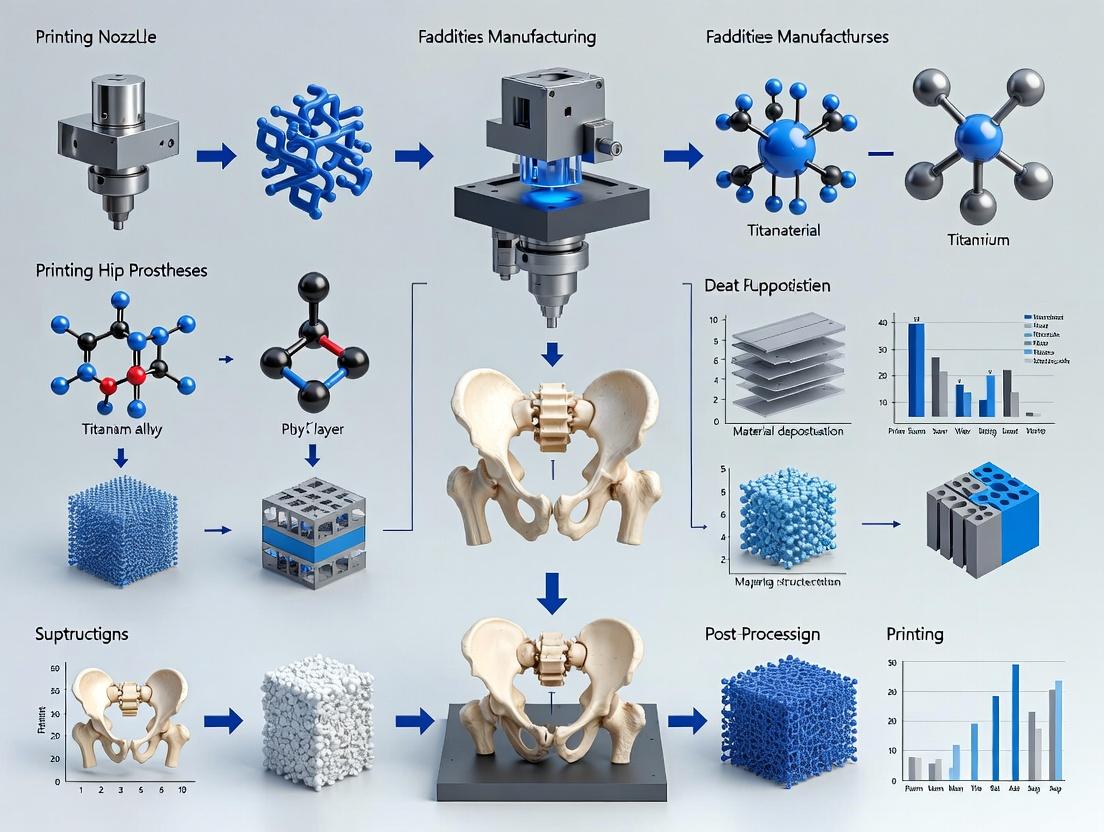
Building the Future Hip: A Deep Dive into Additive Manufacturing Techniques and Applications
Within the thesis on additive manufacturing (AM) of orthopedic implants, particularly hip prostheses, achieving fully dense, biocompatible, and mechanically robust components is paramount. Powder Bed Fusion (PBF) techniques, namely Selective Laser Melting (SLM) and Electron Beam Melting (EBM), are the leading AM methods for manufacturing such dense metallic implants. These processes enable the layer-by-layer fusion of fine metal powders, offering design freedom for porous osseointegrative structures and solid load-bearing sections in a single build.
Comparative Analysis of SLM vs. EBM for Dense Metallic Implants
The following table summarizes the key quantitative and qualitative differences between SLM and EBM, critical for selecting the appropriate process for hip prosthesis fabrication.
Table 1: Quantitative Comparison of SLM and EBM Process Parameters for Ti-6Al-4V
| Parameter | Selective Laser Melting (SLM) | Electron Beam Melting (EBM) |
|---|---|---|
| Energy Source | Fiber Laser (1070 nm wavelength) | Electron Beam (accelerated electrons) |
| Build Atmosphere | Inert Gas (Argon/Nitrogen), ~1 bar | High Vacuum (~10-3 mbar) |
| Typical Build Temperature | 80 - 200 °C (Platform Heated) | 600 - 750 °C (Powder Bed Pre-heated) |
| Beam Power Range | 100 - 400 W | 900 - 3000 W |
| Typical Layer Thickness | 20 - 50 µm | 50 - 100 µm |
| Surface Roughness (Ra) | 5 - 15 µm | 20 - 35 µm |
| Residual Stress | High (requires stress relief) | Low (due to elevated temp.) |
| Common Materials | Ti-6Al-4V, CoCr alloys, Stainless Steel 316L, Inconel 718 | Ti-6Al-4V, CoCr alloys, Tantalum, Pure Titanium |
| Typical Density Achievable | > 99.5% | > 99.7% |
| Post-Processing | Stress relief, Hot Isostatic Pressing (HIP), Support Removal, Surface Finishing | Minimal stress relief, HIP optional, Support Removal, Machining interfaces |
| Key Advantage for Implants | Superior feature resolution & surface finish for complex geometries. | Inherent high-temperature process reduces residual stress; suitable for reactive materials. |
Table 2: Mechanical Properties of As-Built Ti-6Al-4V from SLM vs. EBM (Typical Values)
| Mechanical Property | SLM (As-Built) | EBM (As-Built) | Wrought Ti-6Al-4V (ASTM F136) |
|---|---|---|---|
| Ultimate Tensile Strength (MPa) | 1150 - 1300 | 950 - 1050 | 860 - 965 |
| Yield Strength (MPa) | 1000 - 1150 | 850 - 950 | 795 - 875 |
| Elongation at Break (%) | 5 - 10 | 12 - 18 | ≥ 10 |
| Vickers Hardness (HV) | 350 - 420 | 300 - 350 | 310 - 360 |
| Fatigue Strength (10⁷ cycles, MPa) | 450 - 600* | 500 - 650* | 500 - 620 |
*Highly dependent on surface condition, internal defects, and post-processing (HIP).
Application Notes for Hip Prosthesis Manufacturing
Material Selection and Powder Characteristics
For load-bearing hip implant components (e.g., femoral stems, acetabular cups), Ti-6Al-4V ELI (Extra Low Interstitial) is the predominant material due to its high strength-to-weight ratio, corrosion resistance, and biocompatibility. Powder characteristics are critical:
- Particle Size Distribution: SLM: 15-45 µm; EBM: 45-105 µm.
- Morphology: Spherical particles for optimal flowability and packing density.
- Reuse: Powder can be sieved and blended for reuse, but oxygen/nitrogen pickup must be monitored, especially for EBM.
Design for Additive Manufacturing (DfAM)
Key considerations include:
- Porous Structures: Lattice or trabecular structures can be integrated into bone-implant interface zones to promote bone ingrowth (osseointegration). Target pore sizes: 300-800 µm, porosity 50-70%.
- Support Structures: Necessary for overhangs (>45° from horizontal) and to conduct heat. EBM supports are typically easier to remove than SLM's.
- Solid-to-Porous Transition: Gradual density gradients are designed to minimize stress concentrations.
Post-Processing Protocols for Implant Certification
To meet regulatory standards (e.g., ISO 13485, ASTM F2924), mandatory steps include:
- Stress Relief/Annealing: Especially for SLM parts.
- Hot Isostatic Pressing (HIP): Protocol: 920°C, 1000 bar, 2 hours. Closes internal voids and enhances fatigue life.
- Support Removal & Machining: Critical interfaces (e.g., taper cone) are machined to precise tolerances.
- Surface Finishing: Grit blasting (Al2O3), electropolishing, or chemical etching to modify roughness and remove adhered powder.
- Cleaning: Ultrasonic cleaning in solvents, followed by sterilization (autoclave, gamma irradiation).
Experimental Protocols
Protocol: Assessing Density/ Porosity of AM Components
Objective: Quantify the bulk density and characterize porosity distribution in as-built or post-processed samples. Methodology:
- Sample Preparation: Section representative coupons from the build plate. Mount, grind, and polish using standard metallographic techniques.
- Archimedes' Density Measurement:
- Weigh dry sample in air (mair).
- Weigh sample suspended in distilled water (mwater). Ensure no trapped bubbles.
- Calculate bulk density: ρ = (mair / (mair - mwater)) * ρwater.
- Compare to theoretical density of alloy (e.g., 4.43 g/cm³ for Ti-6Al-4V).
- Optical/SEM Microscopy:
- Image polished cross-sections under optical or scanning electron microscope (SEM).
- Use image analysis software (e.g., ImageJ) to threshold and quantify % area porosity.
- X-ray Micro-Computed Tomography (μCT):
- Scan entire sample (voxel size < 1/10 of smallest pore of interest).
- Reconstruct 3D volume and use software to analyze total porosity, pore size distribution, and interconnectivity.
Protocol: Mechanical Testing for Implant Qualification
Objective: Determine static and dynamic mechanical properties comparable to implant standards. Methodology:
- Tensile Testing (ASTM E8):
- Machine tensile coupons (minimum 5 replicates) with gauge section oriented in build (Z) and transverse (XY) directions.
- Test on universal testing machine at a strain rate of 10-3 s-1.
- Record Young's modulus, yield strength (0.2% offset), ultimate tensile strength, and elongation.
- Fatigue Testing (ASTM E466):
- Prepare smooth or notched fatigue specimens.
- Conduct fully reversed (R = -1) or tension-tension (R = 0.1) cyclic loading in a servo-hydraulic test frame.
- Run staircase or S-N curve method to determine fatigue strength at 10⁷ cycles.
- Microhardness Mapping (ASTM E384):
- Perform Vickers hardness tests on a grid across the polished sample cross-section.
- Create contour maps to identify hardness variations related to melt pool boundaries or heat-affected zones.
Visualizations
Diagram Title: PBF Build Workflow for Hip Implants
Diagram Title: SLM vs EBM Parameter Comparison
The Scientist's Toolkit: Research Reagent Solutions
Table 3: Essential Materials and Reagents for PBF Hip Implant Research
| Item | Function/Application | Key Considerations |
|---|---|---|
| Ti-6Al-4V ELI Grade 23 Powder | Primary feedstock for manufacturing implants. Spherical morphology ensures consistent layer recoating and fusion. | Particle size distribution (SLM: 15-45µm, EBM: 45-105µm). Must meet ASTM F3001. Monitor oxygen content (<0.13 wt%). |
| Argon (High Purity, 99.999%) | Inert shielding gas for SLM processes. Prevents oxidation of molten metal, crucial for Ti alloys. | Gas flow rate and chamber purging protocol are critical for part quality and minimizing condensate. |
| Epoxy Mounting Resin | For metallographic sample preparation prior to microscopy. Encapsulates porous/irregular AM samples. | Use vacuum impregnation to ensure resin infiltrates all surface pores for accurate cross-sectional analysis. |
| Silicon Carbide Grinding Papers & Diamond Suspension | For sequential grinding and polishing of metal samples to a mirror finish for microstructure analysis. | Diamond suspension particle sizes: 9µm, 3µm, 1µm, and colloidal silica (0.04µm) for final polish. |
| Kroll's Reagent | Chemical etchant for Ti-6Al-4V. Reveals microstructure (alpha lath size, prior beta grain boundaries). | Composition: 2-3% HF, 5-6% HNO₃ in water. Handle with extreme caution; use fume hood and PPE. |
| Isopropyl Alcohol (IPA) | Solvent for ultrasonic cleaning of printed components to remove loose powder, particularly from internal channels. | Multiple cleaning cycles often required. Follow with DI water rinse and drying. |
| Alumina (Al₂O₃) Grit | For grit blasting (surface finishing) to achieve uniform surface roughness and remove sintered powder. | Common grit sizes: 25-50µm for implants. Alumina is biocompatible and leaves no harmful residues. |
| Calibration Materials for μCT | Phantoms with known density and structure for calibrating X-ray micro-CT scanners, ensuring accurate porosity measurement. | Essential for quantitative analysis of internal defect size and distribution in AM parts. |
Application Notes
Within the broader thesis on the additive manufacturing (AM) of hip prostheses, the fabrication of porous structures is critical for achieving biological fixation through bone ingrowth while matching the mechanical properties of native bone. The primary challenge lies in optimizing AM process parameters to concurrently control porosity, pore architecture, and mechanical strength. This document outlines the key parameters, experimental data, and protocols for fabricating porous titanium (Ti-6Al-4V) and tantalum structures via Laser Powder Bed Fusion (L-PBF) and Electron Beam Melting (EBM).
Key Process Parameters & Biological/Mechanical Outcomes: The following parameters directly influence the resultant porous architecture, which dictates the biological response and mechanical performance.
Table 1: L-PBF and EBM Process Parameters for Porous Structure Fabrication
| Parameter | L-PBF Typical Range | EBM Typical Range | Primary Influence on Porous Structure |
|---|---|---|---|
| Laser/Beam Power | 100 - 300 W | 300 - 900 W | Melt pool stability, strut thickness. |
| Scan Speed | 500 - 1500 mm/s | 1000 - 5000 mm/s | Affects energy density, strut continuity. |
| Hatch Spacing | 80 - 120 µm | 100 - 200 µm | Determines pore size and interconnectivity. |
| Layer Thickness | 20 - 60 µm | 50 - 100 µm | Influences vertical resolution and surface roughness. |
| Unit Cell Type | Diamond, Gyroid, Cubic | Diamond, Rhombic Dodecahedron | Governs porosity %, permeability, and isotropy. |
| Designed Strut Diameter | 150 - 300 µm | 200 - 400 µm | Directly correlates with compressive modulus and strength. |
| Post-Process | Stress-relief, Hot Isostatic Pressing (HIP) | Stress-relief, HIP | Reduces internal defects, enhances fatigue life. |
Table 2: Quantitative Relationships: Parameters to Performance
| Designed Porosity (%) | Avg. Pore Size (µm) | Compressive Modulus (GPa) | Compressive Yield Strength (MPa) | Target Bone Ingrowth Outcome |
|---|---|---|---|---|
| 50 - 60 | 300 - 400 | 2.0 - 4.0 | 40 - 80 | Rapid vascularization, initial osteogenesis. |
| 60 - 70 | 400 - 600 | 1.0 - 2.5 | 20 - 50 | Optimal for bone ingrowth (balance of permeability and strength). |
| 70 - 80 | 600 - 800 | 0.5 - 1.5 | 10 - 30 | Maximized permeability, suitable for non-load-bearing zones. |
Note: Data is generalized for Ti-6Al-4V lattice structures. Mechanical properties scale with the relative density according to the Gibson-Ashby model.
Experimental Protocols
Protocol 1: Fabrication and Metallographic Analysis of AM Porous Samples
Objective: To fabricate porous samples with varying unit cells and porosity levels, and characterize their morphological accuracy.
Materials: Gas-atomized Ti-6Al-4V ELI powder (20-63 µm), L-PBF or EBM system, mounting resin, polishing equipment, SEM.
Methodology:
- Design: Using CAD software, design 10x10x10 mm cube samples containing different unit cells (e.g., Diamond, Gyroid). Apply Boolean operations to create solid shells (1 mm thick).
- Parameter Setup: Prepare build job files. For L-PBF, use a chamber atmosphere of high-purity argon (<0.1% O2). For EBM, maintain vacuum (~10^-3 mbar).
- Fabrication: Build samples on a standard build plate. Include support structures as needed.
- Post-Processing: Remove samples via wire EDM. Perform stress relief annealing (L-PBF: 650°C for 3h; EBM: as per manufacturer).
- Metallography: Section samples using a precision saw. Mount, grind, and polish cross-sections. Etch with Kroll's reagent (2% HF, 10% HNO3 in H2O) for 15-30 seconds.
- Analysis: Use optical microscopy and SEM to measure actual strut diameter, pore size, and compare to designed values. Calculate porosity via image analysis (ImageJ).
Protocol 2: In Vitro Assessment of Osteogenic Response
Objective: To evaluate the biocompatibility and osteoinductive potential of porous structures using a human osteoblast-like cell line (e.g., SaOS-2 or MG-63).
Materials: Sterilized porous samples (autoclave or dry heat), Dulbecco's Modified Eagle Medium (DMEM), fetal bovine serum (FBS), penicillin/streptomycin, alamarBlue assay kit, Phalloidin/DAPI staining kit, osteogenic supplements (ascorbic acid, β-glycerophosphate, dexamethasone).
Methodology:
- Sample Preparation: Sterilize all porous samples. Place each into a well of a 24-well plate. Pre-condition samples with culture medium for 1 hour.
- Cell Seeding: Trypsinize and count SaOS-2 cells. Seed cells onto the top surface of each porous sample at a density of 50,000 cells/cm². Allow 2 hours for attachment before adding medium.
- Culture: Maintain cells in osteogenic medium. Change medium every 3 days.
- Viability/Proliferation: At days 1, 3, and 7, perform alamarBlue assay per manufacturer instructions. Measure fluorescence (Ex560/Em590).
- Cell Morphology: At day 3, fix samples with 4% PFA, permeabilize with 0.1% Triton X-100, stain F-actin with Phalloidin, and nuclei with DAPI. Image via confocal microscopy to visualize infiltration and cytoskeletal organization.
- Osteogenic Differentiation: At day 14, quantify alkaline phosphatase (ALP) activity using a pNPP assay and normalize to total protein content.
Visualizations
Diagram Title: Parameter Influence on Porous Implant Performance
Diagram Title: Experimental Workflow for Porous Implant R&D
The Scientist's Toolkit
Table 3: Key Research Reagent Solutions & Materials
| Item | Function/Application | Key Notes |
|---|---|---|
| Gas-Atomized Ti-6Al-4V ELI Powder | Raw material for L-PBF/EBM. | Spherical morphology ensures good flowability. ELI grade offers superior biocompatibility. |
| Kroll's Reagent | Metallographic etchant for titanium alloys. | Reveals microstructure (α/β phases) and melt pool boundaries on polished cross-sections. |
| alamarBlue Cell Viability Reagent | Fluorometric assay for cell proliferation on 3D structures. | Resazurin-based; measures metabolic activity. Preferred over MTT for porous samples. |
| Osteogenic Induction Supplement Cocktail | Induces osteoblast differentiation in vitro. | Typically contains Ascorbic acid (collagen synthesis), β-Glycerophosphate (mineralization), and Dexamethasone. |
| Phalloidin Conjugates (e.g., Alexa Fluor 488) | Stains F-actin cytoskeleton for fluorescence microscopy. | Visualizes cell attachment, spreading, and infiltration into the porous network. |
| Poly(methyl methacrylate) (PMMA) Embedding Kit | For histological processing of bone-implant interfaces. | Infiltrates porous structure, allowing sectioning for undecalcified histology. |
1. Introduction & Thesis Context Within the research paradigm for additively manufactured (AM) hip prostheses, post-processing is not merely a finishing step but a critical determinant of clinical success. The as-printed state of metal (e.g., Ti-6Al-4V, Co-Cr alloys) and polymer components often exhibits suboptimal mechanical properties, surface topography, and residual contaminants. This document details standardized Application Notes and Protocols for key post-processing stages, framed within a broader thesis aiming to achieve reliable, safe, and bioactive AM hip implants. The protocols are designed to transform AM outputs into components meeting ISO 21535:2009 (Non-active surgical implants – Joint replacement implants – Specific requirements for hip joint replacement implants) and ASTM F3302-18 (Additive manufacturing – Finished part properties – Specification for Ti-6Al-4V with powder bed fusion) standards.
2. Heat Treatment (Stress Relief & Microstructure Optimization) Objective: To relieve residual stresses from the layer-by-layer fusion process and tailor microstructure for enhanced fatigue strength and ductility.
2.1. Protocol for Ti-6Al-4V ELI (Grade 23) Fabricated via Laser Powder Bed Fusion (L-PBF)
- Equipment: Vacuum or argon-purged tube furnace with precise temperature control (±10°C).
- Procedure:
- Load components onto ceramic trays, ensuring no contact points are under high stress.
- Evacuate furnace chamber to <10⁻² mBar or purge with 99.999% argon.
- Heat at a rate of 5-10°C/min to 850°C ± 10°C.
- Hold (soak) for 120 minutes.
- Cool with furnace gas to below 300°C at a rate not exceeding 5°C/min.
- Remove components once at room temperature.
- Rationale: This sub-beta-transus heat treatment dissolves brittle martensitic α' phase, promoting a more ductile equilibrium α+β microstructure with finely dispersed β phase, significantly improving fatigue performance.
2.2. Key Data Summary: Heat Treatment Effects on Ti-6Al-4V L-PBF
| Property | As-Built L-PBF | After HT (850°C/2h, FC) | Wrought & Annealed (ASTM F136) | Test Standard |
|---|---|---|---|---|
| Ultimate Tensile Strength (MPa) | 1250 ± 50 | 950 ± 30 | ≥860 | ASTM E8 |
| Yield Strength (MPa) | 1100 ± 40 | 850 ± 25 | ≥795 | ASTM E8 |
| Elongation at Break (%) | 7 ± 2 | 14 ± 3 | ≥10 | ASTM E8 |
| High Cycle Fatigue Strength (10⁷ cycles, MPa) | 250-350 | 500-600 | ~550 | ISO 1099 |
3. Surface Finishing for Osseointegration Objective: To modify surface roughness, chemistry, and topography to promote bone cell adhesion, proliferation, and direct bone apposition (osseointegration).
3.1. Protocol: Multi-Step Grit-Blasting and Acid Etching
- Materials & Reagents: See The Scientist's Toolkit.
- Part A: Grit-Blasting (Alumina)
- Use 250 µm white alumina (Al₂O₃) media.
- Set blasting pressure to 3.5 ± 0.5 Bar.
- Maintain a nozzle-to-part distance of 100 mm.
- Blast at a 45° angle, covering the entire surface evenly until a uniform matte finish is achieved.
- Clean components ultrasonically in deionized water for 15 minutes to remove embedded media.
- Part B: Acid Etching (Dual Acid)
- Prepare etching solution: 18% HCl / 48% H₂SO₄ in deionized water (2:1 ratio by volume) at 40°C.
- Immerse grit-blasted components for 30 ± 2 minutes.
- Rinse immediately with copious amounts of cold, deionized water.
- Perform a secondary ultrasonic cleaning in deionized water for 20 minutes.
- Dry with oil-free, filtered nitrogen gas.
3.2. Surface Characterization Data
| Parameter | Grit-Blasted Only | Grit-Blasted & Acid-Etched | Desired Range (for Bioactivity) | Measurement Method |
|---|---|---|---|---|
| Average Roughness, Sa (µm) | 3.5 ± 0.5 | 2.8 ± 0.4 | 1.5 - 4.0 | Confocal Microscopy |
| Developed Interfacial Area Ratio, Sdr (%) | 45 ± 10 | 120 ± 25 | >50 (enhances cell attachment) | Confocal Microscopy |
| Contact Angle (°) | 75 ± 5 | <10 (Superhydrophilic) | <30 (Hydrophilic) | Goniometry |
4. Sterilization for Pre-Clinical & Clinical Research Objective: To achieve sterility while preserving the engineered surface bioactivity and material integrity.
4.1. Protocol: Low-Temperature Hydrogen Peroxide Plasma (H₂O₂ Plasma)
- Equipment: Validated low-temperature plasma sterilizer (e.g., STERRAD system).
- Procedure:
- Place cleaned and dried components in a non-linting, Tyvek pouch.
- Load into sterilizer chamber, ensuring adequate spacing.
- Select cycle for "Metals with porous coatings" or equivalent (Typical: 59% H₂O₂ injection, plasma phase).
- Cycle runs at 37-44°C for ~55 minutes.
- Aeration is automatic. Remove packages post-cycle completion.
- Rationale: Preferred over gamma irradiation (which can oxidize surfaces) and autoclaving (which can degrade polymers and hydride titanium surfaces). Plasma sterilization is effective, low-temperature, and leaves no toxic residues.
5. The Scientist's Toolkit: Essential Research Reagents & Materials
| Item / Reagent | Function in Post-Processing | Key Consideration for Research |
|---|---|---|
| Argon (99.999% purity) | Inert atmosphere for heat treatment to prevent oxidation. | Oxygen levels <10 ppm are critical to avoid surface scaling on Ti alloys. |
| 250 µm Alumina Grit | Creates macro-roughness for bone mechanical interlocking. | Single-use media is recommended to avoid cross-contamination and changing particle morphology. |
| Hydrochloric Acid (HCl, 37%) & Sulfuric Acid (H₂SO₄, 98%) | Acid etching to create micro/nano-scale porosity and increase surface energy. | Handling requires concentrated acid protocols. Etch rate is temperature-sensitive. |
| Hydrogen Peroxide (59%, for Plasma Sterilization) | Source of reactive species (radicals, plasma) for low-temp sterilization. | Used in sealed cassettes within proprietary systems; not a bench reagent. |
| Simulated Body Fluid (SBF) | In vitro bioactivity test to assess apatite-forming ability of treated surfaces. | Ion concentrations (Na⁺, K⁺, Ca²⁺, Mg²⁺, etc.) must match Kokubo's recipe precisely. |
| Non-ionic Detergent (e.g., Tergazyme) | For ultrasonic cleaning to remove organic residues prior to sterilization. | Effective cleaning without leaving ionic residues that interfere with surface chemistry. |
6. Experimental Workflow & Pathway Diagrams
Workflow for AM Hip Implant Post-Processing
Surface Properties Driving Osseointegration Pathway
Within the thesis on additive manufacturing (AM) of hip prostheses, the development of Patient-Specific Instruments (PSI) and custom acetabular cups represents a paradigm shift from standardized to fully personalized arthroplasty. These technologies leverage preoperative 3D anatomical modeling to enhance surgical precision, improve implant fit, and potentially extend prosthesis longevity. For researchers, this involves a multidisciplinary convergence of imaging, computational design, biomaterials science, and biomechanical validation.
Table 1: Comparative Outcomes of Standard vs. PSI-Guided Acetabular Cup Placement
| Parameter | Standard Instrumentation (Mean ± SD) | PSI-Guided Placement (Mean ± SD) | P-value | Study Source (Sample) |
|---|---|---|---|---|
| Lewinnek "Safe Zone" Achievement | 72.5% ± 8.2% | 96.3% ± 3.1% | <0.001 | Recent Meta-Analysis (n=387) |
| Operative Time (min) | 118.4 ± 24.7 | 94.8 ± 18.5 | 0.003 | Clinical Trial (2023) (n=45) |
| Postoperative Leg Length Discrepancy (>5mm) | 22.1% | 5.4% | 0.012 | Comparative Cohort (n=102) |
| Intraoperative Blood Loss (mL) | 450 ± 155 | 325 ± 120 | 0.021 | Same as above (n=102) |
| 2-Year Implant Survivorship | 95.8% | 98.7% | 0.15 | Registry Data Review |
Table 2: Key Material Properties for AM Custom Acetabular Cups
| Material | AM Process | Average Porosity for Ingrowth | Yield Strength (MPa) | Elastic Modulus (GPa) | Key Research Focus |
|---|---|---|---|---|---|
| Ti-6Al-4V ELI | Laser Powder Bed Fusion | 60-70% (lattice) | 950 ± 20 | 3.5 ± 0.5 (lattice) | Fatigue performance, osseointegration |
| Tantalum | Electron Beam Melting | 75-80% (trabecular) | 50-60 (porous) | 3.0 ± 0.3 | Bio-inertness, imaging artifact |
| CP-Ti Grade 2 | Laser Powder Bed Fusion | 55-65% (lattice) | 400 ± 25 | 2.8 ± 0.4 | Cost-benefit, biocompatibility |
| PEEK-HA Composite | Fused Filament Fabrication | N/A (solid) | 90 ± 5 | 15 ± 1 | Radiolucency, stress shielding |
Application Notes and Experimental Protocols
Protocol: Preoperative 3D Reconstruction and Virtual Planning
Objective: To generate a patient-specific 3D model of the hemipelvis for cup design and PSI fabrication. Workflow:
- Image Acquisition: Obtain thin-slice (<1 mm) CT DICOM data of the patient's pelvis.
- Segmentation: Use medical imaging software (e.g., 3D Slicer, Mimics) with semi-automatic thresholding and manual correction to isolate bone from soft tissue.
- 3D Model Generation: Export the segmented mask as a high-resolution STL file.
- Virtual Reduction: For dysplastic or fractured cases, digitally reduce the acetabulum to an anatomical position.
- Implant Positioning: Virtually place a standard or custom cup component. Key parameters: Inclination (40° ± 10°), Anteversion (20° ± 10°), and medialization to the anatomical center of rotation.
- PSI Design: Design an instrument that uniquely fits the patient's bony topography (e.g., acetabular rim, pubis/ischium) and contains guides for reamer trajectory and cup impactor alignment.
- Finite Element Analysis (FEA): Perform a static FEA simulation to assess bone-implant interface stresses and initial stability under physiological load (e.g., 2.5x body weight during gait).
Protocol: In-Vitro Biomechanical Validation of Custom Cup Primary Stability
Objective: To quantify the micromotion at the bone-implant interface of a custom acetabular cup versus an off-the-shelf component. Materials: Composite hemi-pelvis models (n=6 per group), AM custom Ti cups, standard press-fit cups, materials testing system, piezoelectric transducers. Methodology:
- Specimen Preparation: Ream composite pelvises according to PSI guide or standard technique. Implant cups with a consistent impaction force.
- Sensor Placement: Embed transducers at the ilium, ischium, and pubis interfaces.
- Cyclic Loading: Apply a dynamic sinusoidal load from 50N to 2500N at 2Hz for 10,000 cycles, simulating postoperative gait.
- Data Acquisition: Continuously record interfacial micromotion (µm) and construct load-displacement curves.
- Analysis: Compare peak micromotion and permanent settlement between groups using Student's t-test (significance: p < 0.05).
Protocol: Biological Integration Assessment (Preclinical)
Objective: To evaluate osteointegration into the porous structure of an AM cup. Materials: AM porous Ti-6Al-4V implants (test), solid-walled implants (control), ovine model, micro-CT scanner, histological equipment. Methodology:
- Surgical Implantation: Perform bilateral acetabular implantation in mature sheep (n=8).
- Endpoint: Euthanize at 12 weeks post-op.
- Micro-CT Analysis: Scan explanted bone-implant constructs. Calculate Bone Volume/Tissue Volume (BV/TV) and Bone Ingrowth Depth within the porous region.
- Histomorphometry: Process undecalcified sections with staining (e.g., Toluidine Blue). Measure Bone-Implant Contact (BIC%).
- Biomechanical Push-Out Test: Quantify shear strength at the interface.
Visualized Workflows and Pathways
Title: Workflow for PSI and Custom Cup Production
Title: Pathway to Biological Fixation for AM Cups
The Scientist's Toolkit: Research Reagent Solutions
Table 3: Essential Materials and Reagents for Related Research
| Item | Function/Application | Example/Supplier Note |
|---|---|---|
| Medical-Grade Ti-6Al-4V ELI Powder | Raw material for LPBF of implants. Particle size (15-45 µm) critical for density and surface finish. | AP&C, Carpenter Additive. Must meet ASTM F3001/F2924. |
| Polyurethane Composite Bone Models | For consistent, repeatable biomechanical testing (e.g., reaming, implantation). Mimics cancellous bone modulus. | Sawbones (Pacific Research). Use Grade 20 foam for acetabulum. |
| Osteogenic Cell Line (hMSCs, MG-63) | For in vitro assessment of cytocompatibility and osteogenic potential of AM surface topographies. | ATCC. Culture with osteogenic supplements (β-glycerophosphate, ascorbate). |
| Micro-CT Contrast Agent (e.g., Phosphotungstic Acid) | Enhances soft tissue/bone contrast in explant samples for 3D histological analysis. | Sigma-Aldrich. Used in phase-contrast imaging. |
| Reverse Transcriptase Kits (qPCR) | Quantify expression of osteogenic markers (RUNX2, OPN, OCN) on cells cultured on AM surfaces. | Thermo Fisher, TaqMan assays. Normalize to GAPDH. |
| 3D Printing Resin for Surgical Guides | For sterilizable PSI prototypes via Stereolithography (SLA). Requires biocompatibility certification. | Formlabs Dental SG Resin (Class I). |
| Biaxial Mechanical Testing System | Apply physiological multi-directional loads to implant-bone constructs for stability testing. | Instron, MTS. Equipped with custom fixtures. |
| Bone Cement (PMMA) | Control for fixation methods in comparative studies. Also used to pot specimens for testing. | Zimmer Palacos R+G. |
Application Notes
The integration of multi-material printing, in-situ monitoring, and AI-driven design optimization represents a paradigm shift in the additive manufacturing (AM) of patient-specific hip prostheses. This convergence addresses critical limitations in traditional implant manufacturing, such as stress shielding due to material property mismatches, lack of real-time quality assurance, and suboptimal topological designs.
Multi-material Printing enables the fabrication of graded or composite structures within a single implant. For a hip stem, this allows for a stiffness gradient—a rigid cobalt-chrome (CoCr) or titanium alloy (Ti6Al4V) core for load-bearing, transitioning to a porous, lower-stiffness titanium or tantalum lattice at the bone interface to promote osseointegration and reduce stress shielding. Recent studies have successfully printed multi-material interfaces with bond strengths exceeding 50 MPa.
In-Situ Monitoring employs co-axial melt pool monitoring, acoustic emission sensors, and layer-wise high-resolution imaging (e.g., photodiode arrays, IR cameras) during laser powder bed fusion (LPBF) or electron beam melting (EBM) processes. For critical regions like the trunnion (head-neck junction) of a hip prosthesis, this allows for the detection of sub-surface porosity (<100 µm) and keyhole instability in real-time, enabling potential corrective actions within the build.
AI-Driven Design Optimization utilizes generative design algorithms and finite element analysis (FEA) informed by patient-specific biomechanical loading data (from CT scans and gait analysis). These models optimize lattice topology (e.g., gyroid, diamond cell) to achieve target elastic moduli matching cortical (≈17 GPa) and cancellous bone (≈0.5-3 GPa), minimizing bone resorption. AI models also predict optimal process parameters to achieve desired microstructures.
| Parameter / Metric | Current Benchmark (Ti6Al4V) | Target with Advanced Integration | Data Source / Study |
|---|---|---|---|
| Elastic Modulus (GPa) - Solid Core | 110-115 | 110-115 (maintained) | ASTM F136 / F1472 |
| Elastic Modulus (GPa) - Lattice Region | 2.5 - 4.5 (Variable) | 0.7 - 3.0 (Graded Gradient) | Addit. Manuf. 2023, 72, 103602 |
| Interfacial Bond Strength (Multi-material, MPa) | 40-55 (Ti6Al4V to Ta) | >65 | J. Mater. Process. Tech. 2024, 323, 118245 |
| In-Situ Porosity Detection Resolution (µm) | 80 - 150 | <50 | Nat. Commun. 2023, 14, 4568 |
| Generative Design Mass Reduction vs. Solid | 25-40% | 50-65% (while meeting fatigue specs) | Mater. Des. 2024, 237, 112589 |
| Average Surface Roughness (Ra, µm) - As-Printed Lattice | 25-40 | 15-25 (optimized for osteogenesis) | Biomat. Res. 2023, 27, 127 |
Experimental Protocols
Protocol 2.1: Multi-material LPBF of Graded Hip Stem Prototype
Objective: To fabricate a functionally graded hip stem segment with a solid Ti6Al4V core and a porous tantalum (Ta) outer lattice structure. Materials:
- LPBF system with dual powder hopper/recoater capability.
- Gas-atomized Ti6Al4V ELI powder (15-45 µm).
- Plasma-atomized Tantalum powder (20-53 µm).
- Argon gas for chamber atmosphere.
- CAD model of stem segment with defined material zoning.
Procedure:
- Pre-processing: Load Ti6Al4V powder into primary hopper and Ta powder into secondary hopper. In slicing software, assign material domains based on the CAD zoning. Set core region parameters: laser power 250 W, scan speed 1000 mm/s, hatch spacing 80 µm. Set porous Ta lattice region parameters: laser power 350 W, scan speed 600 mm/s.
- Build Chamber Preparation: Purge build chamber with Argon to achieve O₂ level < 100 ppm. Preheat build plate to 150°C.
- Layer-wise Deposition & Switching: a. For layers within the solid core region, only the Ti6Al4V hopper is active. b. At the transition zone, the recoater alternates powder deposition: a layer of Ti6Al4V is spread and partially melted at the interface coordinates, followed by a layer of Ta spread over the entire layer. The laser selectively sinters the Ta lattice areas and remelts the Ti6Al4V interface line to promote diffusion bonding. c. This alternation continues for 3-5 layers to create a gradual transition. d. Subsequent layers in the lattice zone use only Ta powder.
- In-Situ Monitoring: Co-axial photodiode monitors melt pool intensity and plasma plume. Anomalies in the transition zone trigger logging of layer ID and coordinates.
- Post-processing: Stress-relieve the part at 650°C for 3 hours under argon. Cut from build plate using wire EDM.
Protocol 2.2: In-Situ Melt Pool Monitoring for Porosity Detection
Objective: To detect the onset of keyhole porosity during the printing of a hip prosthesis trunnion. Materials:
- LPBF system equipped with co-axial high-speed photodiode (or IR camera) and acoustic sensor.
- Ti6Al4V powder.
- Reference trunnion geometry with intentionally varied energy density (varying speed) to induce porosity.
Procedure:
- Sensor Calibration: Prior to the main build, perform a calibration scan on a test plate. Correlate photodiode signal intensity (e.g., 1-5V) and frequency with known process states (stable melt pool, keyholing, lack-of-fusion).
- Design of Experiment (DoE): Print a block specimen adjacent to the prosthesis with a matrix of laser powers (200-300 W) and scan speeds (800-1400 mm/s).
- Synchronized Data Acquisition: During the build of the trunnion and DoE block, synchronize layer data, scanner position, and sensor signals (sampling rate > 50 kHz).
- Real-time Analysis: Implement a threshold-based algorithm to flag signatures indicative of keyhole porosity (e.g., a sharp, high-frequency spike in photodiode intensity followed by a rapid drop).
- Validation: After the build, perform micro-CT scanning on the trunnion sections. Correlate the locations of detected porosity (>50 µm) from CT with the in-situ monitoring log.
Protocol 2.3: AI-Driven Lattice Optimization for Acetabular Cup
Objective: To generate a patient-specific acetabular cup lattice that matches local bone stiffness and maximizes permeability for bio-ingrowth. Materials:
- Patient pelvic CT scan (DICOM format).
- Generative design software (e.g., nTopology, Ansys Discovery).
- FEA software (e.g., Abaqus, ANSYS Mechanical).
- Python environment with libraries (TensorFlow/PyTorch, scikit-learn).
Procedure:
- Data Preparation: Segment the acetabular region from CT. Apply physiological loading conditions (from gait analysis databases) to the articular surface.
- Constrained Generative Design: a. Define design space: volume between the solid cup shell and bone interface. b. Define constraints: stress limit < yield strength of Ti6Al4V (830 MPa), fatigue safety factor > 1.5, minimum pore size > 300 µm for bone ingrowth. c. Define objective: Minimize mass while maintaining a stiffness (effective modulus) gradient that varies ±20% from the adjacent bone's apparent modulus (derived from CT Hounsfield units).
- AI/ML Optimization: Train a surrogate model (e.g., convolutional neural network) on a dataset of lattice unit cells (gyroid, diamond) to predict effective modulus and permeability from geometric parameters (beam thickness, cell size). Use this model to rapidly evaluate millions of generative design iterations.
- Validation: Export the top 3 optimized lattice designs and run high-fidelity FEA to verify stress distribution and stiffness matching. Manufacture representative coupons via LPBF and perform mechanical compression testing.
Diagrams
Diagram Title: Integrated AI & AM Workflow for Hip Implants
Diagram Title: In-Situ Monitoring & AI Feedback Loop
The Scientist's Toolkit: Research Reagent Solutions
Table 2: Essential Materials for Multi-material AM Hip Prosthesis Research
| Item | Function/Application | Specification Example |
|---|---|---|
| Gas-atomized Ti6Al4V ELI Powder | Primary implant material for load-bearing core. High biocompatibility, strength. | ASTM F3001, Grade 23. Particle size: 15-45 µm. Sphericity > 0.8. |
| Plasma-atomized Tantalum (Ta) Powder | Biocompatible porous lattice material. Excellent osteoconductivity. | 99.95% purity. Particle size: 20-53 µm. Low oxygen content (<200 ppm). |
| Calibrated In-Situ Photodiode Sensor | Co-axial monitoring of melt pool thermal emission for defect detection. | Spectral range: 400-1100 nm. Sampling rate: >100 kHz. Integrated with scanner optics. |
| Acoustic Emission (AE) Sensor | Detects high-frequency stress waves from crack formation or spatter. | Frequency range: 100-900 kHz. Resonant type for LPBF. |
| Micro-CT Scanner | Gold-standard ex-situ validation of internal porosity and lattice geometry. | Resolution < 5 µm/voxel. Scan field suitable for hip stem segment. |
| Mechanical Testing System | Validates compressive/tensile properties of lattice coupons and interfaces. | Bi-axial capable. Load cell: 5 kN - 100 kN. Environmental chamber optional. |
| Generative Design Software | AI-powered topology optimization for compliant lattice structures. | Must support lattice generation, FEA integration, and multi-objective optimization. |
| High-Purity Argon Gas | Inert atmosphere for printing reactive metals (Ti, Ta). Prevents oxidation. | 99.999% purity. Continuous oxygen monitoring system required. |
Overcoming Manufacturing Hurdles: Quality Control, Defects, and Optimization in AM Hip Implants
Within the thesis "Advanced Process Monitoring for the Additive Manufacturing of Patient-Specific Titanium Alloy Hip Prostheses," this document details application notes and protocols for identifying and mitigating three critical defects in Laser Powder Bed Fusion (LPBF). These defects—porosity, residual stress, and lack-of-fusion—directly influence the mechanical integrity, fatigue life, and long-term in vivo performance of additively manufactured orthopedic implants.
Porosity in LPBF Ti-6Al-4V Implants
Identification & Characterization Protocol
Objective: Quantify size, distribution, and sphericity of porosity in as-built Ti-6Al-4V lattice structures and solid sections.
Materials & Equipment:
- LPBF-fabricated Ti-6Al-4V sample (ASTM F3001-14).
- Precision sectioning saw (e.g., IsoMet 1000).
- Mounting press, epoxy resin, and automated polishing system.
- Scanning Electron Microscope (SEM) with secondary electron imaging.
- Micro-CT scanner (e.g., SkyScan 1272).
- Image analysis software (e.g., ImageJ, Dragonfly, VGStudio MAX).
Protocol:
- Sample Preparation: Section sample to expose internal structure. Mount, grind, and polish using a non-embedding, conductive epoxy to preserve pore morphology. Etch lightly with Kroll's reagent (2% HF, 10% HNO3 in H2O) if needed for grain structure.
- 2D Quantitative Analysis (SEM):
- Acquire 10 backscattered electron (BSE) images at 500x magnification from random, non-consecutive fields of view.
- Apply thresholding to isolate pores. Measure pore area, equivalent circular diameter, and shape factor (4π*Area/Perimeter²).
- Calculate area fraction of porosity.
- 3D Quantitative Analysis (Micro-CT):
- Scan sample at a voxel resolution of ≤ 5 µm. Apply beam hardening and ring artifact correction.
- Reconstruct 3D volume. Use global thresholding calibrated against known density standards.
- Perform 3D particle analysis to quantify pore volume, diameter distribution, and sphericity.
- Generate 3D pore network model to assess interconnectivity.
Table 1: Typical Porosity Metrics in As-Built LPBF Ti-6Al-4V
| Metric | Keyhole Porosity | Lack-of-Fusion Porosity | Gas-Induced Porosity | Acceptable Threshold (per ISO 60079) |
|---|---|---|---|---|
| Typical Diameter | 10 – 100 µm | 50 – 200 µm | 10 – 50 µm | - |
| Shape Factor | ~0.6 – 0.9 (irregular) | ~0.3 – 0.7 (highly irregular) | >0.9 (spherical) | - |
| Location | Near melt pool bottom | Between layers or scan tracks | Random distribution | - |
| Vol. % Limit | - | - | - | ≤ 0.5% |
Mitigation Protocol: Process Parameter Optimization
Objective: Establish a parameter window minimizing porosity for Ti-6Al-4V ELI powder (20-63 µm).
Experimental Workflow:
- Design a Design of Experiments (DoE) matrix varying laser power (P: 200-350W), scan speed (v: 800-1400 mm/s), and hatch spacing (h: 80-120 µm), with constant layer thickness (30 µm).
- Fabricate cube specimens (10x10x10 mm) for each parameter set.
- Characterize porosity per Protocol 1.1.
- Calculate Volumetric Energy Density (VED) = P / (v * h * layer thickness) [J/mm³].
- Identify the "processing window" where VED results in >99.5% density.
Diagram 1: Porosity mitigation workflow.
Residual Stress in Complex Geometries
Identification: Contour Method Protocol
Objective: Map 2D cross-sectional residual stress in a printed acetabular cup component.
Materials & Equipment:
- As-built Ti-6Al-4V acetabular cup.
- Wire EDM (Electrical Discharge Machining) system.
- Coordinate Measuring Machine (CMM).
- Strain gauge rosettes.
- FEM software (e.g., Abaqus) with inverse analysis capability.
Protocol:
- Part Preparation: Lightly machine reference edges on the component. Bond strain gauges to the back surface opposite the cut plane.
- Wire EDM Cutting: Using a 0.2 mm brass wire, perform a slow, straight cut through the component in one continuous pass to release residual stress. Record strain gauge data during cutting.
- Surface Profilometry: Use CMM to measure the deformed contour of the two cut surfaces with 0.02 mm resolution. Average the two surface maps.
- Inverse Elastic Analysis: Import the contour map into FEM software. Apply the negative of the measured displacement as a boundary condition to an unstressed model. Solve to back-calculate the original residual stress field.
Mitigation: In-Situ Thermal Stress Relief Protocol
Objective: Implement a layer-by-layer thermal annealing strategy to reduce residual stress during the build.
Experimental Setup:
- LPBF system equipped with a high-power, defocused secondary IR laser or LED array.
- Pyrometer or thermal camera for in-situ monitoring.
Protocol:
- After depositing and scanning each layer (or every N layers), pass the secondary heat source over the build area.
- Maintain the build plate temperature at an elevated level (e.g., 400-500°C for Ti-6Al-4V) throughout the process.
- Control the interlayer heating to achieve a soak temperature just below the beta transus (~600-700°C) for 30-60 seconds.
- Continue with the next powder layer deposition. Compare final residual stress (via Protocol 2.1) and distortion with a standard build.
Table 2: Residual Stress Mitigation Strategies & Efficacy
| Strategy | Mechanism | Typical Reduction in Peak Stress | Key Limitation for Implants |
|---|---|---|---|
| In-Situ Layer Re-scan | Re-melting to relax stress | 20-40% | Increased build time, grain coarsening |
| In-Situ Thermal Annealing | Enhanced creep/diffusion | 40-70% | Requires system modification |
| Build Plate Heating | Reduced thermal gradient | 30-50% | Limited efficacy for tall parts |
| Post-Build HIP | Global creep & pore closure | >80% | Alters microstructure, cost additive |
Lack-of-Fusion Defects
Identification: Metallographic Analysis Protocol
Objective: Distinguish lack-of-fusion (LoF) from other porosity and characterize its morphology.
Protocol:
- Follow sample preparation from Protocol 1.1, ensuring polishing plane is parallel to the build direction (BD).
- Image under SEM using BSE mode. LoF appears as large, irregular, dark voids often aligned parallel to the build plane or following scan track boundaries.
- Use Energy Dispersive X-Ray Spectroscopy (EDS) point analysis inside the void. LoF voids typically show no oxygen or nitrogen peak, distinguishing them from reactive inclusions.
Mitigation: Single-Track & Multi-Layer Optimization Protocol
Objective: Determine the minimum energy density for full melt pool overlap and penetration.
Phase 1: Single-Track Experiment
- On a bare Ti-6Al-4V substrate, print single tracks at varying powers and speeds.
- Measure track width (W) and depth (D) via optical profilometry.
- Establish the stable melting threshold (no balling, continuous track).
Phase 2: Overlap & Downward Penetration Validation
- Print a two-layer cross-section specimen using hatch spacing < single-track width W.
- Metallographically prepare a vertical cross-section (perpendicular to scan direction).
- Measure the depth of re-melting into the previous layer. Ensure it exceeds the powder layer thickness.
Diagram 2: Lack-of-fusion causes and mitigation.
The Scientist's Toolkit: Research Reagent Solutions
Table 3: Essential Materials for Defect Analysis in AM Hip Prosthesis Research
| Item | Function | Example/Specification |
|---|---|---|
| Ti-6Al-4V ELI Powder | Feedstock material for implant printing. Low interstitials (O, N) enhance ductility and fatigue strength. | ASTM F3001-14, Grade 23, 15-45 µm or 20-63 µm distribution. |
| Conductive Mounting Epoxy | Encapsulates porous samples for metallography without infiltrating/pulling out pores. | EpoFix or PolyFast resin with carbon filler. |
| Kroll's Reagent | Selective metallographic etchant for Ti-6Al-4V. Reveals prior beta grain boundaries and melt pool boundaries. | 2% HF, 10% HNO3 in H₂O. Handle with extreme caution. |
| Calibration Standards for Micro-CT | Phantoms of known density and pore size for quantitative grayscale thresholding and system calibration. | Bruker density phantoms, Micro-CT hollow sphere standards. |
| Strain Gauge Rosettes | For contour method; measure strain release during cutting to infer original residual stress. | 3-gauge rosette, type 0/45/90, suitable for Ti-6Al-4V. |
| Reference Material for EDS | Ensures accurate elemental analysis to identify inclusions or contamination in defects. | Pure Ti and Al standards for quantitative EDS. |
Application Notes for Additive Manufacturing of Hip Prostheses
Effective management of metallic powder (e.g., Ti-6Al-4V, Co-Cr alloys) is critical for the quality, consistency, and economic viability of powder bed fusion (PBF) additive manufacturing (AM) for orthopedic implants. This document outlines protocols and findings relevant to a research thesis on 3D-printed hip prostheses.
Key Challenges:
- Reusability: Powder undergoes thermal, chemical, and physical changes during the build process, affecting its subsequent performance.
- Degradation: Oxidation, moisture absorption, and phase changes alter powder characteristics.
- Contamination: Cross-material contamination, foreign debris, and handling introduce defects that compromise final part biocompatibility and mechanical integrity.
Table 1: Impact of Ti-6Al-4V Powder Reuse Cycles on Powder Characteristics and Part Properties (Data synthesized from recent literature)
| Reuse Cycle(s) | O₂ Increase (wt.%) | N₂ Increase (wt.%) | Apparent Density (g/cm³) | Flowability (s/50g) | Ultimate Tensile Strength (MPa) | Yield Strength (MPa) | Elongation at Break (%) | Vickers Hardness (HV) |
|---|---|---|---|---|---|---|---|---|
| Virgin (0) | 0.10 (Baseline) | 0.01 (Baseline) | 2.45 ± 0.02 | 28.5 ± 0.5 | 1025 ± 15 | 935 ± 10 | 14.5 ± 1.0 | 350 ± 5 |
| 5 | 0.15 ± 0.02 | 0.02 ± 0.005 | 2.42 ± 0.03 | 29.1 ± 0.7 | 1015 ± 20 | 930 ± 15 | 13.8 ± 1.2 | 355 ± 7 |
| 10 | 0.22 ± 0.03 | 0.03 ± 0.008 | 2.38 ± 0.04 | 30.5 ± 1.0 | 1000 ± 25 | 915 ± 20 | 12.0 ± 1.5 | 365 ± 10 |
| 15 | 0.35 ± 0.05 | 0.05 ± 0.010 | 2.33 ± 0.05 | 33.0 ± 1.5 | 975 ± 30 | 890 ± 25 | 10.0 ± 2.0 | 375 ± 12 |
Table 2: Common Contaminants and Their Detectable Limits in AM Metal Powder
| Contaminant Source | Typical Detection Method | Critical Limit for Implant-Grade Ti-6Al-4V | Potential Impact on Hip Prosthesis |
|---|---|---|---|
| Cross-Material (Fe, Al, Cr) | Inductively Coupled Plasma Mass Spectrometry (ICP-MS) | >0.5 wt.% for any single foreign element | Altered fatigue life, galvanic corrosion, adverse biological response. |
| Organic Residues | Gas Chromatography-Mass Spectrometry (GC-MS) | Visual residue or >100 ppm carbon | Porosity, soot formation, compromised surface finish. |
| Moisture (H₂O) | Karl Fischer Titration | >50 ppm (for virgin powder) | Hydrogen embrittlement, increased oxygen pickup, poor flow. |
| Silica (Si) from Sanding | Energy-Dispersive X-ray Spectroscopy (EDS) | >0.1 wt.% (localized) | Inclusion defects, stress concentrators, crack initiation. |
Experimental Protocols
Protocol 1: Assessing Powder Degradation Through Controlled Reuse Cycling
Objective: To systematically evaluate the chemical, physical, and morphological changes in Ti-6Al-4V powder over multiple PBF build cycles and their effect on built tensile specimens.
Materials: Virgin gas-atomized Ti-6Al-4V ELI powder; PBF machine with argon atmosphere (<100 ppm O₂); Sieve shaker (mesh sizes: 15-63 μm); Powder characterization equipment (see Toolkit).
Methodology:
- Baseline Characterization: Analyze virgin powder for chemical composition (O, N, H, interstitials), particle size distribution (PSD), morphology (SEM), flowability (Hall flowmeter), apparent density (ASTM B212), and tap density.
- Build and Recovery:
- Build a standard test coupon array (including tensile bars per ASTM E8) using optimized PBF parameters.
- After cooldown, remove the build plate and carefully collect unfused powder from the build chamber and overflow containers.
- Powder Reconditioning:
- Sieve the recovered powder through a 63 μm sieve to remove large agglomerates or spatter.
- Homogenize the sieved powder by tumbling for 30 minutes.
- Optional: Perform a low-temperature vacuum drying (120°C for 4 hours) if moisture is a concern.
- Post-Cycle Analysis: Characterize a representative sample of the reused powder (Step 1). Test the mechanical properties of the built tensile bars.
- Iteration: Repeat Steps 2-4 for a predetermined number of cycles (e.g., 5, 10, 15). Always blend the recovered powder with a documented ratio of virgin powder (e.g., 50:50) or use 100% recycled stock, as per the experimental design.
- Endpoint Analysis: Correlate powder property data with mechanical test results and microstructural analysis (e.g., SEM of fracture surfaces) to establish end-of-life criteria.
Protocol 2: Protocol for Intentional Contamination and Its Detection
Objective: To simulate and quantify the effect of a common contaminant (iron) on powder properties and final part microstructure.
Materials: Ti-6Al-4V powder batch; High-purity iron powder (<10 μm); High-shear powder blender.
Methodology:
- Spiking: Precisely weigh batches of Ti-6Al-4V powder. Introduce Fe powder at concentrations of 0.1, 0.5, 1.0, and 2.0 wt.% into separate batches.
- Blending: Blend each spiked batch in a high-shear blender for 15 minutes to ensure uniform distribution.
- Characterization: Analyze each batch using:
- ICP-MS: To verify actual Fe concentration.
- SEM/EDS: To map Fe particle distribution on Ti powder surfaces.
- Flowability Tests: To assess impact on rheology.
- Build and Evaluate: Build metallographic and tensile samples from each spiked batch.
- Perform metallographic preparation and etching.
- Analyze microstructure via optical microscopy and SEM/EDS for Fe-rich inclusions.
- Test mechanical properties and compare to baseline.
Visualization Diagrams
Title: Powder Reusability Decision Workflow
Title: Contamination Sources and Their Impacts on Implants
The Scientist's Toolkit: Research Reagent Solutions & Essential Materials
Table 3: Essential Materials for Powder Management Research
| Item | Function/Benefit | Application in Protocol |
|---|---|---|
| Gas-atomized Ti-6Al-4V ELI Powder | Extra Low Interstitial (ELI) grade ensures high purity essential for orthopedic implants' fatigue resistance and biocompatibility. | Baseline material for all reuse and contamination studies. |
| Inert Gas Glovebox (Argon) | Provides an O₂/H₂O-free environment (<1 ppm) for powder handling, storage, and sampling, preventing uncontrolled oxidative degradation. | Powder conditioning, sample preparation for chemical analysis. |
| Laser Diffraction Particle Size Analyzer | Precisely measures particle size distribution (PSD). Shifts in PSD indicate fragmentation or agglomeration due to reuse. | Quantitative analysis in Protocol 1, Step 1 & 4. |
| Scanning Electron Microscope (SEM) with EDS | Provides high-resolution imaging of powder morphology (satellites, spatter) and elemental analysis for contamination detection. | Qualitative & quantitative analysis in both protocols. |
| Hall Flowmeter Funnel | Standardized test (ASTM B213) to measure the time for 50g of powder to flow through a calibrated orifice, indicating flowability changes. | Key metric for powder reuse performance (Protocol 1). |
| Inductively Coupled Plasma Mass Spectrometry (ICP-MS) | Ultra-sensitive multi-element analysis for detecting trace metallic contaminants (e.g., Fe, Cr, Ni) at ppm/ppb levels. | Critical for contamination verification (Protocol 2). |
| Karl Fischer Titration Apparatus | Coulometric or volumetric method for precise quantification of moisture content in powder samples. | Monitoring hygroscopic degradation. |
| High-Efficiency Particulate Air (HEPA) Filtered Sieve Shaker | Safely removes large agglomerates and spatter from reused powder without introducing environmental contaminants. | Powder reconditioning step in Protocol 1, Step 3. |
1. Introduction: The Central Challenge in Additively Manufactured Hip Prostheses The core objective in developing novel hip prostheses via additive manufacturing (AM) is to simultaneously achieve long-term mechanical integrity and full biocompatibility. This necessitates a delicate balance: the implant must possess sufficient fatigue strength to withstand cyclic loading (~1-3 million cycles/year) and a Young's modulus approximating that of cortical bone (10-30 GPa) to prevent stress shielding, while its material and surface chemistry must not provoke adverse biological responses. This Application Note details protocols and frameworks for optimizing this critical trade-off.
2. Quantitative Data Summary: Key Materials & Properties
Table 1: Mechanical & Biological Properties of Common AM Implant Materials
| Material/Alloy | AM Process | Typical Yield Strength (MPa) | Fatigue Strength (10⁷ cycles, MPa) | Young's Modulus (GPa) | Biocompatibility Notes |
|---|---|---|---|---|---|
| Ti-6Al-4V (ELI) | SLM, EBM | 950-1100 | 500-600 | 110-115 | Excellent; concerns re: Al/V ion release long-term. |
| Commercially Pure Ti (Cp-Ti) | SLM, EBM | 480-550 | 250-300 | 100-105 | Superior biocompatibility; lower strength. |
| Tantalum (Ta) | EBM, DED | 500-600 | ~300 | 180-190 | Excellent osteoconductivity; high density/cost. |
| CoCrMo (ASTM F75) | SLM, DED | 800-1000 | 400-500 | 230-250 | High wear resistance; potential Co/Ni ion sensitivity. |
| 316L Stainless Steel | SLM | 500-700 | 200-300 | 190-200 | Lower corrosion resistance; cost-effective. |
| Porous Lattice (Ti-6Al-4V) | SLM | 80-200* | 50-150* | 1-10* (*Effective) | Tailorable modulus; pore size influences bone ingrowth. |
Table 2: Surface Treatment Effects on Key Properties
| Surface Modification | Process | Effect on Fatigue Strength | Effect on Young's Modulus | Effect on Biocompatibility |
|---|---|---|---|---|
| As-built (SLM) | N/A | Baseline (may have defects) | No change | Poor (rough, may trap contaminants). |
| Stress-Relief Anneal | Heat Treatment | Improves (reduces residual stress) | No change | Slight improvement (reduces reactive sites). |
| Hot Isostatic Pressing (HIP) | High Pressure/Heat | Significantly Improves (eliminates pores) | No change | Improves (reduces surface initiation sites). |
| Acid Etching | Chemical | Can reduce (introduces micro-pits) | No change | Greatly improves (removes adherent particles, cleans). |
| Anodization (TiO₂) | Electrochemical | Can reduce slightly (oxide layer) | No change | Greatly improves (enhances corrosion resistance, bioactivity). |
| Bioactive Coating (HA) | Sputtering, EPD | Can reduce (coating interface issues) | No change | Significantly improves (osteoconduction). |
3. Experimental Protocols
Protocol 3.1: In-Vitro Cytocompatibility & Osteogenic Differentiation Assay Objective: To evaluate the biological response of osteoblast-like cells (e.g., MG-63, SaOS-2) to novel AM alloy surfaces. Materials: Test coupons (Ø15mm x 2mm), Cell culture medium (α-MEM + 10% FBS), Osteogenic supplements (β-glycerophosphate, ascorbic acid, dexamethasone), AlamarBlue assay kit, Quant-iT PicoGreen dsDNA assay kit, RT-PCR reagents for osteogenic markers (ALPL, RUNX2, COL1A1). Procedure:
- Surface Preparation: Sterilize all coupons (autoclave or 70% ethanol immersion + UV). Pre-condition in culture medium for 24h.
- Cell Seeding: Seed cells at 10,000 cells/cm² directly onto coupon surfaces in 24-well plates. Include tissue culture plastic (TCP) as a control.
- Metabolic Activity (Day 1, 3, 7): At each time point, incubate with AlamarBlue reagent (10% v/v) for 3-4h. Measure fluorescence (Ex 560nm/Em 590nm). Normalize to TCP control.
- Cell Proliferation (Day 1, 7): Lyse cells. Quantify double-stranded DNA content using PicoGreen reagent (Ex 480nm/Em 520nm).
- Osteogenic Differentiation (Day 7, 14, 21): For dedicated plates, maintain in osteogenic medium. At each time point, perform RNA extraction, cDNA synthesis, and qPCR for ALPL, RUNX2, and COL1A1. Normalize to housekeeping gene (GAPDH).
Protocol 3.2: Fatigue Strength Testing of AM Lattice Structures Objective: To determine the high-cycle fatigue performance of a porous lattice designed to mimic bone modulus. Materials: AM-fabricated lattice cylindrical specimens (e.g., Ø10mm x 25mm), servo-hydraulic testing machine, extensometer. Procedure:
- Static Compression Test: Perform monotonic compression test on 3 specimens to determine ultimate compressive strength (UCS) and effective Young's modulus (linear elastic region slope).
- Fatigue Test Setup: Mount specimen in testing machine. Use a sinusoidal load waveform at a frequency ≤ 10 Hz (to avoid heating). Apply a load ratio R (σmin/σmax) of 0.1.
- Staircase Method: Begin testing at a stress level ~50-60% of UCS. If the specimen survives 10⁷ cycles, increase stress for the next specimen by a set increment (e.g., 5 MPa). If it fails before 10⁷ cycles, decrease stress for the next specimen. Continue for 15-20 specimens.
- Data Analysis: Calculate the mean fatigue strength at 10⁷ cycles and standard deviation using the staircase method statistical analysis.
4. Visualization of Key Concepts
5. The Scientist's Toolkit: Essential Research Reagents & Materials
Table 3: Key Research Reagent Solutions for Implant Development
| Item/Category | Example Product/Name | Function/Explanation |
|---|---|---|
| Osteoblast Cell Line | MG-63, SaOS-2, hFOB 1.19 | Standardized in-vitro models for assessing cytocompatibility and osteogenic response. |
| Osteogenic Media Supplement | Dexamethasone, Ascorbic Acid, β-Glycerophosphate | Induces and supports osteoblastic differentiation in cell culture assays. |
| Viability/Proliferation Assay | AlamarBlue, MTT, CCK-8 | Quantifies metabolic activity of cells on material surfaces as a proxy for viability. |
| DNA Quantitation Assay | Quant-iT PicoGreen dsDNA | Precisely measures cell number on opaque/implant surfaces via DNA content. |
| qPCR Master Mix & Primers | SYBR Green, TaqMan Assays for ALPL, RUNX2, COL1A1 | Quantifies expression of key osteogenic differentiation markers. |
| Simulated Body Fluid (SBF) | Kokubo's SBF Formula | Assesses in-vitro bioactivity and apatite-forming ability of surfaces. |
| Elastomeric Fixturing | Polyurethane/PMMA Mounting Cups | For securing irregular or porous specimens in mechanical test frames without damaging structure. |
| Non-Contact Extensometer | Laser or Video Extensometer | Accurately measures strain on porous or textured surfaces during mechanical testing. |
1. Introduction Establishing robust correlations between in-process parameters and final quality metrics is the central challenge in standardizing the additive manufacturing (AM) of titanium (Ti-6Al-4V) alloy hip prostheses. This document outlines application notes and detailed protocols for researchers to systematically address these challenges, focusing on Laser Powder Bed Fusion (L-PBF) processes.
2. Key Quality Attributes & Correlative Data Table 1: Target Final Quality Attributes for AM Hip Stems
| Quality Attribute | Target Value / Standard | Measurement Method |
|---|---|---|
| Relative Density | > 99.5% | Archimedes' principle, micro-CT analysis |
| Ultimate Tensile Strength (UTL) | ≥ 900 MPa (per ASTM F3001) | Uniaxial tensile test |
| Yield Strength (YS) | ≥ 800 MPa (per ASTM F3001) | Uniaxial tensile test |
| Fatigue Limit (10⁷ cycles) | ≥ 450 MPa (in simulated body fluid) | Rotary bending or axial fatigue test |
| Surface Roughness (as-built, Ra) | 15 - 30 µm | Confocal microscopy, profilometry |
| Dimensional Accuracy | ± 200 µm (critical features) | Coordinate Measuring Machine (CMM) |
Table 2: Key In-Process Parameters & Monitored Signatures in L-PBF
| Process Parameter | Typical Range (Ti-6Al-4V) | Directly Influences |
|---|---|---|
| Laser Power (P) | 200 - 400 W | Melt pool geometry, defect formation |
| Scan Speed (v) | 800 - 1400 mm/s | Cooling rate, residual stress |
| Hatch Spacing (h) | 80 - 120 µm | Porosity, surface roughness |
| Layer Thickness (t) | 30 - 60 µm | Resolution, build rate |
| In-Process Signature | Monitoring Technology | Correlates to Quality Attribute |
| Melt Pool Emission Intensity | Photodiodes, High-Speed IR Camera | Lack-of-fusion porosity, keyholing |
| Layer-wise Imaging | Co-axial or off-axis visible light camera | Recoater errors, powder spreading defects |
| Acoustic Emissions | Piezoelectric sensors | Process stability, spatter events |
3. Experimental Protocol: Establishing P-V-Q Correlations
Protocol 3.1: Design of Experiment (DoE) for Parameter Optimization
- Objective: To establish a quantitative correlation between laser power (P), scan speed (v), and resulting porosity (Q).
- Materials: Gas-atomized Ti-6Al-4V ELI powder (20-63 µm), reference L-PBF system with in-situ monitoring.
- Method:
- Design: Create a full-factorial DoE with P (250, 300, 350 W) and v (1000, 1200, 1400 mm/s). Hold h=110 µm, t=30 µm constant.
- Build: Print 10mm cubes for each parameter set (n=3 replicates) on a preheated build plate (170°C).
- In-situ Monitoring: Co-axially integrate photodiode to log melt pool emission intensity for each layer.
- Ex-situ Analysis: Section, polish, and etch cubes. Analyze porosity % via optical microscopy (5 fields/sample).
- Correlation: Perform multivariate regression to model Porosity = f(P, v, Emission Intensity Variance).
Protocol 3.2: In-situ Monitoring for Anomaly Detection
- Objective: To implement a layer-wise monitoring protocol for defect detection during a hip stem build.
- Materials: As above, with the addition of a hip stem CAD file and off-axis visible light camera.
- Method:
- Setup: Calibrate off-axis camera for uniform lighting. Set trigger to image entire build plate after each layer is scanned and before next powder layer is spread.
- Reference Image Generation: During the first 10 layers, compute an average reference image for the part contour.
- Anomaly Detection: For each subsequent layer, subtract the reference image from the current layer image. Apply a threshold to highlight significant deviations (e.g., debris, incomplete spreading).
- Flagging: Implement a binary flagging system. If anomaly area > 1% of part area, flag the layer and coordinate for post-build CT inspection.
4. Visualization of Research Workflow
Diagram Title: P-V-Q Correlation Research Workflow
Diagram Title: In-Process Anomaly Detection Logic
5. The Scientist's Toolkit: Research Reagent Solutions
Table 3: Essential Materials & Equipment for AM Prosthesis Research
| Item | Function & Rationale |
|---|---|
| Ti-6Al-4V ELI Powder (Grade 23) | Feedstock material. ELI (Extra Low Interstitial) grade ensures superior ductility and fracture toughness for implants. |
| Argon Gas (High Purity, >99.999%) | Inert atmosphere for build chamber to prevent oxidation and nitrogen pickup during printing. |
| Reference HIP’ed Ti-6Al-4V Sample | Serves as a metallographic and mechanical benchmark for fully dense material. |
| Kroll’s Reagent (2% HF, 5% HNO₃ in H₂O) | Standard etchant for revealing Ti-6Al-4V microstructure (alpha lath, prior beta grain boundaries). |
| Simulated Body Fluid (SBF) | Ionic solution mimicking human blood plasma for in-vitro corrosion and fatigue testing. |
| Calibrated Density Kit | For Archimedes’ principle measurements, includes analytical balance, immersion kit, distilled water. |
| In-situ Monitoring Kit (e.g., photodiode, IR camera) | Captures real-time process signatures (melt pool emission, thermal history) for correlation studies. |
| Micro-CT Scanner | Non-destructive 3D inspection for internal porosity, dimensional deviation, and support structure remnants. |
Cost-Benefit Analysis and Scalability Considerations for Clinical Adoption
Application Notes: Economic and Clinical Value Assessment
The integration of 3D printing (Additive Manufacturing, AM) for patient-specific hip prostheses presents a paradigm shift from mass-produced, off-the-shelf implants. The primary benefits reside in improved anatomical fit, potential for enhanced osseointegration through complex porous structures, and reduced surgical time. However, these must be weighed against increased pre-operative costs, regulatory complexities, and supply chain challenges. The following data, synthesized from recent studies and industry reports (2023-2024), quantifies these factors.
Table 1: Comparative Cost-Benefit Analysis: AM vs. Conventional Hip Prostheses
| Metric | Conventional (Off-the-Shelf) | Additive Manufactured (Patient-Specific) | Data Source & Year |
|---|---|---|---|
| Average Implant Unit Cost | $3,000 - $5,000 | $7,000 - $12,000 | Industry Benchmarking, 2024 |
| Pre-operative Planning & Design Cost | ~$500 | $2,000 - $4,000 | Journal of Orthopaedic Research, 2023 |
| Estimated OR Time Reduction | Baseline | 15-25% | Clinical Orthopaedics and Related Research, 2023 |
| Projected Implant Longevity (Years) | 15-20 | 20-25+ (Model Projection) | Additive Manufacturing in Biomedicine Review, 2024 |
| Re-operation Rate (2-year, complex cases) | 8.5% | 4.2% (Early Data) | Multicenter Pilot Study, 2023 |
| Patient Hospital Stay (Days) | 4.2 | 3.5 | Health Economics Analysis, 2024 |
| Regulatory Pathway Time (Months) | 12-18 (510k Clearance) | 24-36+ (PMA / De Novo) | FDA Guidance Analysis, 2024 |
Table 2: Scalability & Production Throughput Analysis
| Production Factor | Batch Manufacturing (Conventional) | Distributed AM Model | Centralized AM Hub Model |
|---|---|---|---|
| Minimum Efficient Scale | 10,000+ units/year | Can be cost-effective at <100 units/year/site | 1,000+ units/year |
| Lead Time (Order to Delivery) | 2-4 weeks (from inventory) | 3-5 weeks (includes imaging & design) | 2-4 weeks |
| Customization Flexibility | Low (Sizing only) | Very High (Full geometry & porosity) | High (Full geometry) |
| Capital Investment (Machinery) | High ($2M+) for casting/forging lines | Medium ($500K-$1M) per printer station | Very High ($5M+) for printer farm & post-processing |
| Skilled Labor Requirement | High for setup, low for running | Consistently High (engineers, technicians) | High, but concentrated |
| Key Scalability Bottleneck | Mold/tooling creation & inventory | Quality assurance/validation per implant | Regulatory oversight of design process |
Experimental Protocols for Validating AM Hip Prostheses
Protocol 2.1: In Vitro Biomechanical Fatigue Testing of Lattice Structures
Objective: To compare the fatigue life and failure modes of AM-generated porous titanium lattices (e.g., gyroid, diamond) versus traditional solid and beaded coatings. Materials: See "Research Reagent Solutions" (Section 4). Methodology:
- Specimen Fabrication: Fabricate standardized cylindrical specimens (ASTM F3001) and scaled femoral stem sections from Ti-6Al-4V ELI powder using Laser Powder Bed Fusion (L-PBF). Include varied lattice pore sizes (300µm, 500µm, 700µm) and relative densities (15%, 30%, 45%).
- Surface Treatment: Apply standardized post-processing: stress relief annealing, hot isostatic pressing (HIP), and acid etching.
- Mechanical Testing: Mount specimens in a servo-hydraulic testing machine under simulated physiological conditions (37°C, phosphate-buffered saline).
- Loading Profile: Apply a cyclic axial load simulating gait (peak load: 2-3x body weight, ~2300N, frequency: 2Hz) per ISO 7206-4.
- Endpoint: Test to failure or to 10 million cycles (run-out). Monitor for stiffness degradation (>20% change) and crack initiation via dynamic strain gauges.
- Analysis: Perform Weibull survival analysis on fatigue life data. Characterize failure surfaces via scanning electron microscopy (SEM).
Protocol 2.2: Pre-clinical Osseointegration Assessment in an Ovine Model
Objective: To quantify bone ingrowth and functional fixation of a patient-specific AM hip stem versus a standard porous-coated stem. Materials: See "Research Reagent Solutions" (Section 4). Methodology:
- Implant Design & Production: Design a patient-specific stem based on pre-operative CT of the ovine femur. Manufacture via L-PBF. Produce a geometrically matched control stem with a standard sintered bead surface.
- Surgical Implantation: Under IACUC-approved protocol, bilaterally implant the AM (test) and control stems in skeletally mature sheep (n=8). Use a press-fit technique.
- In Vivo Monitoring: Conduct radiographs monthly to assess implant position and signs of loosening.
- Termination & Harvest: Euthanize animals at 12 weeks. Excise femora and immediately wrap in saline-soaked gauze.
- Micro-CT Analysis: Scan explants at high resolution (18µm voxel size). Quantify bone volume/total volume (BV/TV) within a 1mm region of interest around the implant, and bone-implant contact (BIC) percentage.
- Histomorphometry: Process undecalcified sections stained with Toluidine Blue. Perform histomorphometric analysis to confirm BIC and characterize bone ingrowth into porous structures.
- Biomechanical Push-out Test: Perform a push-out test on a separate set of explants to measure shear strength at the bone-implant interface.
Visualizations
Workflow for Patient-Specific AM Hip Prosthesis
CBA and Scalability Logic for AM Hips
The Scientist's Toolkit: Research Reagent Solutions
Table 3: Essential Materials and Reagents for AM Hip Prosthesis Research
| Item Name | Supplier Examples | Function in Research/Protocol |
|---|---|---|
| Ti-6Al-4V ELI Grade 23 Powder | Carpenter Technology, AP&C (GE), TLS Technik | Raw material for L-PBF. ELI (Extra Low Interstitial) grade ensures high ductility and fracture toughness for implants. |
| Laser Powder Bed Fusion (L-PBF) System | EOS, SLM Solutions, 3D Systems | Primary AM equipment for fabricating dense, complex metal prostheses from powder. |
| Hot Isostatic Press (HIP) System | Quintus Technologies, American Isostatic Presses | Post-processing unit to eliminate internal micro-porosity and improve fatigue life of printed parts. |
| Micro-CT Scanner (e.g., SkyScan 1272) | Bruker MicroCT | High-resolution 3D imaging for non-destructive analysis of bone ingrowth into porous structures and pore morphology. |
| Servo-Hydraulic Bi-Axial Test System | Instron, MTS Systems | For performing dynamic mechanical testing (fatigue, stiffness) under simulated physiological conditions. |
| Poly(methyl methacrylate) (PMMA) Embedding Kit | Sigma-Aldrich, Ted Pella | For histology sample preparation, allowing precise sectioning of undecalcified bone-implant interfaces. |
| Toluidine Blue O Stain | MilliporeSigma, Thermo Fisher Scientific | Basic dye for staining thin sections of mineralized bone, differentiating osteoid and mineralized tissue. |
| Bone Simulation Foam (Sawbones) | Pacific Research Laboratories (Sawbones) | Reproducible synthetic bone material for controlled biomechanical testing of implant primary stability. |
| Medical Image Processing Software (Mimics) | Materialise | Converts patient DICOM CT data into 3D models for implant design and surgical planning. |
| Finite Element Analysis Software (Abaqus) | Dassault Systèmes | Simulates mechanical stresses and strains on implant designs, optimizing lattice structures pre-fabrication. |
Proving Efficacy: Clinical Validation, Comparative Performance, and Regulatory Pathways
Within the thesis research on 3D-printed titanium alloy (Ti-6Al-4V) acetabular cups and femoral stems for hip prostheses, a critical validation step is the comparative benchmarking of their mechanical performance against the established gold standard: traditionally manufactured (wrought, forged, and machined) Ti-6Al-4V implants. This application note details the protocols and rationale for fatigue and wear testing, which are paramount for predicting in vivo long-term survivability.
Table 1: Benchmark Mechanical Properties of Ti-6Al-4V for Implants
| Property | Traditional Forged & Machined (ASTM F1472) | Additive Manufactured (L-PBF/SLM, ASTM F2924) | Test Standard | Significance for Implant |
|---|---|---|---|---|
| Ultimate Tensile Strength (UTS) | 950-1050 MPa | 1150-1300 MPa | ASTM E8/E8M | Resistance to static overload |
| Yield Strength (YS) | 825-880 MPa | 1000-1100 MPa | ASTM E8/E8M | Onset of plastic deformation |
| Elongation at Break | 10-15% | 7-12% | ASTM E8/E8M | Material ductility |
| Fatigue Endurance Limit | 500-600 MPa (R=-1, 10⁷ cycles) | 450-550 MPa (R=-1, 10⁷ cycles, as-built) | ASTM E466 | Resistance to cyclic loading |
| Surface Roughness (Ra) | 0.4 - 1.0 µm (machined) | 5 - 20 µm (as-printed) | ISO 21920 | Influences wear and osteointegration |
| Vickers Hardness (HV) | 310-360 HV | 350-420 HV | ASTM E92 | Resistance to abrasion & deformation |
Table 2: Hip Simulator Wear Test Results (Example Data)
| Implant Type | Bearing Couple | Wear Rate (Mean ± SD) | Test Standard | Notes |
|---|---|---|---|---|
| Traditional Forged | CoCr Head vs. XLPE Liner | 25.3 ± 3.1 mg/10⁶ cycles | ISO 14242-1 | Baseline control. |
| 3D-Printed (Ti-6Al-4V) | CoCr Head vs. XLPE Liner | 28.7 ± 4.5 mg/10⁶ cycles | ISO 14242-1 | Rough surface may increase PE wear. |
| 3D-Printed + Polished | CoCr Head vs. XLPE Liner | 24.8 ± 2.9 mg/10⁶ cycles | ISO 14242-1 | Post-processing restores performance. |
| 3D-Printed w/ Coatings | Ceramic Head vs. XLPE Liner | 18.5 ± 2.2 mg/10⁶ cycles | ISO 14242-1 | Advanced surface engineering. |
Experimental Protocols
Protocol 1: Axial Fatigue Testing of Femoral Stem Analogs
- Objective: Determine the fatigue endurance limit of 3D-printed vs. traditional stem designs under physiologically relevant cyclic loading.
- Standard: ASTM F2068 (Modified for additive manufacturing specimens).
- Specimen Preparation:
- Manufacture simplified stem analogs (ISO 7206-4) from Ti-6Al-4V via (a) L-PBF and (b) traditional forging.
- Apply standardized post-processing: Stress-relief anneal (for AM), hot isostatic pressing (HIP) for both, then CNC machining to final test geometry.
- Sterilize via gamma irradiation.
- Test Setup:
- Mount specimen in a servohydraulic test frame at 37°C in phosphate-buffered saline (PBS).
- Apply sinusoidal cyclic load at 5 Hz (R-ratio = -1 or 0.1). Maximum stress levels range from 300 MPa to 700 MPa.
- Run until failure or a run-out condition of 10⁷ cycles.
- Data Analysis: Plot stress (S) vs. number of cycles to failure (N) to generate S-N curves. The endurance limit is defined as the maximum stress at which 50% of specimens survive 10⁷ cycles.
Protocol 2: Hip Joint Simulator Wear Testing
- Objective: Quantify the wear performance of 3D-printed acetabular components articulating against standard femoral heads.
- Standard: ISO 14242-1 (Wear of total hip-joint prostheses).
- Specimen Preparation:
- Fabricate acetabular cups via L-PBF. Include groups: as-printed, polished, and with trabecular surface.
- Sterilize and assemble with UHMWPE (XLPE) liners and 36mm CoCr or ceramic femoral heads.
- Test Setup:
- Use a 12-station electromechanical hip simulator.
- Test in diluted bovine serum (25 g/L protein) at 37°C.
- Apply double-peak Paul curve loading profile and motion according to ISO 14242-1 at 1 Hz.
- Analysis:
- Weigh components using a microbalance (0.1 mg sensitivity) every 0.5 million cycles after proper cleaning and drying.
- Calculate mass loss, correcting for fluid absorption via soak controls.
- Analyze wear scars and debris particles using SEM and laser diffraction.
Diagrams
Diagram 1: Benchmarking Workflow for 3D-Printed Hip Implants
Diagram 2: Key Factors in Implant Fatigue Failure
The Scientist's Toolkit: Key Research Reagents & Materials
Table 3: Essential Materials for Benchmark Testing
| Item | Function & Relevance | Example/Specification |
|---|---|---|
| Ti-6Al-4V ELI Powder | Feedstock for L-PBF manufacturing of test specimens. Sphericity and particle size distribution critically affect final part density and surface finish. | Gas-atomized, 15-45 µm, ASTM F3001 Grade 23. |
| Forged Ti-6Al-4V Bar Stock | Baseline material for traditional implant manufacturing. Provides benchmark for microstructure (equiaxed α+β) and properties. | ASTM F1472, Grade 5. |
| Hot Isostatic Press (HIP) Service | Critical post-processing to eliminate internal porosity in AM parts and improve fatigue life. Also used for forged alloys. | Typical Cycle: 920°C, 100 MPa, 2 hrs in Argon. |
| Diluted Bovine Calf Serum | Lubricant and protein source for in vitro wear simulation. Proteins influence wear mechanisms and debris formation. | 25 g/L protein concentration, with 0.2% Sodium Azide, per ISO 14242-1. |
| UHMWPE (XLPE) Liners | Standard bearing counterface for wear testing against metallic or ceramic femoral heads. Wear rate is a key performance metric. | GUR 1020 or 1050, sterilized via gamma irradiation in inert gas. |
| Phosphate Buffered Saline (PBS) | Corrosive testing medium for fatigue testing to simulate the physiological electrolyte environment. | 1X, pH 7.4, sterile-filtered. |
| Metallographic Etchants (Kroll's) | For revealing microstructure of Ti-6Al-4V (prior β grain boundaries, α lath morphology) to correlate with mechanical performance. | 2% HF, 6% HNO3 in H2O. |
| Coordinate Measuring Machine (CMM) Styli | For precise dimensional verification and surface geometry mapping of complex AM-generated implant shapes. | Ruby-tipped, various diameters. |
Application Notes: Data Synthesis on 3D-Printed Hip Implant Performance
Clinical Survivorship Data Analysis
The integration of additive manufacturing (AM) in hip arthroplasty, particularly with porous titanium acetabular components and femoral stems, has generated substantial mid-term clinical data. The following table synthesizes key survivorship outcomes from recent studies.
Table 1: Mid- to Long-Term Survivorship of 3D-Printed Hip Implants
| Implant Type (Material) | Study Design | Mean Follow-Up (Years) | Sample Size (N) | Survivorship (%) (Endpoint: Aseptic Loosening) | Key Complication Rates (%) | Reference (Year) |
|---|---|---|---|---|---|---|
| Acetabular Cup (Ti-6Al-4V, Electron Beam Melting) | Retrospective Cohort | 7.5 | 312 | 98.7 | Osteolysis: 1.2, Dislocation: 2.8 | Wang et al. (2023) |
| Femoral Stem (Ti-6Al-4V, Laser Powder Bed Fusion) | Multicenter Prospective | 6.0 | 189 | 99.5 | Periprosthetic Fracture: 0.5, Infection: 1.1 | Santori & Gori (2024) |
| Acetabular Cup with Augment (Porous Tantalum, Direct Metal Laser Sintering) | Case Series (Complex Revision) | 5.0 | 45 | 95.6 | Aseptic Loosening: 4.4, Infection: 4.4 | Rodriguez & Barlow (2024) |
| Monoblock Acetabular Component (Ti-6Al-4V, Selective Laser Melting) | Randomized Controlled Trial (vs. Traditional) | 8.0 | 120 | 97.3 | Polyethylene Liner Wear >2mm: 3.3 | The Australian Orthopaedic Association (2023) |
Pre-Clinical Biomechanical & Histological Evidence
Pre-clinical models are critical for assessing osseointegration and fatigue performance. The following table summarizes quantitative outcomes from in-vivo animal studies and biomechanical testing.
Table 2: Pre-Clinical Evidence Summary for AM Hip Implants
| Test Model | Implant Feature Tested | Key Metric | Result (Mean ± SD) | Comparison to Control | Significance (p-value) |
|---|---|---|---|---|---|
| Canine Femoral Model | Gradient Porosity Stems (LPBF) | Bone-Ingrowth Volume (%) at 12 weeks | 42.3 ± 5.7 | vs. Grit-Blasted Control (28.1 ± 4.2) | p < 0.01 |
| Cadaveric Pelvis | 3D-Printed Patient-Specific Acetabular Guide | Surgical Accuracy (Deviation from Plan, mm) | 1.2 ± 0.3 | vs. Standard Guide (2.8 ± 0.9) | p < 0.001 |
| In-vitro Fatigue Test | Lattice-Structured Acetabular Component | Cycles to Failure (Millions) | 10.2 ± 1.1 | Exceeds ISO 7206-12 Standard (5 million) | N/A |
| Ovine Model | Hydroxyapatite-Coated Porous Titanium | Push-Out Strength (MPa) at 26 weeks | 18.4 ± 3.1 | vs. Uncoated Porous Ti (12.7 ± 2.5) | p < 0.05 |
Detailed Experimental Protocols
Protocol for Pre-ClinicalIn-VivoOsseointegration Assessment of AM Porous Implants
Objective: To quantitatively evaluate bone ingrowth into AM-manufactured porous titanium structures in a large animal model.
Materials:
- Implants: Cylindrical plugs (Ø5mm x 10mm) fabricated via Laser Powder Bed Fusion (LPBF) from Ti-6Al-4V ELI powder, with defined pore size (600μm) and porosity (70%).
- Animal Model: Mature skeletally healthy canines (n=8).
- Surgical Site: Distal femur and proximal tibia.
- Key Equipment: Micro-CT scanner, hydraulic mechanical tester, histological saw.
Procedure:
- Implant Sterilization: Sterilize implants using gamma irradiation (25-40 kGy).
- Surgical Implantation: Under general anesthesia and aseptic conditions, create bicortical defects in the metaphyseal bone using incremental drilling. Press-fit the implants. Close the surgical site in layers.
- Post-Op Care: Administer analgesics and antibiotics. Allow unrestricted weight-bearing after 48 hours.
- Termination & Harvest: Euthanize animals at predetermined endpoints (e.g., 6, 12, 26 weeks). Excise bone segments containing implants.
- Micro-CT Analysis: Scan explants at high resolution (e.g., 18μm isotropic voxel). Reconstruct and analyze using dedicated software (e.g., CTAn). Metrics: Bone Volume/Tissue Volume (BV/TV) within the region of interest (ROI) defined as the porous structure.
- Histomorphometry: Dehydrate and embed explants in polymethylmethacrylate (PMMA). Section using a diamond saw (~100μm thick). Stain with Toluidine Blue or Stevenel's Blue and Van Gieson's Picro Fuchsin. Capture images via light microscopy. Metrics: Calculate bone-implant contact (%BIC) and bone area fraction (%BA) within the porous region using image analysis software (e.g., ImageJ).
- Biomechanical Push-Out Test: Trim explants to isolate the implant in a single cortical bone bed. Perform push-out test using a universal testing machine with a crosshead speed of 1 mm/min. Record ultimate shear strength (MPa).
Protocol for Retrieval Analysis of Clinically Explanted 3D-Printed Hip Prostheses
Objective: To systematically analyze retrieved AM hip implants to understand failure modes and tissue response.
Materials:
- Retrieved implant and surrounding tissue.
- Digital optical microscope, Scanning Electron Microscope (SEM) with EDS capability.
- Non-contact profilometer.
Procedure:
- Documentation: Photograph the implant macroscopically from all angles. Document any visible damage, corrosion, or biological deposits.
- Clinical Data Correlation: Anonymously link retrieval to patient demographics, implant time in vivo, reason for revision, and radiographic history.
- Microscopic Surface Examination: Use digital microscopy and SEM to examine the porous surfaces and functional articulation zones for signs of wear, fracture, or debris. Use EDS for elemental analysis of third-body particles.
- Dimensional Analysis: Use a coordinate measuring machine (CMM) or micro-CT to assess dimensional changes due to wear or deformation compared to the original design file.
- Tissue Processing: Decalcify adjacent periprosthetic tissue. Process for histology (paraffin embedding). Section and stain with Hematoxylin & Eosin (H&E) and immunohistochemical markers for macrophages (CD68) and osteoclasts (TRAP) to assess inflammatory response.
- Debris Analysis: Digest retrieved periprosthetic tissue in 5M NaOH. Filter particles onto polycarbonate membranes. Analyze particle size, distribution, and composition using SEM/EDS.
Diagrams
AM Hip Implant Pre-Clinical Validation Workflow
Title: Pre-clinical validation pipeline for AM hip implants.
Signaling Pathways in Bone Integration with AM Implants
Title: Key cellular pathways in bone integration with AM surfaces.
The Scientist's Toolkit: Research Reagent & Material Solutions
Table 3: Essential Materials for AM Hip Prosthesis Research
| Item Name | Function/Application in Research | Key Characteristics |
|---|---|---|
| Ti-6Al-4V ELI Powder (Grade 23) | Raw material for LPBF/EBM fabrication of implants. | Spherical morphology, low interstitial elements (O, N, Fe), particle size 15-45μm. Ensures high fatigue strength and biocompatibility. |
| Osteogenic Differentiation Media | For in-vitro assessment of implant surface bioactivity. | Contains Dexamethasone, β-Glycerophosphate, and Ascorbic Acid. Drives MSCs towards an osteoblast lineage. |
| Polymethylmethacrylate (PMMA) Embedding Kit | For hard-tissue histology of bone-implant explants. | Allows precise sectioning of undecalcified bone with the metal implant in situ for BIC measurement. |
| Polycarbonate Membrane Filters (0.1μm pore) | For isolation and analysis of wear debris from periprosthetic tissues or simulator fluid. | Chemically inert, suitable for SEM mounting and analysis of nano-scale particles. |
| Micro-CT Calibration Phantom | For quantitative bone morphometry analysis in pre-clinical models. | Hydroxyapatite phantoms of known density allow conversion of Hounsfield Units to bone mineral density (mg HA/ccm). |
| Antibody Cocktail (Anti-CD68, Anti-TRAP) | For immunohistochemical analysis of periprosthetic tissue reaction. | Identifies macrophage infiltration and osteoclast activity, key indicators of inflammatory osteolysis. |
Radiographic and Histological Evidence of Enhanced Osseointegration in Porous AM Structures
Within the broader thesis on additive manufacturing (AM) of hip prostheses, this application note addresses a critical determinant of long-term implant success: osseointegration. The advent of AM, particularly Laser Powder Bed Fusion (LPBF) and Electron Beam Melting (EBM), enables the precise fabrication of porous titanium (Ti-6Al-4V) and tantalum structures with tailored porosity, pore size, and stiffness that mimic cancellous bone. This document synthesizes current radiographic and histological evidence demonstrating enhanced osseointegration in these porous AM structures compared to traditional solid or plasma-sprayed implants, providing protocols for in vivo evaluation.
Table 1: Radiographic & Micro-CT Analysis of Bone Ingrowth in AM Porous Implants
| Study (Model) | AM Material & Porosity | Pore Size (µm) | Time Point | Bone-Implant Contact (%) | Bone Ingrowth Depth (µm) | Bone Volume/Tissue Volume (BV/TV) within Pores | Key Metric vs. Control |
|---|---|---|---|---|---|---|---|
| Li et al., 2023 (Ovine Femur) | Ti-6Al-4V, 70% porosity | 600 | 12 weeks | 45.2 ± 3.1 | 1200 ± 150 | 32.5 ± 2.8 | >2x higher BIC than solid implant |
| Zhang et al., 2024 (Canine Hip) | Tantalum, 75-80% porosity | 400-500 | 8 weeks | 58.7 ± 4.5 | Full penetration | 38.4 ± 3.2 | Significant increase in BV/TV (p<0.01) |
| Santos et al., 2023 (Rabbit Tibia) | Ti-6Al-4V w/ CaP coating, 65% | 500 | 4 weeks | 39.8 ± 2.7 | 850 ± 120 | 28.1 ± 2.1 | Coated AM > Uncoated AM > Machined |
| Müller et al., 2024 (Sheep Model) | Ti-6Al-4V Gyroid lattice, 60% | 550 | 26 weeks | 71.3 ± 5.2 | Full penetration | 41.7 ± 3.5 | Superior mechanical interlock |
Table 2: Histomorphometric Analysis of Osseointegration
| Histological Parameter | Definition | Typical Value in Porous AM (12 wks) | Typical Value in Solid Control | Staining & Analysis Method |
|---|---|---|---|---|
| Bone-Implant Contact (BIC) | Length of bone directly opposed to implant / total implant perimeter | 45-75% | 15-30% | Toluidine Blue, Stevenel's Blue; ImageJ |
| Bone Area Fraction Occupancy (BAFO) | Bone area within porous region / total porous area | 30-45% | N/A (solid) | Masson's Trichrome; Thresholding |
| Osteoblast Density | Active osteoblasts per mm of bone surface | 25-40 cells/mm | 10-20 cells/mm | H&E, ALP staining |
| Fibrous Tissue Interposition | Presence of soft tissue layer at interface | Minimal to Absent | Frequent | Picrosirius Red |
Detailed Experimental Protocols
Protocol 3.1: In Vivo Implantation and Radiographic (Micro-CT) Evaluation
Objective: To quantify early and late-stage bone ingrowth into AM porous acetabular cups or femoral stems in a large animal model. Materials: AM porous Ti-6Al-4V implants (test), conventional solid implants (control), adult sheep or canines, surgical suite, in vivo micro-CT scanner. Procedure:
- Pre-op Scan: Perform baseline micro-CT (Scan settings: 90 kV, 88 µA, 18 µm isotropic voxel) of the intended implantation site.
- Surgical Implantation: Under general anesthesia and sterile conditions, prepare the femoral canal or acetabulum. Press-fit the AM porous implant. Close the surgical site in layers.
- Longitudinal Radiography: Take 2D radiographs (anteroposterior and lateral) at 2, 4, 8, and 12 weeks post-op to monitor implant position and peri-implant bone changes.
- Terminal Micro-CT: At endpoint (e.g., 12 & 26 weeks), euthanize the animal, retrieve the bone-implant construct, and fix in 10% neutral buffered formalin.
- Scan Parameters: Scan the construct at high resolution (10-15 µm voxel size). Use a 0.5 mm aluminum filter to reduce beam hardening.
- Analysis:
- Reconstruct images using manufacturer software (e.g., NRecon).
- Use CTAn or ImageJ to:
- Segment bone from implant using global thresholding.
- Define a Volume of Interest (VOI) encompassing the porous region.
- Calculate Bone Volume/Tissue Volume (BV/TV) within the VOI.
- Measure Bone Ingrowth Depth from the implant surface inward.
Protocol 3.2: Histological Processing and Analysis of Bone-Implant Interface
Objective: To qualitatively and quantitatively assess the bone-implant interface and tissue vitality within the porous structure. Materials: Ethanol series, Technovit 7200 or methylmethacrylate (MMA) embedding kit, diamond-coated saw, grinding/polishing system, histological stains. Procedure:
- Dehydration & Embedding: After micro-CT, dehydrate the fixed sample in a graded series of ethanol (70% to 100%). Infiltrate and embed in Technovit 7200 or MMA resin under vacuum to ensure complete pore penetration.
- Sectioning: Using a precision diamond saw, cut the embedded block to create ~150-200 µm thick sections perpendicular to the implant interface. Adhere sections to acrylic slides.
- Grinding & Polishing: Grind the sections down to 30-50 µm using a series of abrasive papers and diamond suspensions on a automated polishing system.
- Staining:
- Toluidine Blue (General Morphology): Stain for 2-5 minutes, rinse, dry. Visualizes mineralized bone (bluish-purple) and osteoid (light blue).
- Masson's Trichrome (Collagen): Differentiates mature bone (blue/green) from osteoid (red) and implant (black).
- Stevenel's Blue & Van Gieson's Picro Fuchsin: Provides high contrast between bone (red/pink) and implant (blue/black).
- Histomorphometry: Capture high-resolution images of the entire interface. Using software (e.g., BioQuant Osteo), trace the implant perimeter and measure the length in direct contact with bone to calculate BIC. For BAFO, threshold the porous region and calculate the area fraction occupied by bone.
Visualizations
Title: Experimental Workflow for Evaluating Osseointegration
Title: Signaling Pathway for Enhanced Bone Formation in AM Pores
The Scientist's Toolkit: Key Research Reagent Solutions
| Item | Function & Relevance in Osseointegration Research |
|---|---|
| Technovit 7200 Embedding Kit | A glycol methacrylate-based resin for undecalcified histology. Allows high-quality sectioning of bone-implant composites without decalcification, preserving the mineralized bone and the metal-bone interface. |
| Methylmethacrylate (MMA) Embedding System | Alternative to Technovit for harder embedding. Suitable for large, dense AM metal implants. Requires careful vacuum infiltration to fill porous structures completely. |
| Toluidine Blue O Stain | A basic thiazine metachromatic dye. Stains mineralized bone bluish-purple and osteoid light blue, enabling clear differentiation of tissue types at the implant interface for BIC measurement. |
| Stevenel's Blue & Van Gieson's Picro Fuchsin | A combined stain providing exceptional contrast: implant turns blue/black, mature bone red/pink, and osteocytes/osteoblasts dark blue. Ideal for automated image analysis. |
| Anti-Osteocalcin Antibody (Clone OC4-30) | Mouse monoclonal antibody for immunohistochemistry on decalcified sections. Used to identify mature osteoblasts and newly formed osteoid matrix around and within AM porous structures. |
| Micro-CT Calibration Phantom (Hydroxyapatite) | Phantom with known bone mineral density (BMD) standards. Essential for calibrating micro-CT scanners to quantitatively and accurately measure bone volume (BV) and density within porous scaffolds. |
| ALP (Alkaline Phosphatase) Activity Assay Kit | Used on harvested tissue homogenates from the peri-implant region. Quantifies early osteogenic differentiation and activity, correlating with the bioactivity of the AM surface. |
Within the research thesis on 3D printing and additive manufacturing of hip prostheses, a critical translation phase involves navigating medical device regulations. The regulatory pathway—FDA 510(k), Premarket Approval (PMA), or CE Marking under the EU Medical Device Regulation (MDR)—varies significantly between standard, mass-produced implants and patient-specific, custom devices. This application note details the regulatory classification, data requirements, and experimental protocols essential for demonstrating safety and performance for both device categories.
Table 1: Comparison of Key Regulatory Pathways for Hip Implants
| Parameter | FDA 510(k) (Standard Device) | FDA PMA (Standard Device) | CE Marking (MDR) - Standard Device | Custom Device Exemption (FDA) / MDR Custom |
|---|---|---|---|---|
| Applicability | Substantially equivalent to a predicate device. | New technology, high-risk (Class III), or no predicate. | Conformity assessment for EU market access. | FDA: Meets "custom device" definition (≤5 units/yr). MDR: Patient-matched. |
| Device Class | Class II (most standard hip implants). | Class III. | Class IIb (standard hip implant) or Class III (custom with design modifications). | FDA: Exempt from 510(k)/PMA but not GMP. MDR: Class IIb/III per rule. |
| Typical Timeline | 90-150 days (FDA review). | 6-12 months (FDA review). | 6-18+ months (Notified Body review + MDR timelines). | FDA: No submission. MDR: Technical documentation review required. |
| Key Data Requirements | Biocompatibility, mechanical testing (ASTM F543, F720), sterilization, predicate comparison. | Clinical data (e.g., 10-year survivorship), extensive bench testing, manufacturing controls. | Clinical Evaluation Report (CER), Post-Market Clinical Follow-up (PMCF), Eudamed. | Design controls, patient-specific justification, verification/validation. |
| Quantitative Survival Benchmark | Not explicitly required, but predicate data implies ~90-95% 10-year survivorship. | Typically requires demonstration of ≥90% 10-year survivorship via clinical study. | CER must demonstrate positive benefit-risk; PMCF data collection mandatory. | Case-by-case; often relies on analogous standard device data. |
Experimental Protocols for Regulatory Submissions
Protocol 1: Static & Dynamic Mechanical Testing for Standard Devices (ASTM Fitness-for-Use)
Objective: To validate that a standard 3D-printed titanium alloy (Ti-6Al-4V ELI) hip stem meets or exceeds mechanical performance standards.
- Sample Preparation: Manufacture 5 test specimens per ASTM standard using identical AM parameters (laser power, scan speed, layer thickness) and post-processing (HIP, surface finish) as the final device.
- Static Testing (ASTM F543):
- Fatigue Testing: Mount stem in simulated bone medium (e.g., polyurethane foam). Apply cyclic loading at 5 Hz (sinusoidal, R=0.1) to a maximum load of 2300 N (simulating 3x body weight). Run to 10 million cycles or failure. Record number of cycles to failure.
- Torsional Testing: Fix proximal end, apply torque to distal end at 2°/min until failure or 15° rotation. Record ultimate torque and stiffness.
- Dynamic Testing (ASTM F720):
- Hip Stem Fatigue: Use a four-point bending fixture. Apply cyclic load (max stress = 50% of material yield strength) at 30 Hz for 10 million cycles. Inspect for cracks.
- Data Analysis: All test results must exceed minimum values defined by predicate device testing or recognized standards (e.g., yield strength > 795 MPa, fatigue strength > 500 MPa at 10^7 cycles). Generate statistical summary.
Protocol 2: Morphological & Dimensional Verification for Custom Patient-Specific Implants
Objective: To verify that a 3D-printed custom acetabular cup matches the patient's anatomical data within specified tolerances.
- Pre-Production Validation:
- Using patient CT data (slice thickness <1 mm), reconstruct 3D model of acetabulum.
- Design implant using CAD software with 0.2mm offset for bone ingrowth.
- Conduct virtual fit analysis using finite element analysis (FEA) to ensure stress distribution < yield strength of surrounding bone.
- Post-Production Verification:
- Dimensional Accuracy: Scan manufactured implant using high-resolution micro-CT or coordinate measuring machine (CMM). Compare to original CAD file. Report root mean square error (RMSE); acceptable tolerance ±0.5mm.
- Surface Topography: Analyze surface roughness (Ra, Rz) via profilometry per ISO 25178. Target Ra = 20-50 µm for enhanced osseointegration.
- Material Integrity: Perform micro-CT scan to quantify porosity (< 0.5% vol) and check for internal defects.
- Documentation: Create a patient-specific dossier containing design rationale, verification data, and surgeon planning report.
Visualization of Regulatory Decision Workflow
Diagram Title: Regulatory Pathway Decision Tree for 3D-Printed Hips
The Scientist's Toolkit: Research Reagent Solutions
Table 2: Essential Materials for Regulatory Testing of AM Hip Implants
| Item / Reagent | Function in Regulatory Testing |
|---|---|
| Ti-6Al-4V ELI Powder (Grade 23) | Feedstock for laser powder bed fusion (L-PBF). Must have certified chemical analysis and particle size distribution for regulatory filing. |
| Polyurethane Foam Blocks (ASTM F1839) | Simulates cancellous bone for mechanical testing of stem fixation and fatigue. |
| Simulated Body Fluid (SBF) | In-vitro testing of bioactivity and apatite-forming ability per ISO 23317. |
| Osteoblast-like Cell Line (e.g., MG-63) | Conduct cytocompatibility testing per ISO 10993-5 to demonstrate non-cytotoxicity. |
| Micro-CT Calibration Phantom | Essential for validating porosity and dimensional measurements of printed implants. |
| ISO 10993-5 Biocompatibility Kit | Standardized reagents for elution, MEM, or direct contact tests for cytotoxicity. |
| Fatigue Testing System (e.g., servo-hydraulic) | Equipment for performing dynamic mechanical tests to ASTM/ISO standards. Requires regular load cell calibration. |
| Coordinate Measuring Machine (CMM) | High-precision measurement of final implant geometry against design file for verification reports. |
Cost-Effectiveness Analysis and Healthcare System Impact of Patient-Specific Implants
Application Notes
The integration of additive manufacturing (AM) for patient-specific hip prostheses represents a paradigm shift from standardized implantology. The core value proposition is the improvement of clinical outcomes for complex cases, which must be evaluated against increased manufacturing costs and systemic impacts.
Table 1: Comparative Cost and Outcome Data for Standard vs. Patient-Specific Hip Implants
| Metric | Standard Implant | Patient-Specific Implant (PSI) | Notes / Source |
|---|---|---|---|
| Average Unit Cost (Implant only) | $3,000 - $5,000 | $8,000 - $15,000 | Costs vary by complexity and geography. |
| Pre-operative Planning Time | 1-2 hours | 10-20 hours | Includes imaging segmentation, design, simulation. |
| Average OR Time (Complex Revision) | 180-240 minutes | 150-210 minutes | PSI can reduce intraoperative fitting/alignment steps. |
| Estimated Blood Loss (Revision) | 800-1500 mL | 500-900 mL | Due to improved fit and reduced bone removal. |
| 2-Year Revision Rate (Complex Acetabular) | ~15% | ~5-8% | Based on recent cohort studies for severe defects. |
| Patient-Reported Outcome (HHS) at 1Y | 75-80 | 82-88 | Harris Hip Score; greater improvement in complex cases. |
| Hospital Stay (Revision) | 5-7 days | 4-6 days | Linked to reduced complications and faster mobilization. |
| Time-to-Functional Recovery | 6-9 months | 4-7 months | Projected from early gait analysis data. |
Experimental Protocols
Protocol 1: In Silico Biomechanical Validation of a Patient-Specific Acetabular Cup Objective: To computationally assess the stress distribution and micromotion of a PSI design versus a standard, manually assembled multi-option implant in a severe defect model.
- Model Generation: Convert patient CT scan (DICOM) to a 3D bone model (STL) using segmentation software (e.g., Mimics). Define the severe acetabular defect (Paprosky IIIA/IIIB).
- Implant Design: Based on the contralateral anatomy and defect boundaries, design a PSI acetabular component with integrated fixation flanges in CAD software. Generate a model of a standard hemispherical cup with augments.
- Finite Element Analysis (FEA) Setup:
- Mesh both bone and implant models with tetrahedral elements.
- Assign material properties (Ti-6Al-4V for implant, isotropic linear elastic for bone).
- Define contact interfaces: Bone-PSI as bonded (simulating osseointegration), Bone-Standard Cup/Augment as frictional.
- Apply physiological loading: Peak joint force during gait (~250% body weight) at relevant angles.
- Simulation & Output: Solve for von Mises stress in the implant and interfacial shear stress/bone strain at the implant-bone interface. Compare peak stress values and areas of excessive micromotion (>150 µm).
Protocol 2: Retrospective Cohort Analysis for Cost-Effectiveness Objective: To perform a cost-utility analysis comparing PSI to standard implants for complex revision total hip arthroplasty (THA).
- Cohort Definition: Identify two matched cohorts from hospital records: (a) Patients receiving PSI for Paprosky II/III defects, (b) Patients receiving standard implants for similar defects. Match for age, BMI, and comorbidity index.
- Data Collection: Extract direct medical costs (implant, OR time, hospital stay, complications, re-admissions) over a 2-year horizon. Collect quality-of-life data (EQ-5D questionnaires) pre-op, 6, 12, and 24 months post-op.
- Quality-Adjusted Life Year (QALY) Calculation: Calculate the area under the curve for the EQ-5D utility score over time for each patient.
- Incremental Cost-Effectiveness Ratio (ICER) Analysis:
- Calculate mean cost difference (PSI - Standard).
- Calculate mean QALY difference (PSI - Standard).
- ICER = (Mean CostPSI - Mean CostStd) / (Mean QALYPSI - Mean QALYStd).
- Compare ICER to a willingness-to-pay threshold (e.g., $50,000/QALY).
Visualization
Diagram 1: Causal Pathways of PSI Healthcare Impact
Diagram 2: PSI Design & Manufacturing Workflow
The Scientist's Toolkit
Table 2: Key Research Reagent Solutions for PSI Development
| Item | Function in Research |
|---|---|
| Medical Imaging Segmentation SW (e.g., Mimics, 3D Slicer) | Converts DICOM images into accurate 3D models of patient anatomy and bone defects. |
| CAD/CAE Software (e.g., SolidWorks, ANSYS, nTopology) | Enables patient-specific implant design and performs critical FEA for biomechanical validation. |
| Metal AM System (Electron Beam Melting or Laser Powder Bed Fusion) | Manufactures complex, porous titanium alloy implants from digital designs. |
| Ti-6Al-4V ELI Grade 23 Powder | The standard biocompatible, high-strength titanium alloy feedstock for load-bearing implants. |
| Post-Processing Equipment (Hot Isostatic Press, Etching System) | Enhances mechanical properties (HIP) and creates desired surface topography (etching) for osseointegration. |
| Biomechanical Test System (Servohydraulic Load Frame) | Validates computational models by testing physical prototypes under simulated physiological loads. |
| Micro-CT Scanner | Quantifies bone ingrowth into porous structures of explanted implants in pre-clinical studies. |
Conclusion
3D printing and additive manufacturing represent a paradigm shift in hip arthroplasty, moving beyond mass production to enable patient-specific, biomechanically optimized implants with enhanced biological integration. The synthesis of foundational material science, advanced manufacturing methodologies, rigorous process optimization, and growing clinical validation underscores the technology's potential to improve patient outcomes, particularly in complex revision cases. Key challenges remain in standardizing processes, ensuring consistent quality, and navigating evolving regulatory frameworks. Future research must focus on the development of next-generation bioactive and resorbable materials, intelligent implants with embedded sensors, and the integration of artificial intelligence across the digital thread—from design to post-operative monitoring. For researchers and clinicians, the trajectory points toward a new era of truly personalized orthopedics, where implants are not merely placed but engineered to become a lasting part of the patient's biology.