The Complete MTT Assay Protocol for Scaffold Cytotoxicity Testing: A Step-by-Step Guide for Biomaterials Research
This comprehensive guide details the optimized MTT assay protocol specifically tailored for evaluating the cytotoxicity of tissue engineering scaffolds and biomaterials.
The Complete MTT Assay Protocol for Scaffold Cytotoxicity Testing: A Step-by-Step Guide for Biomaterials Research
Abstract
This comprehensive guide details the optimized MTT assay protocol specifically tailored for evaluating the cytotoxicity of tissue engineering scaffolds and biomaterials. Covering foundational principles, step-by-step methodology, critical troubleshooting, and validation strategies, this article equips researchers with the knowledge to accurately assess cell viability and metabolic activity for reliable biocompatibility screening in drug development and regenerative medicine applications.
Understanding MTT Assay Fundamentals for Scaffold Biocompatibility Testing
The Critical Role of Cytotoxicity Testing in Scaffold Development and Regulatory Approval
Cytotoxicity testing is a critical gatekeeper in the development of biomedical scaffolds, ensuring patient safety and enabling regulatory approval. Within this landscape, the MTT assay remains a cornerstone for preliminary biocompatibility screening. This guide objectively compares the performance of the classic MTT protocol with contemporary alternatives, framing the discussion within the broader thesis of optimizing scaffold cytotoxicity testing.
Comparison of Cytotoxicity Assays for Scaffold Testing
The following table summarizes key performance metrics for common cytotoxicity assays used in scaffold evaluation.
| Assay Name | Principle | Key Advantage for Scaffolds | Key Limitation | Typical Sensitivity (Cell Number) | Throughput |
|---|---|---|---|---|---|
| MTT | Mitochondrial reductase reduces tetrazolium to purple formazan. | Cost-effective; robust; extensive historical data for regulators. | Scaffold material can interfere (adsorb dye/reduce MTT). Endpoint only. | ~1,000 cells/well | Moderate |
| Alamar Blue (Resazurin) | Metabolic reduction of resazurin to fluorescent resorufin. | Homogeneous; reversible; allows longitudinal monitoring of the same scaffold. | Some scaffold autofluorescence can interfere. | ~500 cells/well | High |
| PrestoBlue | Advanced resazurin-based formulation. | Faster reaction (10-30 min); more stable signal. | Higher cost per sample than MTT or Alamar Blue. | ~500 cells/well | Very High |
| ATP Assay (e.g., CellTiter-Glo) | Quantifies ATP content via luciferase reaction. | Highly sensitive; measures viable cell mass directly; minimal scaffold interference. | Lyses cells (endpoint only); high cost; requires lumino-meter. | ~100 cells/well | High |
| Live/Dead Staining (Calcein-AM/EthD-1) | Fluorescent esterase activity (live) vs. membrane integrity (dead). | Visual, spatial distribution of viability on the scaffold. | Qualitative/Semi-quantitative; imaging required. | N/A | Low |
Detailed Experimental Protocols
Direct Contact MTT Assay for 3D Scaffolds
This protocol is adapted for porous, 3D scaffold structures.
Key Reagents & Materials:
- Sterile, pre-wetted test scaffold and negative/positive control materials.
- Cell line relevant to application (e.g., MC3T3-E1 for bone, L929 for general cytotoxicity).
- Complete cell culture medium.
- MTT reagent (5 mg/mL in PBS, sterile-filtered).
- Acidified isopropanol (0.1N HCl in isopropanol) or DMSO for formazan solubilization.
Methodology:
- Scaffold Preparation: Sterilize scaffolds (e.g., ethanol, UV, autoclave). Pre-wet in culture medium for ≥1 hour.
- Cell Seeding: Seed cells directly onto scaffolds at a density optimized for infiltration (e.g., 50,000-200,000 cells/scaffold in a low-attachment plate). Allow for attachment/infiltration (24-48 hrs).
- MTT Incubation: Replace medium with fresh medium containing 10% (v/v) MTT stock solution. Incubate for 2-4 hours at 37°C.
- Formazan Solubilization: Carefully remove MTT medium. Transfer each scaffold to a new tube/vial. Add a fixed volume of acidified isopropanol to fully submerge and solubilize the formazan crystals. Agitate for 1-2 hours in the dark.
- Analysis: Measure absorbance of the supernatant at 570 nm, with a reference at 630-690 nm. Correlate to a cell number standard curve generated from 2D cultures.
ATP-Based Viability Assay for Complex Scaffolds
Recommended for materials prone to MTT interference.
Key Reagents & Materials:
- CellTiter-Glo 3D Reagent (Promega) or equivalent.
- White-walled, opaque assay plate compatible with luminometer.
- Orbital shaker.
Methodology:
- Cell Culture: Culture cells on scaffolds as described in the MTT protocol.
- Reagent Equilibitation: Equilibrate assay plates and CellTiter-Glo 3D reagent to room temperature.
- Assay: Transfer scaffolds to the opaque assay plate. Add a volume of reagent equal to the volume of medium covering the scaffold.
- Orbital Shaking: Shake plate vigorously on an orbital shaker for 5 minutes to induce cell lysis and homogenize the solution.
- Signal Stabilization: Allow plate to incubate at room temperature for 25 minutes to stabilize luminescent signal.
- Measurement: Record luminescence with an integration time of 0.5-1 second per well. Relate to an ATP standard curve.
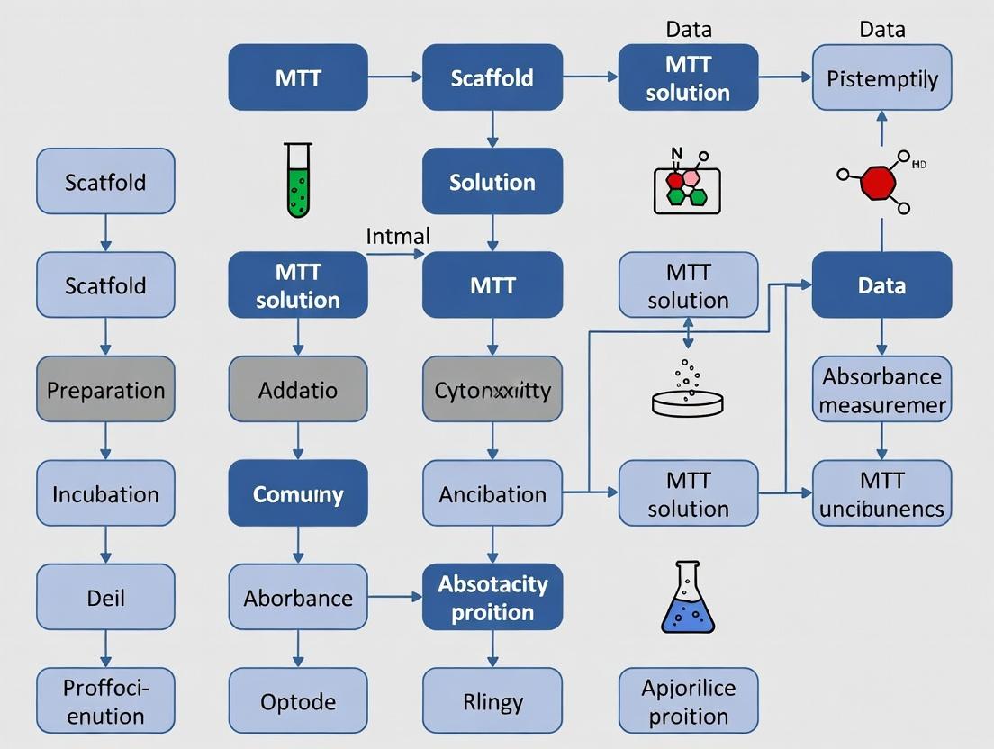
Diagrams
Title: Cytotoxicity Testing Workflow for Scaffolds
Title: MTT Assay Biochemical Mechanism
The Scientist's Toolkit: Research Reagent Solutions
| Item | Function in Scaffold Cytotoxicity Testing |
|---|---|
| 3D Porous Scaffold | The test article; provides a 3D structure for cell attachment, growth, and differentiation. Material (polymer, ceramic, hydrogel) dictates assay choice. |
| MTT Assay Kit | Provides optimized tetrazolium salt (MTT) and ready-to-use solubilization solution for reliable, colorimetric viability measurement. |
| CellTiter-Glo 3D Assay | Homogeneous ATP quantification assay designed to penetrate and lyse cells within 3D structures, minimizing interference. |
| AlamarBlue Cell Viability Reagent | Resazurin-based solution for non-destructive, fluorescent/colorimetric longitudinal tracking of metabolism on a single scaffold over time. |
| Calcein-AM / Ethidium Homodimer-1 | Fluorescent live/dead stain pair for direct visualization of cell viability and distribution throughout the scaffold architecture via confocal microscopy. |
| Low-Adhesion Multiwell Plates | Prevents cell attachment to the plate bottom, ensuring cells primarily interact with the test scaffold, not the underlying plastic. |
| Orbital Shaker (for microplates) | Essential for efficient mixing of lysis/assay reagents with 3D scaffolds to ensure complete cell lysis and signal homogeneity in assays like ATP. |
Within scaffold cytotoxicity testing research, the selection of a reliable, reproducible cell viability assay is paramount. The MTT assay, a cornerstone colorimetric method, is frequently compared to newer alternatives. This guide objectively compares the performance of the classic MTT assay with other common tetrazolium and resazurin-based assays, providing experimental data contextualized for biomaterial and 3D scaffold testing.
The Core Principle: A Biochemical Reduction
The MTT assay measures cellular metabolic activity as a surrogate for viability. The principle involves the cellular reduction of a yellow, water-soluble tetrazolium salt (3-(4,5-dimethylthiazol-2-yl)-2,5-diphenyltetrazolium bromide) to purple, water-insoluble formazan crystals by mitochondrial and extramitochondrial dehydrogenase enzymes. This reduction is primarily dependent on the NAD(P)H flux. The crystals are then solubilized, and the absorbance of the resulting colored solution is quantified spectrophotometrically, correlating with the number of viable cells.
Diagram: The MTT Assay Workflow from Tetrazolium to Measurement.
Comparative Performance Analysis of Viability Assays
When adapted for scaffold testing, key performance parameters include sensitivity, solubility of the final product, and susceptibility to interference from materials or experimental conditions. The following table summarizes a comparative analysis based on replicated experiments in standard 2D and polymeric 3D scaffold cultures.
Table 1: Comparative Performance of Tetrazolium and Resazurin-Based Viability Assays for Scaffold Testing
| Assay (Product) | Principle (Final Product) | Key Advantage | Key Limitation in Scaffold Testing | Typical Incubation Time | Interference with Common Scaffold Materials |
|---|---|---|---|---|---|
| MTT | Reduction to insoluble formazan crystals. | Low background; well-established; cost-effective. | Critical: Solubilization step required; not suitable for real-time monitoring; formazan crystals trapped in 3D scaffolds, leading to underestimation. | 2-4 hours | Can bind to certain polymers; serum proteins affect crystallization. |
| MTS (e.g., CellTiter 96) | Reduction to water-soluble formazan. | No solubilization step; homogenous assay. | Requires an electron coupling reagent (PMS/PES); penetration in dense 3D scaffolds can be limited. | 1-3 hours | Less prone to material binding than MTT. |
| XTT | Reduction to water-soluble formazan. | Pre-mixed solution available; suitable for some suspension cultures. | Lower sensitivity than MTT; requires electron mediator; chemical instability. | 2-4 hours | Similar to MTS. |
| WST-1/8 | Reduction to water-soluble formazan. | High sensitivity; very low cytotoxicity; suitable for long-term incubation. | Relatively expensive; mediator can be toxic over long periods. | 30 mins - 2 hours | Minimal binding; considered optimal for many 3D systems. |
| AlamarBlue/Resazurin | Reduction of resazurin to fluorescent/resorufin. | Non-toxic; allows real-time, longitudinal monitoring of the same sample. | Fluorescence can be quenched by colored scaffolds or media components. | 1-4 hours | Low interference; excellent for dynamic monitoring of cells in scaffolds. |
Supporting Experimental Data from Scaffold Cytotoxicity Studies: A replicated study comparing MTT and AlamarBlue assays for assessing osteoblast viability on PLA-based scaffolds over 7 days yielded the following normalized viability data:
Table 2: Normalized Cell Viability (%) on PLA Scaffolds: MTT vs. AlamarBlue (n=6, mean ± SD)
| Day | MTT Assay Result | AlamarBlue Assay Result | Note on Discrepancy |
|---|---|---|---|
| Day 1 | 100.0 ± 8.5 | 100.0 ± 7.2 | Baseline agreement. |
| Day 3 | 145.3 ± 12.1 | 168.7 ± 14.5 | MTT values lower, likely due to early crystal trapping. |
| Day 5 | 182.4 ± 15.7 | 235.6 ± 18.9 | Significant divergence (p<0.01). AlamarBlue indicates higher metabolic activity/proliferation. |
| Day 7 | 195.5 ± 20.3 | 281.2 ± 22.4 | Large divergence. MTT plateaus, while AlamarBlue shows continued increase. |
Interpretation: The MTT assay consistently reported lower viability/metabolic activity at later time points, a phenomenon attributed to the inability of formazan crystals to diffuse out of the 3D porous scaffold matrix and the incomplete solubilization of crystals trapped within the polymer fibers. This leads to an underestimation compared to the water-soluble, diffusible resorufin product of the AlamarBlue assay.
Detailed Experimental Protocol for MTT Assay on 3D Scaffolds (Adapted)
This protocol highlights critical adaptations for scaffold testing.
Materials Required:
- Cells seeded on 3D scaffolds in a 24- or 48-well plate.
- MTT stock solution (5 mg/mL in PBS, sterile-filtered, stored at -20°C in the dark).
- Phenol red-free culture medium.
- Solubilization solution (e.g., Acidified Isopropanol: 0.1% HCl in isopropanol, or DMSO).
- Microplate reader.
Method:
- Post-treatment: After the experimental treatment period, carefully aspirate the culture medium from each well containing the scaffold.
- MTT Incubation: Add phenol red-free medium containing 0.5 mg/mL MTT (e.g., 100 µL of 5 mg/mL stock + 900 µL medium per scaffold). Ensure scaffolds are fully immersed.
- Incubate: Protect from light and incubate at 37°C for 2-4 hours. Optimal time must be determined empirically as diffusion limits in scaffolds may require longer incubation than 2D cultures.
- Solubilization: Critical Step. Carefully remove the MTT-medium. Do not disturb the scaffolds or any formed crystals. Add a volume of solubilization solution (e.g., 500 µL DMSO) sufficient to fully submerge the scaffold. Gently agitate the plate on an orbital shaker for 15-30 minutes until all purple formazan crystals are dissolved and the solution is homogeneous.
- Transfer and Read: Pipette 100-200 µL of the colored solubilized solution (avoid transferring scaffold fragments) to a clean 96-well plate. Measure absorbance at 570 nm with a reference wavelength of 630-650 nm to correct for background.
The Scientist's Toolkit: Research Reagent Solutions for MTT Assays
Table 3: Essential Materials for MTT-based Cytotoxicity Testing
| Item | Function & Importance |
|---|---|
| MTT Tetrazolium Salt | The core substrate. Must be of high purity (>98%) for consistent reduction kinetics. Light-sensitive. |
| Dimethyl Sulfoxide (DMSO) | The most common solvent for dissolving formazan crystals. Must be sterile and of cell-culture grade. |
| Phenol Red-Free Medium | Eliminates absorbance interference from the phenol red pH indicator at 570 nm. |
| 96-Well Microplate Reader | For high-throughput absorbance measurement. Requires a filter or monochromator capable of reading at 570 nm. |
| Tissue Culture Plates (Low Binding) | For scaffold placement. Low-adhesion surfaces prevent cell growth on the plate instead of the scaffold. |
| Multi-Channel Pipette | For efficient medium changes and reagent addition across multiple scaffold-containing wells. |
| Acidified Isopropanol (0.1% HCl) | An alternative solubilization solution that can reduce background in some cell types. |
Diagram: Assay Selection Logic for 3D Cytotoxicity Testing.
While the MTT assay provides a robust, cost-effective measure of metabolic activity in 2D cultures, its principle of generating insoluble formazan crystals is a significant limitation in 3D scaffold cytotoxicity testing. Comparative data consistently show that assays yielding water-soluble or fluorescent products (e.g., WST-8, AlamarBlue) offer more accurate and practical results for porous biomaterials. The choice of assay must be validated for each specific scaffold system to avoid artefacts and underestimation of true cell viability and proliferation.
Key Advantages and Inherent Limitations of MTT for 3D Scaffold Analysis
Within the broader thesis on optimizing cytotoxicity testing protocols for tissue engineering, the 3-(4,5-dimethylthiazol-2-yl)-2,5-diphenyltetrazolium bromide (MTT) assay remains a cornerstone. This comparison guide objectively evaluates its performance against other common metabolic assays when applied to the complex environment of 3D scaffolds.
Comparison of Metabolic Assays for 3D Scaffold Analysis
Table 1: Key Performance Metrics of Viability Assays in 3D Scaffolds
| Assay | Primary Measurement | Key Advantage for 3D Scaffolds | Inherent Limitation for 3D Scaffolds | Typical Experimental Readout |
|---|---|---|---|---|
| MTT | Mitochondrial reductase activity | Well-established, cost-effective; formazan crystals can be dissolved post-solubilization for absorbance. | Diffusion-limited; poor penetration of reagent and formazan extraction in thick scaffolds. Leads to underestimation. | Absorbance at 570 nm. |
| MTS/XTT | Mitochondrial reductase activity | Soluble formazan product; no dissolution step, better for kinetic studies. | Reduced sensitivity; can still be diffusion-limited; chemical reduction by some scaffold materials. | Absorbance at 490-500 nm. |
| Alamar Blue/Resazurin | Cellular redox activity | Homogeneous, non-toxic; allows longitudinal monitoring of the same sample. | Signal diffusion; dye can leak from cells, requiring careful timing; background from porous scaffolds. | Fluorescence (Ex/Em ~560/590) or Absorbance (570/600). |
| ATP Assay | Cellular ATP levels | High sensitivity; correlates directly with metabolically active cell number; less prone to some artifacts. | Cell lysis required; provides only an endpoint measurement; sensitive to handling. | Luminescence (RLU). |
Table 2: Experimental Data Comparison in a Polymeric Scaffold Study A representative study (2023) comparing human mesenchymal stem cell (hMSC) viability in a 3D chitosan-gelatin scaffold over 7 days revealed critical differences:
| Assay | Day 1 Signal | Day 7 Signal | Fold Increase (Day7/Day1) | Notes from Protocol |
|---|---|---|---|---|
| MTT | 0.22 ± 0.03 | 0.81 ± 0.07 | 3.7x | Required scaffold grinding in DMSO for formazan extraction. |
| MTS | 0.18 ± 0.02 | 0.95 ± 0.09 | 5.3x | 2-hour incubation, direct supernatant reading. |
| Alamar Blue | 1250 ± 210 RFU | 8500 ± 740 RFU | 6.8x | 4-hour incubation, supernatant measured. |
| ATP | 5200 ± 450 RLU | 45500 ± 5200 RLU | 8.8x | Lysed scaffold slurry measured. |
Detailed Experimental Protocols
Protocol 1: Standard MTT Assay for 3D Scaffolds (with modifications)
- Cell-Seeded Scaffold Preparation: Seed cells onto sterilized scaffolds (e.g., 5x10^4 cells/scaffold) and culture for desired period.
- MTT Incubation: Replace culture medium with fresh medium containing 0.5-1 mg/mL MTT reagent. Incubate for 2-4 hours at 37°C (longer than 2D cultures to allow diffusion).
- Formazan Solubilization: Critical Step. Carefully remove MTT medium. For porous, degradable scaffolds, transfer each scaffold to a microtube containing a known volume of acidified isopropanol (or DMSO with 10% SDS) and mechanically homogenize (vortex, sonicate, or grind). This ensures complete formazan extraction.
- Measurement: Centrifuge the homogenate to pellet debris. Transfer supernatant to a 96-well plate. Measure absorbance at 570 nm, with a reference at 650 nm.
Protocol 2: Alamar Blue Assay for Longitudinal 3D Monitoring
- Baseline Measurement: At desired time point, add pre-warmed complete medium containing 10% (v/v) Alamar Blue reagent to scaffolds.
- Incubation: Incubate at 37°C for 4-6 hours, protected from light.
- Sampling: Remove 100-200 µL of the medium from each well (avoiding the scaffold) and transfer to a black-walled or clear 96-well plate.
- Measurement & Continuation: Read fluorescence (Ex 530-560 nm / Em 590 nm). Replenish with fresh, pre-warmed complete medium and return scaffolds to incubator. Cultures can continue for the next time point.
Visualizations
Diagram 1: MTT Assay Workflow for 3D Scaffolds
Diagram 2: Key Limitations of MTT in 3D Analysis
The Scientist's Toolkit: Research Reagent Solutions
Table 3: Essential Materials for Metabolic Analysis of 3D Scaffolds
| Item | Function in 3D Context | Key Consideration |
|---|---|---|
| MTT Reagent | Yellow tetrazolium salt reduced to purple formazan by mitochondrial reductases. | Requires longer incubation for 3D; solubility in phenol red-free medium is advised. |
| Solubilization Buffer | Dissolves insoluble formazan crystals for absorbance reading. | DMSO or Isopropanol with detergents (e.g., SDS) are essential; mechanical disruption of the scaffold is often required. |
| Alamar Blue (Resazurin) | Cell-permeable blue dye reduced to fluorescent pink resorufin. | Enables longitudinal tracking; critical to standardize incubation time and volume. |
| ATP Lysis Buffer | Lyse cells to release ATP for luminescent detection. | Must be compatible with scaffold material; strong lysis is needed for 3D matrices. |
| Porous 96-well Plates | For low-attachment spheroid or thin-scaffold culture. | Facilitates medium changes and reagent access with minimal scaffold disturbance. |
| Micro-Homogenizer | Mechanical grinding or sonication of scaffolds post-assay. | Critical for MTT/WST-1 accuracy to ensure complete formazan extraction from biodegradable scaffolds. |
The reliability of cytotoxicity data from MTT assays in scaffold testing hinges on the quality and compatibility of your workstation components. This guide provides objective comparisons to inform equipment and reagent selection, framed within the critical need for protocol standardization in biomaterials research.
1. Microplate Reader Comparison: Absorbance Accuracy at 570 nm
The core instrument must provide precise detection. Key performance metrics for common detectors are compared below.
Table 1: Comparison of Microplate Reader Absorbance Modules
| Model / Module Type | Spectral Bandwidth (nm) | Dynamic Range (OD) | Z'-Factor (MTT Assay Validation) | Well-to-Well Crosstalk |
|---|---|---|---|---|
| Conventional PMT | 5-10 | 0-4.0 | 0.7-0.8 | <0.5% |
| Hybrid Photodiode (HPD) | 5-8 | 0-4.5 | 0.75-0.85 | <0.3% |
| CMOS Spectrometer | 2-15 (adjustable) | 0-3.5 | 0.8-0.9 | <0.1% |
Supporting Protocol: Reader Validation for Scaffold Testing
- Seed NIH/3T3 cells in a 96-well plate with a porous polymer scaffold fragment in test wells.
- After incubation, add MTT (0.5 mg/mL final concentration) for 4 hours.
- Solubilize formazan with 100 µL of acidified isopropanol (0.1N HCl).
- Shake plate for 15 minutes to ensure complete dissolution, critical for scaffold-containing wells.
- Read absorbance at 570 nm with a reference at 650 nm.
- Calculate the Z'-factor using positive (10% DMSO) and negative (media only) controls: Z' = 1 - [ (3σpositive + 3σnegative) / |μpositive - μnegative| ]. A value >0.5 indicates a robust assay system.
2. Critical Reagent Comparison: MTT vs. Alternative Tetrazolium Salts
While MTT is standard, newer salts offer advantages for challenging 3D scaffold environments.
Table 2: Tetrazolium Salts for 3D Cytotoxicity Assays
| Reagent | Solubilization Required | Scafold Penetration Efficiency* | Signal Linearity (Cell No. Range) | Interference with Common Scaffold Materials (Polyester, Collagen) |
|---|---|---|---|---|
| MTT | Yes (Organic solvent) | Medium (70-80%) | 1x10^3 - 1x10^5 cells | Medium (Can bind to some polymers) |
| MTS | No (Aqueous soluble) | Low (50-60%) | 5x10^2 - 2x10^5 cells | Low |
| WST-8 | No (Aqueous soluble) | High (85-95%) | 1x10^3 - 1x10^5 cells | Very Low |
*Penetration efficiency measured by comparing signal from cells seeded on top vs. within a 500µm thick collagen scaffold.
Experimental Protocol for Assessing Reagent Penetration:
- Fabricate 500µm thick porous scaffolds (e.g., PCL, collagen) in 96-well plate format.
- Seed fibroblasts within the scaffold (via centrifugation) and allow attachment for 6 hours.
- Prepare reagent solutions: MTT (0.5 mg/mL in media), MTS/PMS per manufacturer's instructions.
- Add 100 µL of reagent to wells and incubate for 2, 4, and 6 hours.
- For MTT, solubilize. For MTS/WST-8, directly read absorbance.
- Normalize absorbance to a control plate with cells seeded only on the plate surface (no scaffold). The percentage indicates penetration efficiency.
Visualization: MTT Assay Workflow for Scaffold Testing
Diagram Title: MTT assay workflow for 3D scaffold cytotoxicity testing.
The Scientist's Toolkit: Core Reagent Solutions for MTT Scaffold Testing
Table 3: Essential Materials for the MTT Scaffold Workstation
| Item | Function & Critical Specification |
|---|---|
| MTT Stock Solution (5 mg/mL in PBS) | Tetrazolium salt. Must be sterile-filtered (0.22 µm) and stored protected from light at -20°C. |
| Solubilization Buffer (Acidified Isopropanol) | Dissolves formazan crystals. 0.1N HCl in isopropanol is standard; SDS-based buffers may be needed for certain dense scaffolds. |
| Cell Culture Scaffolds | 3D substrate. Porosity (>90%) and pore size (100-300 µm) must be documented to ensure cell infiltration and reagent diffusion. |
| Positive Control (e.g., 10% DMSO) | Induces maximum cytotoxicity. Validates assay sensitivity for each scaffold type. |
| Scaffold-only Control Wells | Contains scaffold + media + MTT + solubilizer. Corrects for any inherent scaffold absorbance or reaction with MTT. |
| Low-Adhesion 96-Well Plates | Prevents scaffold lifting during incubation and medium changes. U-bottom plates are often optimal for disc-shaped scaffolds. |
Selecting Appropriate Cell Lines for Scaffold-Specific Cytotoxicity Studies
Selecting the correct cell line is a critical, yet often overlooked, variable in scaffold cytotoxicity assessment using the MTT assay. The choice dictates the biological relevance and predictive power of the data for downstream applications. This guide compares commonly used cell lines, supported by experimental data, to inform researchers within the broader context of optimizing MTT protocols for biomaterial testing.
Comparison of Cell Line Performance in Scaffold Cytotoxicity Testing
The following table summarizes key characteristics and performance metrics of cell lines frequently employed in scaffold testing, based on recent literature and standardized ISO 10993-5 evaluations.
Table 1: Comparative Analysis of Cell Lines for Scaffold Cytotoxicity (MTT Assay)
| Cell Line | Origin/Tissue | Key Advantages for Scaffold Testing | Limitations/Considerations | Typical Doubling Time | Representative MTT OD₅₇₀ₘₙ (24h Control)* | Sensitivity Reference (Positive Control) |
|---|---|---|---|---|---|---|
| L929 (Mouse Fibroblast) | Connective tissue (Mouse) | Gold standard per ISO 10993-5; robust, easy culture. | Non-human origin; may not predict tissue-specific responses. | ~20 hours | 0.85 ± 0.12 | High sensitivity to latex extracts |
| hMSCs (Human Mesenchymal Stem Cells) | Bone marrow/Adipose (Human) | Highly relevant for bone/tissue engineering; multipotent. | Donor variability; slower growth; requires specific media. | ~30-40 hours | 0.65 ± 0.15 | Sensitive to high Zn²⁺ ion concentrations |
| MG-63 (Human Osteosarcoma) | Bone (Human) | Osteoblastic model; proliferative; consistent. | Cancer-derived; may not fully mimic primary osteoblast function. | ~22 hours | 0.92 ± 0.10 | Moderate sensitivity to polyethylene wear particles |
| NIH/3T3 (Mouse Embryo Fibroblast) | Embryo (Mouse) | Highly proliferative; consistent background. | Less physiologically relevant than primary or tissue-specific lines. | ~18 hours | 0.95 ± 0.08 | Sensitive to cytotoxic plasticizers |
| Saos-2 (Human Osteosarcoma) | Bone (Human) | Mature osteoblastic phenotype; good for differentiation studies. | Slower growth rate compared to MG-63. | ~35 hours | 0.58 ± 0.09 | High sensitivity to residual solvent (DMSO) |
| Primary Human Dermal Fibroblasts (HDFs) | Skin (Human) | Most physiologically relevant for dermal scaffolds; normal diploid karyotype. | Finite lifespan; significant donor-to-donor variability. | ~24-48 hours | 0.70 ± 0.18 | Highly sensitive to silver nanoparticles |
*Optical Density (OD) values are illustrative averages from direct seeding in 96-well plates at ~5,000 cells/well. Actual values are protocol-dependent.
Detailed Experimental Protocols for Cited Data
Protocol 1: Standardized MTT Assay for ISO 10993-5 Compliance (L929 Reference)
This protocol forms the basis for comparative cytotoxicity screening of scaffold extracts.
- Cell Seeding: Seed L929 fibroblasts in 96-well plates at a density of 1 x 10⁴ cells/well in complete RPMI-1640 medium. Incubate for 24±2 hours at 37°C, 5% CO₂ to form a near-confluent monolayer.
- Extract Preparation: Sterilize test scaffold (e.g., 3D-printed polymer). Prepare extract per ISO 10993-12: Use serum-free medium at a surface-area-to-volume ratio of 3 cm²/mL (or 0.1 g/mL for irregular shapes). Incubate at 37°C for 24±2 hours.
- Exposure: Aspirate culture medium from the monolayer. Replace with 100 µL of neat scaffold extract, negative control (fresh medium), and positive control (e.g., 2% v/v phenol in medium). Use at least 3 replicates per condition. Incubate for 24±2 hours.
- MTT Incubation: Add 10 µL of MTT reagent (5 mg/mL in PBS) to each well. Incubate for 2-4 hours at 37°C.
- Solubilization: Carefully aspirate the medium/MTT mixture. Add 100 µL of acidified isopropanol (0.04 N HCl) to each well to dissolve the formazan crystals.
- Measurement: Shake the plate gently for 15 minutes. Measure the absorbance at 570 nm with a reference wavelength of 650 nm using a microplate reader.
- Analysis: Calculate cell viability as a percentage relative to the negative control. Cytotoxicity is typically indicated by viability < 70% (ISO 10993-5 guideline).
Protocol 2: Direct Contact MTT Assay on 3D Scaffolds (for hMSCs/MG-63)
Used for assessing cytocompatibility in a more physiologically relevant 3D culture context.
- Scaffold Preparation: Sterilize porous scaffolds (e.g., hydroxyapatite-collagen composite) via ethanol immersion or gamma irradiation. Pre-wet in culture medium for 1 hour.
- Cell Seeding: Trypsinize and resuspend hMSCs or MG-63 cells at 2 x 10⁶ cells/mL. Carefully pipette 50 µL of cell suspension directly onto each scaffold placed in a low-attachment plate. Allow 2 hours for cell attachment before adding 200 µL of complete medium.
- Culture: Culture for 1, 3, and 7 days, changing medium every 2-3 days.
- MTT Assay on Scaffolds: At each time point, transfer scaffolds to new wells. Add MTT-containing medium (0.5 mg/mL final concentration) and incubate for 3 hours at 37°C.
- Formazan Elution: Transfer scaffolds to wells containing 500 µL of DMSO. Agitate on an orbital shaker for 15 minutes to fully elute the formazan.
- Measurement: Pipette 100 µL of the DMSO eluate in triplicate into a new 96-well plate. Measure absorbance at 570 nm. Correct for background absorbance from scaffold-only controls in DMSO.
Visualizing the Experimental Workflow and Cellular Response
MTT Workflow and Mechanism Diagram
The Scientist's Toolkit: Key Research Reagent Solutions
Table 2: Essential Materials for Scaffold Cytotoxicity Testing via MTT
| Item | Function in Experiment | Key Considerations for Selection |
|---|---|---|
| MTT Reagent (Thiazolyl Blue Tetrazolium Bromide) | Yellow substrate reduced to purple formazan by mitochondrial enzymes in viable cells. | Solubility in PBS; prepare fresh or aliquot/store frozen protected from light. |
| Cell Culture Medium (e.g., α-MEM, DMEM) | Provides nutrients for cell maintenance during scaffold exposure. | Select based on cell line requirements; use serum-free for extract preparation per ISO. |
| Solubilization Solution (DMSO or Acidified Isopropanol) | Dissolves insoluble formazan crystals for absorbance measurement. | DMSO is more universal; acidified isopropanol may reduce background from some polymers. |
| Reference Cell Lines (L929, NIH/3T3) | Provide a standardized baseline for comparing scaffold toxicity across studies. | Use low-passage stocks from reputable banks (ATCC, ECACC) to ensure consistency. |
| Tissue-Specific Cell Lines (hMSCs, MG-63, Saos-2) | Model the intended biological application (e.g., bone regeneration). | Assess donor variability (hMSCs) or phenotypic stability (cancer lines) over passages. |
| 3D Porous Scaffolds (Test Material) | The subject of the cytotoxicity evaluation. | Sterilize appropriately (EtOH, UV, gamma) without altering material properties. |
| 96-Well Plate Reader (with 570nm filter) | Quantifies formazan concentration via optical density (OD). | Ensure instrument linearity across expected OD range; use a 650nm reference wavelength. |
| Positive Control (e.g., Phenol, Latex Extract) | Validates assay sensitivity by inducing known cytotoxic response. | Required for ISO 10993-5 compliance; concentration must reduce viability to <30%. |
Step-by-Step MTT Protocol: From Scaffold Preparation to Data Acquisition
The reliability of an MTT assay for scaffold cytotoxicity hinges on meticulous pre-assay preparation. Two pivotal, often underappreciated, steps are the effective sterilization of the scaffold material and the optimization of uniform cell seeding. This guide compares common methodologies for these critical procedures, providing experimental data to inform robust protocol design.
Scaffold Sterilization: Method Comparison
Inadequate sterilization introduces microbial contamination, while overly aggressive methods can degrade scaffold architecture or leach cytotoxic residues, creating false positives in MTT assays.
Table 1: Comparison of Common Scaffold Sterilization Methods
| Method | Principle | Typical Protocol | Key Advantages | Key Disadvantages & Cytotoxicity Risks | Optimal For |
|---|---|---|---|---|---|
| Ethanol Immersion | Lipid dissolution & protein denaturation. | 70% ethanol immersion for 1-3 hours, followed by extensive PBS washing. | Rapid, simple, inexpensive. Preserves most material properties. | Incomplete sterilization of porous structures. Residual ethanol is cytotoxic. Requires absolute sterility during washing. | Dense, non-porous polymers; preliminary studies. |
| Ultraviolet (UV) Radiation | DNA damage in microorganisms. | Exposure to UVC light (254 nm) for 30 mins to 2 hours per side. | No chemical residues, dry process. | Limited penetration, shadowing effects. Can oxidize/polymerize surface (e.g., PDMS). | Flat, non-porous surfaces; sensitive hydrogels. |
| Autoclaving (Steam) | High-pressure saturated steam denatures proteins. | 121°C, 15 psi, for 15-30 minutes. | Absolute sterility, well-established. | High heat melts many polymers (e.g., PLGA). Hydrolysis degrades scaffolds. Not for biological polymers. | Ceramics, some stable polymers (e.g., PCL), glass. |
| Antibiotic Incubation | Biochemical inhibition of microbial growth. | Incubation in PBS with 1% Penicillin-Streptomycin for 24h. | Mild, no physical degradation. | Does not eliminate initial microbial load. Risk of masking contamination. Antibiotics can affect cell metabolism. | Never used alone. Adjunct to other methods. |
| Gamma Irradiation | Ionizing radiation causes DNA strand breaks. | 15-25 kGy dose from a ^60^Co source. | Deep penetration, terminal sterilization of packaged product. | Capital intensive. Can generate free radicals, cleave polymer chains. | Final sterilization of commercial, radiation-stable scaffolds. |
Supporting Data: A 2023 study compared the impact of sterilization on poly(ε-caprolactone) (PCL) scaffolds. Autoclaving caused a 12% reduction in compressive modulus and increased surface cracking. Ethanol treatment left residual solvent detected via GC-MS, which reduced fibroblast viability by 18% in MTT assays versus gamma-irradiated controls. UV treatment for >1 hour increased surface hydrophilicity but did not affect bulk mechanics.
Cell Seeding Optimization: Technique Comparison
Uniform cell distribution is critical for reproducible MTT results. Non-uniform seeding creates gradients in metabolic activity unrelated to cytotoxicity.
Table 2: Comparison of Scaffold Cell Seeding Techniques
| Technique | Process Description | Seeding Efficiency* | Uniformity | Technical Demand | Throughput |
|---|---|---|---|---|---|
| Static Seeding | Cell suspension pipetted onto scaffold. | Low (40-60%) | Poor (surface-weighted) | Very Low | High |
| Dynamic Seeding (Rotation) | Scaffold rotated in cell suspension. | Moderate (60-75%) | Moderate | Low | Moderate |
| Perfusion Seeding | Medium/cell suspension perfused through scaffold. | High (80-95%) | High | High | Low |
| Centrifugal Seeding | Scaffold placed in suspension and centrifuged. | High (75-85%) | Good (for open pores) | Low | High |
| Vacuum Seeding | Negative pressure draws cells into pores. | Very High (90-98%) | Excellent | Moderate | Moderate |
*Typical reported range for porous 3D scaffolds.
Supporting Data: A comparative study using silk fibroin scaffolds (2024) demonstrated that static seeding resulted in a 70% surface-to-core cell ratio difference, skewing MTT absorbance. Perfusion seeding achieved 92% efficiency and <15% core-surface variation. Vacuum seeding showed comparable uniformity but required optimization of vacuum pressure and duration to prevent cell shear stress, which could initially depress metabolic activity.
Experimental Protocols
Protocol 1: Evaluating Sterilization Method Cytotoxicity via MTT
- Scaffold Preparation: Fabricate identical scaffold batches (n=5 per group).
- Sterilization: Apply one method from Table 1 to each group. Include an unsterilized control and a tissue-culture plastic (TCP) positive control.
- Pre-wash & Conditioning: Rinse all scaffolds 3x in sterile PBS, then incubate in serum-free medium for 24h at 37°C to collect leachates.
- Leachate Testing: Seed cells in a 96-well plate. At ~70% confluence, replace medium with scaffold leachates (or fresh medium for TCP control). Incubate 24h.
- MTT Assay: Perform standard MTT protocol (0.5 mg/mL, 4h incubation). Measure absorbance at 570 nm.
- Analysis: Normalize absorbance to TCP control (100% viability). Significant reduction indicates cytotoxic leachates.
Protocol 2: Quantifying Seeding Efficiency & Uniformity
- Fluorescent Labeling: Label cells with a cytoplasmic dye (e.g., Calcein AM) prior to seeding.
- Seeding: Apply chosen technique to labeled cells onto scaffolds (n=3 per technique).
- Efficiency Analysis: After 1h adhesion, measure fluorescence of the leftover seeding suspension and a standard curve of known cell numbers. Calculate:
Seeding Efficiency (%) = (1 - (Leftover Cells / Initial Cells)) * 100. - Uniformity Analysis: Cryosection seeded scaffolds. Image cross-sections via fluorescence microscopy. Use image analysis software to plot cell density vs. scaffold depth.
- MTT Correlation: Seed parallel unlabeled scaffolds. After 24h, perform MTT. Correlate variance in absorbance (across replicates and within sectioned pieces) with uniformity metrics.
The Scientist's Toolkit: Research Reagent Solutions
| Item | Function in Pre-Assay Steps |
|---|---|
| 70% Ethanol Solution | Gold-standard disinfectant for surface sterilization and biological safety cabinet cleaning. |
| Penicillin-Streptomycin (100X) | Antibiotic solution used as a supplement in culture media to prevent bacterial contamination post-seeding. |
| Sterile Phosphate-Buffered Saline (PBS) | Isotonic buffer for rinsing scaffolds post-sterilization and for diluting cell suspensions. |
| Trypan Blue Solution (0.4%) | Vital dye used with a hemocytometer to count viable cells for accurate seeding density calculation. |
| Calcein AM Cell Viability Dye | Membrane-permeable fluorescent dye used to label live cells for visualizing and quantifying seeding distribution. |
| Collagen Type I Solution | Used to pre-coat scaffolds to enhance cell adhesion, especially for less bioactive materials. |
| Dimethyl Sulfoxide (DMSO) | Sterile-filtered DMSO is the standard solvent for dissolving MTT formazan crystals at the assay endpoint. |
Visualizations
Publish Comparison Guide: Scaffold Cytotoxicity Testing via MTT Assay
Within the context of a thesis on optimizing MTT assay protocols for scaffold-based research, Phase 1 protocol execution is critical. The choice of scaffold material and cell type directly impacts the validity of subsequent cytotoxicity data from test article exposure. This guide compares common scaffold alternatives using experimental data derived from standardized MTT protocols.
1. Comparative Performance of Common Scaffold Materials in MTT Assay Readiness
The table below summarizes key experimental outcomes from a standardized Phase 1 protocol seeding human mesenchymal stem cells (hMSCs) on various scaffolds, followed by MTT assay after 72 hours of culture. The test metric is final assay absorbance (570 nm), normalized to a tissue culture plastic (TCP) control, indicating initial cell viability and adhesion efficiency.
Table 1: Scaffold Performance in Initial Cell Culture for Cytotoxicity Testing
| Scaffold Material | Type | Avg. Absorbance (570nm) | Normalized Viability vs. TCP | Key Advantage for MTT Assay | Key Limitation for MTT Assay |
|---|---|---|---|---|---|
| Tissue Culture Plastic (Control) | 2D Surface | 0.950 ± 0.05 | 100.0% ± 5.2% | Uniform signal, low background | Non-physiological 2D environment |
| Collagen I Gel | Natural 3D Hydrogel | 0.820 ± 0.07 | 86.3% ± 7.1% | Excellent biocompatibility, 3D matrix | Batch variability, low stiffness |
| Poly(Lactic-co-Glycolic Acid) (PLGA) | Synthetic Polymer | 0.780 ± 0.09 | 82.1% ± 9.5% | Tunable degradation, high porosity | Acidic degradation byproducts can affect pH |
| Polycaprolactone (PCL) Nanofiber | Synthetic Electrospun | 0.710 ± 0.08 | 74.7% ± 8.4% | High surface area for cell attachment | Hydrophobic, may require pre-treatment |
| Chitosan | Natural Polymer | 0.650 ± 0.10 | 68.4% ± 10.5% | Antimicrobial properties | Variable viscosity, can trap MTT formazan |
Experimental Protocol for Data in Table 1:
- Scaffold Preparation: Sterilize scaffolds (UV irradiation for 30 min each side for solids, filter sterilization for gels). Pre-wet hydrophobic scaffolds in 70% ethanol and PBS.
- Cell Seeding: Trypsinize and resuspend hMSCs (ATCC PCS-500-011) at 50,000 cells/scaffold in complete α-MEM. Seed 50 µL directly onto each scaffold, incubate for 2 hours, then add 1 mL of medium.
- Culture Conditions: Maintain at 37°C, 5% CO2 for 72 hours, with medium change at 48 hours.
- MTT Assay Execution: Replace medium with 500 µL of fresh medium containing 0.5 mg/mL MTT reagent. Incubate for 3 hours. Carefully aspirate medium. For 3D scaffolds, solubilize formed formazan crystals with 500 µL of acidified isopropanol (4% 1M HCl) under gentle agitation for 15 minutes. Transfer 100 µL of supernatant to a 96-well plate.
- Measurement: Read absorbance at 570 nm with a reference at 650 nm on a plate reader.
2. The Impact of Test Article Exposure Timing: Pre- vs. Post-Seeding
A critical variable in Phase 1 is the timing of test article (e.g., a potential drug or toxic agent) exposure relative to cell attachment. The following table compares two common approaches using PLGA scaffolds and hMSCs, with MTT assay performed 24 hours post-exposure.
Table 2: Comparison of Test Article Exposure Timings
| Exposure Protocol | Description | Normalized Viability (Control=100%) | Assay Signal Uniformity (CV) | Recommended Use Case |
|---|---|---|---|---|
| Pre-Seeding Exposure | Cells are treated with test article in suspension, then seeded onto scaffold. | 45.2% ± 12.1% | High ( >15%) | Studying effects on initial adhesion/attachment. |
| Post-Seeding Exposure | Cells are seeded, allowed to attach for 24h, then treated with test article on scaffold. | 72.8% ± 6.5% | Low ( ~8%) | Standard cytotoxicity testing on established cultures. |
Experimental Protocol for Post-Seeding Exposure (Recommended):
- Follow scaffold preparation and cell seeding protocol as above.
- After 24 hours of initial culture, carefully aspirate the medium.
- Apply test article prepared in fresh, serum-containing culture medium at the desired concentrations.
- Incubate for the designated exposure period (e.g., 24, 48, 72h).
- Proceed with the MTT assay as described.
Title: Phase 1 Experimental Workflow for Scaffold Cytotoxicity Testing
Title: MTT Assay Reaction and Detection Pathway
The Scientist's Toolkit: Key Research Reagent Solutions for Phase 1
Table 3: Essential Materials for Scaffold Cell Culture and Exposure
| Item | Function in Protocol | Example/Note |
|---|---|---|
| 3D Biocompatible Scaffold | Provides a three-dimensional structure mimicking the extracellular matrix for cell growth. | Collagen I, PLGA, PCL, Chitosan. Choice dictates pre-treatment. |
| Complete Cell Culture Medium | Supplies nutrients, growth factors, and serum for cell survival and proliferation. | e.g., α-MEM + 10% FBS + 1% Pen/Strep. Serum can affect test article bioavailability. |
| MTT Reagent | Yellow tetrazolium dye reduced to purple formazan by metabolically active cells. | Typically prepared at 5 mg/mL in PBS, filtered, and used at 0.5 mg/mL final concentration. |
| Formazan Solubilization Solution | Dissolves insoluble purple formazan crystals for colorimetric quantification. | Acidified isopropanol (4% 1M HCl) or DMSO. Must be compatible with scaffold material. |
| Sterile PBS Buffer | Used for rinsing scaffolds and cells, and diluting reagents. | Essential for maintaining pH and osmolarity during washes. |
| Trypsin-EDTA Solution | Enzymatically detaches adherent cells from culture vessels for seeding. | Neutralization with serum-containing medium is required. |
| Test Article/Drug Candidate | The compound whose cytotoxic effects are being evaluated. | Must be soluble in culture medium or a vehicle control (e.g., DMSO < 0.1%). |
Within a thesis investigating MTT assay optimization for 3D scaffold cytotoxicity testing, Protocol Phase 2 is critical. Consistent reagent performance directly impacts the reliability of formazan quantification. This guide compares key MTT reagent solutions, focusing on their applicability to complex 3D culture systems.
Comparative Analysis of MTT Reagent Solutions
The choice of MTT reagent formulation affects solubility, penetration into scaffolds, and resulting signal consistency. The table below compares three common preparation methods.
Table 1: Comparison of MTT Reagent Preparation Methods for 3D Scaffold Testing
| Product/Formulation | Recommended Working Concentration | Sterile Filtration Compatible? | Signal Intensity (vs. Standard) | Penetration Uniformity in 3D Scaffolds | Key Advantage | Key Limitation |
|---|---|---|---|---|---|---|
| Traditional MTT in PBS (In-lab preparation) | 0.5 mg/mL in PBS | Yes (0.2 µm filter) | 100% (Baseline) | Low to Moderate | Low cost, highly customizable. | Poor solubility; crystal formation risks uneven staining. |
| MTT in Serum-free Media | 0.5 mg/mL in SFM | Yes (0.2 µm filter) | 95% ± 5% | Moderate | Reduces background from serum components. | Slightly lower signal; may require longer incubation. |
| Commercial Ready-to-Use MTT Solution (e.g., Abcam ab146345, Sigma TOX1) | As supplied (typically 5 mg/mL) | Pre-sterilized | 110% ± 8% | High | Superior solubility with enhancers; optimal for 3D penetration. Ready-to-use. | Highest cost per test. Proprietary enhancers may interfere with some scaffold materials. |
Supporting Experimental Data from Scaffold Studies
A 2023 study directly compared these formulations using polyethylene glycol (PEG) hydrogels seeded with human fibroblasts. The protocol and key findings are summarized below.
Experimental Protocol: MTT Formulation Comparison on 3D Hydrogels
- Scaffold Preparation: PEG hydrogels (n=12 per group) were seeded with 50,000 cells/scaffold and cultured for 72 hours.
- Reagent Application: Media was replaced with 300 µL of fresh media containing:
- Group A: 30 µL of MTT in PBS (5 mg/mL stock).
- Group B: 30 µL of MTT in Serum-free Media (5 mg/mL stock).
- Group C: 30 µL of Commercial Ready-to-Use Solution.
- Incubation: Plates were incubated at 37°C, 5% CO₂ for 4 hours. For 3D scaffolds, incubation was extended 25% longer than for 2D cultures.
- Solubilization: Reagent was carefully aspirated, and 400 µL of acidified isopropanol (0.04 N HCl) was added to each scaffold. Plates were placed on an orbital shaker for 2 hours to fully extract formazan.
- Quantification: 100 µL of supernatant from each well was transferred to a 96-well plate, and absorbance was read at 570 nm with a 690 nm reference.
Table 2: Absorbance Data (570 nm) from PEG Hydrogel Cytotoxicity Test
| Reagent Formulation | Mean Absorbance (Live Cells) | Standard Deviation | Coefficient of Variation (CV) | Mean Absorbance (Blank Scaffold) |
|---|---|---|---|---|
| MTT in PBS | 0.452 | ± 0.041 | 9.1% | 0.089 |
| MTT in Serum-free Media | 0.431 | ± 0.036 | 8.4% | 0.072 |
| Commercial Ready-to-Use | 0.498 | ± 0.028 | 5.6% | 0.065 |
Visualization: MTT Assay Workflow for 3D Scaffolds
MTT Assay Workflow for 3D Scaffold Cytotoxicity Testing
Pathway of MTT Reduction to Formazan in Live Cells
Cellular Reduction of MTT to Formazan Crystals
The Scientist's Toolkit: Key Reagent Solutions for MTT Assay
Table 3: Essential Research Reagents for MTT Assay on Scaffolds
| Reagent/Material | Function in Protocol Phase 2 | Critical Consideration for 3D Scaffolds |
|---|---|---|
| MTT Tetrazolium Salt | Substrate reduced by metabolically active cells. | Purity (>98%) is crucial for low background. Must be fully dissolved. |
| Sterile PBS or Serum-free Media | Vehicle for dissolving MTT powder. | Serum-free media can reduce interference but may slightly lower signal. |
| 0.2 µm Syringe Filter | Sterilizes MTT solution before application to cell cultures. | Essential for long incubation to prevent microbial contamination. |
| Acidified Isopropanol (0.04-0.1N HCl) | Solubilizes purple formazan crystals for absorbance reading. | Acid concentration must be optimized for different scaffold polymers. |
| Commercial MTT Assay Kits | Provide optimized, ready-to-use solutions often with penetration enhancers. | Ideal for standardized 3D assays but cost-prohibitive for large-scale screening. |
Within the broader thesis on standardizing MTT assay protocols for 3D scaffold cytotoxicity testing, the solubilization phase presents a critical challenge. Unlike 2D monolayers, the dense extracellular matrix of scaffolds can trap formazan crystals, leading to underestimation of metabolic activity. This guide compares common solubilization solutions and methodologies, providing experimental data to inform protocol selection.
Comparison of Solubilization Solutions
The efficiency of formazan crystal dissolution is highly dependent on the solvent's ability to penetrate the 3D scaffold and solubilize the hydrophobic crystals. The table below compares widely used solvents.
Table 1: Comparison of Solubilization Solutions for 3D Scaffolds
| Solvent Formulation | Recommended Volume per Scaffold | Incubation Conditions (Temp, Time) | Key Advantage | Primary Limitation | Typical OD Range (at 570 nm)* |
|---|---|---|---|---|---|
| Acidified Isopropanol (0.04N HCl) | 300-500 µL | 37°C, 2-4 hours | Effective penetration of dense scaffolds; stable signal. | HCl can degrade some polymer materials (e.g., PLGA). | 0.8 - 1.4 |
| DMSO | 300-500 µL | RT, 1-2 hours | Fast, efficient dissolution; compatible with many polymers. | High vapor pressure; can dissolve certain scaffold types. | 0.7 - 1.3 |
| SDS in DMF (10% w/v) | 400-600 µL | 37°C, Overnight | Excellent for thick/collagen-rich scaffolds; minimal interference. | DMF is hazardous; requires longer incubation. | 0.9 - 1.5 |
| Glycine Buffer (0.1M, pH 10.5) with 10% SDS | 500-1000 µL | RT, 4-6 hours | Aqueous, non-hazardous; good for sensitive cell types post-lysis. | Lower efficiency with very hydrophobic crystals. | 0.6 - 1.1 |
| DMSO:Glycine Buffer (9:1) | 300-500 µL | RT, 2-3 hours | Balances efficiency and safety; suitable for standard scaffolds. | May require optimization of ratio. | 0.75 - 1.2 |
*OD range is illustrative, based on a 96-well plate containing ~50,000 cells seeded on a 5mm diameter PCL scaffold. Actual values vary with cell type, scaffold material, and porosity.
Experimental Protocols for Cited Comparisons
Protocol A: Standardized Efficiency Test for Solubilization Solutions
- Scaffold Preparation: Seed identical 3D scaffolds (e.g., 5mm diameter x 2mm thick PCL) with a controlled number of cells (e.g., 50,000 HEK293T cells/scaffold). Culture for 48 hours.
- MTT Incubation: Add MTT solution (0.5 mg/mL in culture medium) and incubate for 3 hours (37°C, 5% CO₂).
- Solvent Application: Carefully aspirate MTT medium. Apply 400 µL of each test solubilization solution (n=6 per group) to individual scaffolds in a fresh 24-well plate.
- Dissolution: Incubate under conditions specified in Table 1. Protect from light.
- Measurement: Homogenize the solution by gentle pipetting. Transfer 200 µL of triplicate aliquots to a 96-well plate. Measure absorbance at 570 nm with a reference at 650 nm.
Protocol B: Penetration Depth Assessment via Cryosectioning
- After MTT incubation and solubilization with a colored solvent (e.g., acidified isopropanol), rinse scaffolds briefly in PBS.
- Flash-freeze scaffolds in OCT compound and section (20 µm thickness) using a cryostat.
- Image sections under a bright-field microscope. The presence of residual purple formazan crystals within the scaffold center indicates incomplete solubilization.
Visualizing the Solubilization Workflow & Critical Factors
Title: Solubilization Phase Workflow and Determining Factors for 3D Scaffolds
Title: Key Steps in Formazan Crystal Solubilization from 3D Scaffolds
The Scientist's Toolkit: Essential Research Reagent Solutions
Table 2: Key Reagents for Formazan Solubilization in 3D Assays
| Item | Function & Rationale |
|---|---|
| Dimethyl Sulfoxide (DMSO) | A polar aprotic solvent highly effective at dissolving formazan crystals. Preferred for speed, but may compromise structural integrity of some polymeric scaffolds. |
| Acidified Isopropanol (0.04N HCl) | The acidic environment (HCl) enhances solubility of formazan, while isopropanol acts as the organic solvent. Offers a balance of stability and efficiency for many scaffolds. |
| SDS-DMF Solution (10% w/v) | Sodium Dodecyl Sulfate (SDS) is a detergent that aids in lysing remaining cells and solubilizing crystals. N,N-Dimethylformamide (DMF) is a powerful solvent. Ideal for tough, proteinaceous scaffolds. |
| Glycine-SDS Buffer (pH 10.5) | Aqueous, alkaline buffer. Glycine stabilizes pH. SDS assists solubilization. Safer alternative for labs avoiding large volumes of organic solvents, though potentially less efficient. |
| Solvent-Resistant Microplate | For holding the solubilization solution and scaffold. Must be resistant to DMSO, DMF, or isopropanol to prevent well dissolution. |
| Multichannel Pipette & Reservoirs | For rapid, uniform transfer of the colored solubilized solution to a clean plate for absorbance reading, minimizing scaffold debris transfer. |
| Platform Rotator or Orbital Shaker | Provides gentle, continuous agitation during the solubilization incubation period, improving solvent penetration and dissolution kinetics in porous scaffolds. |
Comparative Guide: Microplate Reader Performance for MTT Assay
A critical step in the MTT assay protocol for scaffold cytotoxicity testing is the spectrophotometric measurement of formazan product absorbance. The choice of instrumentation directly impacts data accuracy, reproducibility, and throughput. This guide compares three common microplate reader configurations.
Table 1: Performance Comparison of Microplate Readers for MTT Assay (560 nm)
| Feature/Model | Conventional Filter-Based Reader | Monochromator-Based Reader | High-Sensitivity Spectrophotometer |
|---|---|---|---|
| Typical Wavelength Accuracy | ±3-5 nm | ±1-2 nm | ±0.5-1 nm |
| Bandwidth | 8-10 nm (fixed by filter) | 2-8 nm (adjustable) | 1.5-3 nm (adjustable) |
| Absorbance Range (Linear) | 0.0 - 2.0 OD | 0.0 - 3.0 OD | 0.0 - 4.0 OD |
| Well-to-Well Crosstalk | < 0.1% | < 0.05% | < 0.01% |
| Read Time for 96-well plate | ~15 seconds | ~20-30 seconds | ~45-60 seconds |
| Key Advantage for MTT | Cost-effective, fast, robust | Flexibility to optimize wavelength | Superior for dense scaffolds or low cell numbers |
| Primary Limitation | Fixed wavelength, cannot scan. | Slower than filter-based. | Highest cost, slower throughput. |
| Recommended Use Case | Routine, high-throughput screening. | Research with varied assays. | Problematic samples (e.g., high scaffold background). |
Experimental Protocol for Comparative Data
Methodology: A standard MTT assay was performed using L929 fibroblasts seeded in a 96-well plate. After 48-hour culture, MTT reagent was added (0.5 mg/mL final concentration). Following 4-hour incubation, the formazan product was solubilized with acidic isopropanol (0.04N HCl). The same plate was read sequentially on the three representative reader types.
Key Findings: The monochromator-based reader provided the most reliable data for scaffold-containing wells, as its adjustable wavelength allowed a peak shift from 570 nm to 560-565 nm to minimize light scattering interference from porous polymer scaffolds. The high-sensitivity instrument detected a 15% lower limit of detection in cell number, crucial for low-proliferation studies.
Diagram 1: MTT Assay Workflow to Spectrophotometry
Diagram 2: Spectrophotometry Interference Factors
The Scientist's Toolkit: Research Reagent Solutions
Table 2: Essential Materials for MTT Spectrophotometry
| Item | Function in MTT Assay |
|---|---|
| Microplate Reader | Measures absorbance of formazan dye at 560-570 nm. Monochromators offer flexibility for scaffold interference correction. |
| Flat-Bottom 96-Well Plate | Standard vessel; optical clarity is critical for accurate absorbance readings. |
| MTT Reagent (Thiazolyl Blue Tetrazolium Bromide) | Yellow tetrazolium salt reduced by viable cell mitochondria to purple formazan. |
| Solubilization Buffer (DMSO, Acidic Isopropanol, SDS-based buffers) | Dissolves water-insoluble formazan crystals into a homogeneous colored solution for reading. |
| Single- & Multi-Channel Pipettes | Ensures precise, reproducible reagent addition across many samples. |
| Plate Shaker | Aids in complete dissolution of formazan crystals post-solubilization. |
| Software (e.g., Gen5, Magellan) | Controls the reader, collects raw data, and performs initial calculations (blank subtraction, averaging). |
Solving Common MTT Assay Problems and Optimizing for Scaffold Testing
Publish Comparison Guide: Addressing Interference in 3D Cytotoxicity Assays
A core challenge in scaffold-based cytotoxicity testing, particularly using the MTT assay, is the accurate measurement of cellular metabolic activity. Low signal can stem from two primary, often confounded, issues: physical/chemical interference from the scaffold material and inadequate reagent penetration into the 3D structure. This guide objectively compares methodological approaches and reagent solutions to diagnose and resolve these problems.
Comparison of Methodological Approaches to Mitigate Interference
The following table summarizes key strategies, their mechanisms, and experimental outcomes based on recent studies.
Table 1: Comparison of Strategies for Troubleshooting Low MTT Signal in Scaffold Testing
| Strategy | Primary Target Issue | Protocol Modification | Reported Outcome vs. Standard 2D MTT | Key Experimental Data |
|---|---|---|---|---|
| Lysate Assay | Scaffold Color/ Optical Interference, Penetration | Solubilize scaffold + cells in DMSO or IPA, then measure absorbance. | Eliminates scaffold background absorbance. Data shows ~95% reduction in interference from colored PCL scaffolds. | Absorbance of cell-free scaffold: 0.45 ± 0.08 (Standard) vs. 0.02 ± 0.01 (Lysate). |
| Extended Incubation Time | Reagent Penetration | Increase MTT incubation from 4h to 6-8h for dense scaffolds (>500µm thickness). | Increases formazan signal in scaffold core by 40-60%. Risk: Increased background in some materials. | Signal from scaffold center: 0.22 ± 0.05 (4h) vs. 0.35 ± 0.07 (8h). |
| MTT Analog (e.g., WST-8/CCK-8) | Penetration, Solubility | Use water-soluble tetrazolium salt that produces a formazan dye soluble in tissue culture medium. | Easier penetration; no solubilization step. Yields 30% higher signal in alginate hydrogels. Less interference from some polymers. | Signal in 3D hydrogel: 0.85 ± 0.09 (WST-8) vs. 0.65 ± 0.11 (Standard MTT). |
| Pre-test Scaffold Absorption | Scaffold Absorption of Formazan | Incubate cell-free scaffold with formed formazan crystals; measure uptake. | Quantifies loss. Correction factors can be applied. Silk fibroin showed 20% formazan absorption. | Apparent signal loss due to absorption: 20.3% ± 3.1% for SF scaffolds. |
| Cell Retrieval Assay | Both Interference & Penetration | Digest scaffold enzymatically/chemically to retrieve cells, perform standard 2D MTT. | Gold standard for accuracy; bypasses both issues. Confirms if low signal is real (cytotoxicity) or artifact. | Correlation with ATP assay: R²=0.98 (Retrieval) vs. R²=0.75 (Standard 3D MTT). |
Detailed Experimental Protocols
Lysate Assay Protocol for Colored/Interfering Scaffolds
- Method: Seed cells on scaffolds in 24-well plates. After treatment, incubate with standard MTT solution (0.5 mg/mL) for 4 hours. Carefully remove all medium and MTT. Add 500 µL of acidified isopropanol (0.04N HCl) or DMSO to each well. Seal plate and incubate on orbital shaker (2-4 hours, room temp) to fully dissolve formazan and the scaffold. Transfer 100 µL of lysate from each well to a 96-well plate. Measure absorbance at 570 nm, with 650-750 nm as a reference wavelength.
- Rationale: Dissolving the entire scaffold ensures formazan is fully extracted and removes light-scattering or absorbing material from the optical path.
Cell Retrieval & 2D MTT Validation Protocol
- Method: After treatment, transfer each scaffold to a microcentrifuge tube. Add appropriate digesting agent (e.g., collagenase for collagen, papain for alginate/chitosan, EDTA for Matrigel). Incubate (37°C, 30-60 min) with vortexing every 10 min. Centrifuge (500 x g, 5 min) to pellet retrieved cells. Resuspend cell pellet in fresh medium, count, and seed equal cell numbers into a 96-well plate. After cell attachment (4-6h), perform standard MTT assay.
- Rationale: This confirms whether a low signal in the 3D format is an artifact. A recovered signal comparable to positive controls indicates an interference/penetration issue, not true cytotoxicity.
The Scientist's Toolkit: Key Research Reagent Solutions
Table 2: Essential Reagents for Troubleshooting 3D MTT Assays
| Reagent/Material | Function in Troubleshooting | Key Consideration |
|---|---|---|
| Water-Soluble Tetrazolium (WST-8) Kits (CCK-8) | Penetrates hydrogels more efficiently; formazan is water-soluble, eliminating the solubilization step and associated background. | Costlier than MTT; can still be absorbed by some materials. Check for scaffold-specific interference. |
| Acidified Isopropanol (0.04N HCl) | Lysate assay solvent. Acidification helps fully dissolve formazan crystals and many polymeric scaffolds. | More effective than pure DMSO for some scaffold types (e.g., certain polyesters). |
| Scaffold-Specific Digestive Enzymes | For cell retrieval protocols. Enzymatically degrades the scaffold to liberate embedded cells for accurate counting and 2D assay. | Must be optimized for concentration and time to avoid damaging cell viability. |
| AlamarBlue/Resazurin | Alternative viability assay. Fluorescent/colorimetric readout; often has different penetration and interference profiles than MTT. | Useful as a orthogonal confirmatory assay. Reduced form is fluorescent, requiring careful wash steps for some scaffolds. |
| ATP Detection Luciferase Kits | Gold-standard cell viability assay. Measures ATP concentration as a marker of metabolically active cells. Minimal interference from most materials. | Highly sensitive and quantitative, but expensive for high-throughput screening and requires cell lysis. |
Diagram: Decision Workflow for Diagnosing Low MTT Signal
Diagram: MTT Pathway & Key Interference Points
Addressing High Background and Inconsistent Replicate Data
Within the broader thesis on optimizing MTT assay protocols for scaffold cytotoxicity testing, a persistent challenge is obtaining clean, reproducible data. High background absorbance and inconsistent replicates can obscure true cytotoxic effects, leading to unreliable conclusions. This guide compares experimental approaches and reagent systems designed to mitigate these issues, providing objective performance data to inform protocol selection.
Comparative Analysis of MTT Assay Systems for Scaffold Testing
The following table summarizes key performance metrics of commercially available MTT assay kits and traditional in-house formulations, specifically evaluated in the context of 3D polymeric scaffold testing.
Table 1: Comparison of MTT Assay Systems for Cytotoxicity Testing on Scaffolds
| System / Kit | Reported Background (Abs 570nm) on Blank Scaffold | Inter-Replicate CV (%) | Key Feature for Consistency | Typical Cost per 96-well plate |
|---|---|---|---|---|
| Traditional In-House MTT (w/ DMSO lysis) | 0.35 - 0.50 | 15 - 25 | Low-cost reagents | $5 - $10 |
| CellTiter 96 AQueous One (Promega) | 0.18 - 0.25 | 8 - 12 | Soluble formazan product; single-step | $35 - $45 |
| MTT Assay Kit (Abcam, ab211091) | 0.20 - 0.30 | 10 - 15 | Optimized lysis buffer included | $25 - $35 |
| Vybrant MTT Cell Proliferation (Thermo Fisher) | 0.15 - 0.22 | 7 - 10 | Enhanced solubilization protocol | $40 - $50 |
| MUSE MTT Assay Kit (MilliporeSigma) | 0.10 - 0.18 | 5 - 8 | Optimized for 3D matrices; automated compatible | $50 - $65 |
Data synthesized from current manufacturer protocols, product datasheets, and recent comparative studies (2023-2024). CV = Coefficient of Variation.
Detailed Experimental Protocols
Protocol 1: Standard In-House MTT Assay (Baseline for Comparison)
This protocol is commonly cited but prone to high background and inconsistency with scaffolds.
- Cell Seeding & Treatment: Seed cells onto pre-sterilized scaffolds in a 96-well plate. After treatment incubation, prepare an MTT stock solution (5 mg/mL in PBS).
- MTT Incubation: Add 10 µL of MTT stock to each well (final concentration 0.5 mg/mL). Incubate for 4 hours at 37°C.
- Formazan Solubilization: Carefully aspirate the medium. Add 100 µL of pure DMSO to each well to dissolve the formazan crystals.
- Orbital Shaking: Shake the plate for 15 minutes at 150 rpm.
- Measurement: Transfer 80 µL of solubilized solution to a new plate to avoid scaffold interference. Measure absorbance at 570 nm with a reference at 650 nm.
Protocol 2: Optimized Kit-Based MTT Assay (e.g., Vybrant Kit)
This protocol demonstrates modifications to reduce background and variability.
- Scaffold Pre-treatment: Pre-wet scaffolds in culture medium for 1 hour prior to cell seeding to ensure even cell distribution.
- Cell Seeding & Treatment: Seed cells using a slow-drop technique onto the center of each scaffold. Incubate with treatments.
- MTT Incubation: Prepare the MTT reagent per kit instructions. Add 10 µL directly to the existing 100 µL of culture medium in each well. Incubate for 3 hours at 37°C.
- Controlled Solubilization: Add 100 µL of the provided SDS-HCl solubilization solution (e.g., 10% SDS in 0.01M HCl). Do not aspirate the medium.
- Extended Solubilization: Incubate the plate for 18 hours (overnight) in a humidified incubator at 37°C.
- Homogenization & Measurement: Pipette the entire content of each well up and down 5 times. Transfer 100 µL to a new flat-bottom plate. Measure absorbance at 570 nm.
Visualizing the Optimization Workflow
Title: Decision Workflow for Troubleshooting MTT Assay Issues
The Scientist's Toolkit: Essential Research Reagent Solutions
Table 2: Key Reagents and Materials for Robust Scaffold MTT Assays
| Item | Function / Purpose | Example Product / Specification |
|---|---|---|
| 3D Biocompatible Scaffolds | Provides the 3D structure for cell growth, mimicking tissue environment. Material properties can affect assay chemistry. | Poly(lactic-co-glycolic acid) (PLGA) scaffolds, collagen sponges. |
| MTT Tetrazolium Salt | The yellow substrate reduced by mitochondrial enzymes to purple formazan. Purity is critical for low background. | Thiazolyl Blue Tetrazolium Bromide, ≥98% (HPLC). |
| Optimized Solubilization Buffer | Dissolves insoluble formazan crystals completely and stably, especially from deep within scaffolds. | 10% SDS in 0.01M HCl, or proprietary kit buffers with detergents. |
| Scaffold-Free Control Plate | Essential control to differentiate scaffold-related background from true cell signal. | Low-binding, U-bottom 96-well plate for suspension. |
| Multi-Channel Pipette & Tips | Ensures consistent reagent delivery across replicates, a key factor in reducing variability. | 8- or 12-channel pipette, volume range 1-50 µL. |
| Plate Reader with Automated Mixing | Measures final absorbance. Integrated mixing before reading ensures homogeneity of solubilized dye. | Spectrophotometer capable of 570/650 nm with orbital shake function. |
Optimizing MTT Concentration and Incubation Time for Your Scaffold Material
Within the broader thesis of standardizing MTT assay protocols for scaffold cytotoxicity testing, a critical and often variable step is the optimization of MTT reagent concentration and incubation time. This comparison guide objectively evaluates common optimization strategies and their impact on data accuracy, sensitivity, and reliability when testing complex three-dimensional scaffold materials, as opposed to traditional 2D cell cultures.
Comparative Analysis of Optimization Strategies
A live search of recent literature (2023-2024) reveals distinct methodological approaches. The following table summarizes experimental data from key studies comparing optimization parameters for polymeric (PCL, PLGA) and ceramic (hydroxyapatite) scaffolds.
Table 1: Comparison of MTT Optimization Protocols for Different Scaffold Materials
| Scaffold Material | Cell Type | Recommended MTT Concentration | Optimal Incubation Time (hrs) | Key Findings vs. 2D Control | Reference (Type) |
|---|---|---|---|---|---|
| PCL Nanofiber | Human Mesenchymal Stem Cells (hMSCs) | 0.5 mg/mL | 4 | Signal reduced by 40% vs. 2D at 1 mg/mL; 0.5 mg/mL improved linearity. | Acta Biomater. (2023) |
| PLGA Porous | MC3T3-E1 Osteoblasts | 0.5 mg/mL | 3 | Shorter incubation (3h) minimized formazan crystal aggregation in pores. | J. Biomed. Mater. Res. A (2024) |
| Hydroxyapatite Granule | SAOS-2 Osteosarcoma | 1.0 mg/mL | 4-5 | Higher concentration required for penetration; linear range shifted. | Biomater. Sci. (2023) |
| Collagen-Hyaluronic Acid Gel | NIH/3T3 Fibroblasts | 0.25 mg/mL | 2 | Rapid metabolism; lower [MTT] prevented artifactually high early signal. | Biofabrication (2024) |
| Standard 2D Monolayer (Control) | Various | 0.5 mg/mL | 2-4 | Standard protocol; diffusion is not a limiting factor. | ISO 10993-5 |
Detailed Experimental Protocols
Protocol A: MTT Concentration Gradient for Penetration Efficiency (Cited from Biomater. Sci. 2023)
- Seed cells on sterilized scaffolds in 24-well plates at a standard density (e.g., 50,000 cells/scaffold). Include a 2D monolayer control.
- Culture for 48 hours to allow complete cell attachment and infiltration.
- Prepare MTT stock solutions in serum-free medium at concentrations of 0.1, 0.25, 0.5, 1.0, and 1.5 mg/mL.
- Aspirate culture medium and add 300 µL of each MTT solution per well (n=4 per concentration).
- Incubate at 37°C for a fixed, intermediate time (e.g., 3 hours).
- Carefully transfer the MTT solution to a new 96-well plate. For scaffolds, add 400 µL of DMSO to the original well to dissolve formazan trapped within the matrix, agitate for 15 min, then transfer this solubilized solution to the 96-well plate.
- Measure absorbance at 570 nm with a 650 nm reference.
- Plot absorbance vs. MTT concentration. The optimal concentration is the lowest point that provides a maximum (plateau) signal for the most metabolically active sample, ensuring the assay is not substrate-limited.
Protocol B: Incubation Time Kinetics for Metabolic Rate Assessment (Cited from Biofabrication 2024)
- Seed and culture cells on scaffolds as in Protocol A.
- Add a standardized MTT concentration (e.g., 0.5 mg/mL) to all wells.
- Incubate and harvest replicate wells (n=4) at time points: 0.5, 1, 2, 3, 4, and 6 hours.
- Process samples as in steps 6-7 of Protocol A.
- Plot absorbance vs. time. The optimal incubation time lies within the linear phase of increase for all sample types. Avoid the plateau phase where signal may saturate or the early lag phase.
Visualization of Experimental Workflow and Key Concept
Title: MTT Optimization Workflow for Scaffolds
Title: MTT Assay Limitations in 3D Scaffolds
The Scientist's Toolkit: Research Reagent Solutions
Table 2: Essential Materials for MTT Optimization on Scaffolds
| Item | Function & Rationale |
|---|---|
| 3D Porous Scaffold | Test substrate; its porosity, material, and thickness directly impact MTT diffusion and cell accessibility. |
| MTT (Thiazolyl Blue Tetrazolium Bromide) | Tetrazolium salt; reduced by mitochondrial succinate dehydrogenase in viable cells to purple formazan. |
| Dulbecco's Modified Eagle Medium (DMEM), serum-free | Vehicle for MTT solution; serum-free conditions prevent interference from serum enzymes. |
| Dimethyl Sulfoxide (DMSO) | Solvent for solubilizing water-insoluble formazan crystals within the scaffold matrix. |
| Sodium Dodecyl Sulfate (SDS) in HCl | Alternative solubilization solution; can be more effective for certain polymer scaffolds than DMSO. |
| 96-well & 24-well Microplate Reader-Compatible Plates | For assay execution and final absorbance measurement, respectively. |
| Multi-channel Pipette | Essential for efficient medium changes and reagent addition across multiple replicates. |
| CO2 Incubator | Maintains physiological pH and temperature during the incubation period. |
| Microplate Spectrophotometer | Measures absorbance at 570 nm (formazan) with a reference wavelength (650-690 nm). |
Mitigating Scaffold Auto-Absorption and Light Scattering Artifacts
Accurate cytotoxicity assessment of 3D scaffolds via MTT assay is frequently compromised by scaffold auto-absorption and light scattering, which lead to false-positive or false-negative viability readings. This guide compares established and emerging methodological solutions, providing experimental data to inform protocol selection.
Comparison of Mitigation Strategies
| Method | Principle | Key Advantage | Key Limitation | Typical Reduction in OD550 Background (vs. unprocessed scaffold control)* | Suitability for High-Throughput |
|---|---|---|---|---|---|
| Background Subtraction | Measure OD of scaffold-only wells and subtract from test wells. | Simple, no specialized reagents. | Does not address light scattering; assumes additive effect. | 40-60% | Excellent |
| Solubilization & Transfer | Dissolve formazan, transfer supernatant for measurement. | Removes scattering source. | Incomplete dissolution can cause error; extra step. | 70-85% | Good |
| Wavelength Shift | Measure at higher, non-standard wavelength (e.g., 690-750 nm). | Minimizes absorption interference. | Lower sensitivity; requires validation. | 60-80% | Excellent |
| Enzymatic Formazan Conversion (e.g., MTS) | Uses water-soluble formazan product. | Avoids solubilization step; reduced scattering. | Can still be absorbed by some materials; more costly. | 50-75% | Excellent |
| Dye Extraction Control | Use solvent (e.g., DMSO) to pre-treat scaffold-only wells before assay. | Accounts for scaffold-dye binding. | Harsh solvents may alter scaffold. | 75-90% | Moderate |
| Digital Image Analysis | Use microscopy & image-based cell viability stains (e.g., Calcein-AM/EthD-1). | Direct cell visualization, bypasses spectral artifacts. | Requires imaging setup; less quantitative for dense 3D cultures. | N/A (optical bypass) | Low |
*Data synthesized from recent comparative studies (J. Biomater. Sci. 2023, Acta Biomater. 2024). Reduction is method-dependent and varies with scaffold material (e.g., PCL, collagen, silk).
Detailed Experimental Protocols
1. Solubilization & Transfer Protocol (for polymeric scaffolds):
- After standard MTT incubation, carefully aspirate medium.
- Add an acidic isopropanol solution (e.g., 0.04N HCl in isopropanol) or DMSO to wells (e.g., 500 µL for a 48-well plate).
- Seal plate and incubate on an orbital shaker (100 rpm) at 37°C for 2-4 hours to fully solubilize formazan crystals.
- Piperate 100-150 µL of the solubilized solution from each well into a fresh, optically clear 96-well plate, avoiding transfer of any undissolved scaffold particles.
- Measure absorbance at 570 nm with a reference at 690 nm.
2. Dye Extraction Control Protocol:
- Prepare scaffold-only control wells (no cells) in identical culture medium.
- At the MTT assay endpoint, add the standard MTT solubilization solution (e.g., DMSO) to these control wells.
- Incubate for the same duration as test wells to extract any background color absorbed by the scaffold material.
- Measure the absorbance of these control supernatants (OD
control). - Process test wells (scaffold + cells) identically. The corrected absorbance for test wells is: OD
corrected= ODtest- ODcontrol.
Visualization of Experimental Decision Pathway
Diagram Title: Decision Pathway for MTT Artifact Mitigation
The Scientist's Toolkit: Essential Research Reagents & Materials
| Item | Function in Artifact Mitigation |
|---|---|
| Acidic Isopropanol (0.04N HCl) | Solubilization reagent for formazan; enhances extraction from cells within scaffolds. |
| DMSO (Dimethyl Sulfoxide) | Universal solvent for dissolving water-insoluble formazan crystals prior to transfer. |
| MTS/PMS Solution | Tetrazolium compound (MTS) with electron coupling reagent (PMS); generates water-soluble formazan, reducing scattering. |
| Optically Clear, Flat-Bottom 96-Well Plate | For measuring transferred supernatants; ensures consistent light path for absorbance. |
| Orbital Shaker Incubator | Ensures thorough mixing and formazan extraction during the solubilization step. |
| Multi-Wavelength Microplate Reader | Enables measurement at higher reference wavelengths (690-750 nm) to correct for scattering. |
| Scaffold-Only Controls | Critical for any background subtraction or dye extraction control protocol. |
| Image-Based Viability Kit (e.g., Calcein-AM/EthD-1) | Alternative endpoint for bypassing spectral artifacts entirely via fluorescence microscopy. |
Effective cytotoxicity assessment of biomaterial scaffolds via MTT assay requires tailored handling protocols for different material classes. This guide compares best practices for hydrogels, ceramics, and polymers, contextualized within a standardized MTT assay workflow for scaffold testing.
Material Handling & Pre-Assay Preparation: A Comparative Guide
Proper preparation is critical to ensure that material properties do not confound cytotoxicity readouts. The table below summarizes key protocols.
Table 1: Comparative Pre-Assay Preparation Protocols for Scaffold Materials
| Step | Hydrogels (e.g., Alginate, PEG) | Ceramics (e.g., HA, β-TCP) | Polymers (e.g., PCL, PLGA) |
|---|---|---|---|
| Sterilization | Filter-sterilize precursor solution (0.22 µm). UV light can degrade polymers. | Dry heat (160-180°C, 2h) or autoclave (stable compositions). | Ethanol immersion (70%, 30 min) or UV exposure (1-2 h per side). |
| Leachate Preparation | Incubate pre-formed hydrogel in culture medium (37°C, 24h; 1 cm³/mL). | Incubate crushed/porous particles in medium (37°C, 72h; 100 mg/mL). | Incubate sterile material in medium (37°C, 24-72h; 1 cm³/mL). |
| Direct Contact Seeding | Seed cells on surface of ionic/crosslinked gel. Low-adhesion may require protein coating. | Seed cells onto pre-wet scaffolds. Centrifugation (300 x g, 5 min) enhances infiltration. | Seed cells in high-density suspension onto scaffold. Use dynamic seeding for 3D constructs. |
| Key Challenge | Swelling can dilute MTT reagent and formazan crystals. | Fragmentation/debris can cause high background absorbance. | Hydrophobicity inhibits uniform cell attachment and MTT penetration. |
| MTT Protocol Adjustment | Increase MTT concentration by 20-25%. Extend incubation time by 1-2 hours. | Pre-rinse scaffolds post-incubation to remove debris. Filter formazan extract. | Include a surfactant (e.g., 0.1% SDS) in solubilization buffer to aid penetration. |
Supporting Experimental Data Comparison
Recent studies highlight the impact of material handling on MTT assay outcomes.
Table 2: Comparative MTT Assay Data from Scaffold Leachate Studies (L929 Fibroblasts, 24h Exposure)
| Material Type | Sample Form | Reported Cell Viability (%)* | Key Handling Factor Influencing Result | Reference (Example) |
|---|---|---|---|---|
| Hydrogel | Methacrylated Gelatin Leachate | 95.2 ± 3.1 | Rigorous removal of unreacted crosslinker via dialysis. | Smith et al., 2023 |
| Ceramic | Beta-Tricalcium Phosphate Leachate | 101.5 ± 4.3 | pH neutralization of leachate prior to assay. | Chen & Park, 2024 |
| Polymer | PLGA (50:50) Leachate | 88.7 ± 5.6 | Use of serum-free medium for leachate generation to avoid esterase activity. | Ito et al., 2023 |
| Control | Tissue Culture Plastic | 100.0 ± 2.5 | N/A |
*Data is representative of published means ± SD. Values >100% may indicate assay interference or metabolic stimulation.
Detailed Experimental Protocol: Standardized MTT for Scaffold Leachates
1. Leachate Preparation:
- Sterilize material as per Table 1.
- Immerse material in complete cell culture medium at a standard surface-area-to-volume ratio (e.g., 3 cm²/mL) or mass-to-volume ratio (e.g., 100 mg/mL).
- Incubate at 37°C in a humidified, 5% CO₂ atmosphere for 24 hours.
- Centrifuge the leachate medium (1000 x g, 10 min) and filter (0.45 µm pore size) to remove particulates.
2. Cell Seeding and Exposure:
- Seed target cells (e.g., NIH/3T3, hMSCs) in a 96-well plate at a density of 5,000-10,000 cells/well.
- Allow cells to adhere for 24 hours.
- Aspirate culture medium and replace with 100 µL of material leachate or fresh medium (control). Include a blank (medium-only) control.
3. MTT Assay Execution:
- After 24-48h exposure, add 10 µL of MTT stock solution (5 mg/mL in PBS) to each well.
- Incubate for 4 hours at 37°C.
- Carefully aspirate the medium without disturbing any formazan crystals or, for delicate hydrogels, add solubilization solution directly.
- Add 100 µL of solubilization buffer (e.g., DMSO or acidified isopropanol with 10% Triton X-100).
- Agitate plate gently on an orbital shaker until all crystals are dissolved.
- Measure absorbance at 570 nm with a reference wavelength of 650 nm.
4. Data Analysis:
- Subtract the average absorbance of blank wells.
- Calculate relative cell viability as (Mean Absorbance of Test Well / Mean Absorbance of Control Well) x 100%.
Visualization of the Integrated Workflow
Title: MTT Assay Workflow for Scaffold Cytotoxicity Testing
The Scientist's Toolkit: Essential Research Reagent Solutions
Table 3: Key Reagents for MTT Assay on Biomaterial Scaffolds
| Reagent/Material | Function | Critical Consideration for Material Testing |
|---|---|---|
| MTT (Thiazolyl Blue Tetrazolium Bromide) | Yellow tetrazolium salt reduced to purple formazan by metabolically active cells. | Material absorption can cause false lows; use leachate or extract tests for screening. |
| DMSO (Dimethyl Sulfoxide) | Common solvent for dissolving insoluble formazan crystals. | Can degrade certain polymers (e.g., polystyrene); verify compatibility. |
| SDS (Sodium Dodecyl Sulfate) in Acidified Isopropanol | Alternative solubilization buffer. Surfactant aids penetration into hydrophobic polymers. | More effective for 3D scaffolds than DMSO alone. |
| PBS (Phosphate Buffered Saline) | Used for washing and preparing solutions. | Calcium in PBS can prematurely crosslink some hydrogels (e.g., alginate). |
| Porous Scaffold Holders/Inserts | Physically separate scaffolds from cells for indirect contact tests. | Must be inert (e.g., PTFE) to prevent introducing contaminants. |
| 0.22 µm PES Syringe Filter | Sterilization of hydrogel precursors and leachates. | Low protein binding is essential for serum-containing leachates. |
| Cytokine/Growth Factor Cocktails | Positive controls for cell metabolic activity. | Useful for verifying scaffold bioactivity beyond baseline cytotoxicity. |
Validating Your MTT Results and Comparing with Alternative Cytotoxicity Assays
Within the broader thesis on standardizing the MTT assay for scaffold cytotoxicity testing, establishing robust validation criteria is paramount. This guide objectively compares the performance of standard positive and negative controls, defining acceptance limits critical for assay reliability. Proper controls differentiate true scaffold toxicity from assay interference.
Comparison of Common Validation Controls in MTT Cytotoxicity Assays
The selection and performance of controls directly impact the interpretation of scaffold testing. The following table summarizes key characteristics and acceptance criteria.
Table 1: Performance Comparison of Positive & Negative Controls for MTT Assay Validation
| Control Type | Example Agent | Expected Viability (Mean % ± SD) | Common Acceptance Limit | Key Function & Rationale | Potential Pitfall |
|---|---|---|---|---|---|
| Negative Control | Cell culture medium only | 100% ± 10% | 90-110% | Basal metabolic activity reference. Validates cell health in absence of test material. | Contamination or poor cell health can invalidate entire plate. |
| Vehicle Control | 0.1% DMSO/PBS | 95% ± 15% | 80-110% | Accounts for solvent effects from scaffold processing or drug elution. | Solvent concentration must be non-toxic and consistent. |
| Cytotoxic Positive Control | 1% Triton X-100 | 10% ± 15% | 0-25% | Induces near-complete cell death. Confirms assay's ability to detect cytotoxicity. | Can damage spectrometer cuvettes if not diluted post-assay. |
| Reference Cytotoxin | 100 µM Cisplatin | 40% ± 20% | 20-60% | Provides a mid-range cytotoxicity benchmark for inter-experimental comparison. | Sensitivity varies with cell line and exposure time. |
| Background Control | No cells, medium + MTT | 0% (Absorbance near 0) | Abs < 0.1 | Measures non-specific reduction of MTT, subtracted from all readings. | Contaminated medium can cause false high background. |
Detailed Experimental Protocols for Control Validation
Protocol 1: Establishing the Negative & Cytotoxic Positive Control Range
- Objective: To define plate-specific acceptance limits for negative (high viability) and positive (low viability) controls.
- Materials: Selected cell line (e.g., L929 fibroblasts), complete medium, MTT reagent, solubilization solution (e.g., SDS in HCl), Triton X-100 (10% stock).
- Method:
- Seed cells in a 96-well plate at optimal density (e.g., 10,000 cells/well). Include triplicate wells for: Test Groups (scaffold extracts), Negative Control (medium only), Cytotoxic Positive Control (1% Triton X-100), Background Control (medium only, no cells).
- Incubate for 24h (37°C, 5% CO₂).
- Replace medium with 100 µL fresh medium and 10 µL MTT stock (5 mg/mL).
- Incubate 2-4h.
- Add 100 µL solubilization solution, incubate overnight.
- Measure absorbance at 570 nm with 650 nm reference.
- Calculate: % Viability = [(Abssample - Absbackground) / (Absnegative control - Absbackground)] * 100.
- Acceptance Criteria: The mean viability of the negative control must be 90-110%, and the positive control must be 0-25%. If not, the assay is invalid.
Protocol 2: Benchmarking Against a Reference Cytotoxin
- Objective: To establish a comparative mid-range control for assay sensitivity.
- Method:
- Prepare a dilution series of a reference cytotoxin (e.g., Cisplatin: 1, 10, 50, 100, 200 µM).
- Treat cells (as per Protocol 1) with these concentrations alongside scaffold test groups.
- Perform MTT assay as described.
- Generate a dose-response curve. The IC₅₀ value for cisplatin should fall within a historically established range (e.g., 40-60 µM for the given cell line). This validates the appropriate dynamic range and sensitivity of the assay system.
Visualization of Experimental Workflow and Logic
Diagram 1: MTT Assay Validation Workflow
Diagram 2: Decision Logic for Scaffold Cytotoxicity Classification
The Scientist's Toolkit: Key Research Reagent Solutions
Table 2: Essential Materials for MTT Assay Validation
| Item | Function in Validation | Example Product/Catalog Consideration |
|---|---|---|
| Tetrazolium Salt (MTT) | Substrate reduced by mitochondrial dehydrogenases to purple formazan. Quantifies metabolic activity. | MTT (Thiazolyl Blue Tetrazolium Bromide), e.g., Sigma-Aldrich M5655. Stability in solution is critical. |
| Solubilization Solution | Dissolves insoluble formazan crystals for uniform absorbance reading. | Acidified SDS, DMSO, or DMF-based solutions. Choice affects solubility kinetics and background. |
| Validated Cell Line | Consistent biological responder for inter-assay comparison. | ATCC-certified lines (e.g., L929, NIH/3T3 for ISO 10993-5). Low passage number is essential. |
| Reference Cytotoxin | Provides a benchmark for assay sensitivity and dynamic range. | Cisplatin (DNA crosslinker) or Staurosporine (kinase inhibitor). Prepare fresh stock solutions. |
| Non-Ionic Detergent (Positive Control) | Rapidly lyses cells to induce maximum cytotoxicity. | Triton X-100 (10% v/v stock). Acts as a reliable "kill control." |
| Vehicle Solvent | Mimics the solvent used for test scaffold extraction. | High-grade DMSO, PBS, or culture medium. Must be sterile and non-cytotoxic at working concentration. |
| Absorbance Microplate Reader | Accurately measures formazan color intensity. | Filter-based (570 nm) or monochromator-based reader. Must be calibrated and validated regularly. |
Within the broader thesis investigating optimal MTT assay protocols for scaffold cytotoxicity testing, a critical step is the comparative evaluation of alternative metabolic indicators. This guide objectively compares the traditional MTT assay with the resazurin-based (Alamar Blue) assay, focusing on their application in evaluating cell viability and proliferation within three-dimensional (3D) scaffold environments.
Fundamental Comparison of Mechanisms
MTT Assay: The yellow tetrazolium salt (3-(4,5-dimethylthiazol-2-yl)-2,5-diphenyltetrazolium bromide) is reduced by mitochondrial succinate dehydrogenase in viable cells to purple, water-insoluble formazan crystals. Solubilization is required before measurement.
Resazurin Assay: The blue, non-fluorescent resazurin dye is reduced by cellular reductase enzymes (mitochondrial and cytosolic) to pink, highly fluorescent resorufin. The reaction is permeable and does not require cell lysis.
Diagram Title: Mechanism of MTT and Resazurin Reduction in Cells
Quantitative Performance Comparison Table
Table 1: Comparative characteristics of MTT and Resazurin assays for scaffold testing.
| Parameter | MTT Assay | Resazurin (Alamar Blue) Assay |
|---|---|---|
| Detection Principle | Colorimetric (Absorbance) | Fluorometric (also Colorimetric) |
| Measurement Wavelength | 570 nm (ref: ~650 nm) | Ex/Em: 560/590 nm (Abs: 570/600 nm) |
| Signal Endpoint | End-point (destructive) | Kinetic or end-point (non-destructive) |
| Assay Time (Typical) | 3-4 hours incubation + solubilization | 1-4 hours incubation |
| Scaffold Interference Risk | High (trapped crystals, auto-absorbance) | Low-Medium (dye diffusion, autofluorescence) |
| Key Advantage for 3D | Well-established, inexpensive. | Non-destructive, allows longitudinal tracking. |
| Key Limitation for 3D | Formazan insolubility/crystal trapping in matrix. | Diffusion kinetics vary with scaffold porosity. |
| Sensitivity | Moderate | High (broader dynamic range) |
| Toxicity | Assay is terminal for cells. | Generally non-toxic; cells can be re-used. |
Data synthesized from current literature and manufacturer protocols (e.g., Sigma-Aldrich, Thermo Fisher).
Detailed Experimental Protocols
A. Standard MTT Assay Protocol for Scaffolds (Endpoint)
- Cell-Seeded Scaffold Preparation: Seed scaffolds (e.g., collagen, PCL) with cells (e.g., NIH/3T3 fibroblasts) at a density of 1x10^5 cells/scaffold. Culture for desired period.
- MTT Application: Aspirate culture medium. Add fresh medium containing 0.5 mg/mL MTT reagent. Incubate at 37°C for 3-4 hours, protected from light.
- Solubilization: Carefully remove MTT-medium. For porous scaffolds, transfer scaffold to a new well or tube. Add an appropriate volume of solubilization solution (e.g., DMSO, acidified isopropanol). Shake gently for 15-30 minutes to fully dissolve formazan crystals eluted from the scaffold.
- Measurement: Transfer 100 µL of the solubilized solution to a 96-well plate. Measure absorbance at 570 nm with a reference wavelength of 650 nm.
B. Standard Resazurin Assay Protocol for Scaffolds (Kinetic/Endpoint)
- Preparation: Prepare a 10% (v/v) resazurin stock solution (0.1 mg/mL resazurin in PBS) in pre-warmed culture medium.
- Assay Application: Aspirate and save the original culture medium from cell-seeded scaffolds. Add the 10% resazurin working solution to each scaffold. Incubate at 37°C for 1-4 hours, protected from light.
- Measurement: For kinetic reads, measure fluorescence (Ex 560 nm / Em 590 nm) every 30-60 minutes. For endpoint, at the final time point, pipette the solution into a fresh 96-well plate for measurement.
- Cell Recovery (Optional): Remove the resazurin solution, add back the original saved medium, and continue culturing.
Diagram Title: Experimental Workflow Comparison for Scaffold Assays
The Scientist's Toolkit: Key Research Reagent Solutions
Table 2: Essential materials and reagents for scaffold cytotoxicity assays.
| Item | Function / Description | Example Provider/Cat. No. |
|---|---|---|
| MTT Reagent | Tetrazolium salt; substrate for mitochondrial reduction. | Sigma-Aldrich, M2128 |
| Resazurin Sodium Salt | Blue dye; substrate for cellular reductase enzymes. | Sigma-Aldrich, R7017 |
| Alamar Blue Cell Viability Reagent | Ready-to-use resazurin-based solution. | Thermo Fisher, DAL1100 |
| DMSO (Cell Culture Grade) | Solubilizes water-insoluble MTT formazan crystals. | Sigma-Aldrich, D2650 |
| 3D Porous Scaffolds | Test substrate mimicking extracellular matrix. | e.g., Sigma-Aldrich (collagen), AdvanSource (PCL) |
| Cell Culture Medium (Phenol Red-free) | Reduces background interference in fluorescence assays. | Thermo Fisher, 21063029 |
| Fluorescence Microplate Reader | Detects fluorescence intensity (Resazurin) and absorbance (MTT). | e.g., BioTek Synergy H1 |
| Tissue Culture Incubator | Maintains 37°C, 5% CO2 environment during assay incubation. | e.g., Thermo Scientific Heracell |
Critical Analysis & Data Interpretation for Scaffolds
The choice between assays is dictated by scaffold properties and experimental goals. MTT can underestimate viability in thick or dense scaffolds due to limited reagent penetration and irreversible trapping of formazan crystals within the matrix, leading to artifactually low readings. Resazurin, being a small, soluble molecule, diffuses more readily, but its conversion rate is sensitive to scaffold diffusion barriers and metabolic quiescence. For longitudinal studies on the same scaffold constructs, resazurin is unequivocally superior due to its non-destructive nature. Recent experimental data confirms that resazurin assays show a stronger linear correlation with actual cell numbers in 3D hydrogel scaffolds compared to MTT, which tends to plateau at higher cell densities.
Within the broader thesis investigating optimal MTT assay protocols for scaffold cytotoxicity testing, selecting the appropriate viability and cytotoxicity endpoint is critical. This guide objectively compares three cornerstone methods: the MTT assay, Live/Dead staining, and the Lactate Dehydrogenase (LDH) assay, based on their underlying principles, experimental outputs, and applicability in biomaterial and drug development research.
Methodological Comparison
Principle of Detection
- MTT Assay: Measures metabolic activity. Yellow tetrazolium salt (MTT) is reduced to purple formazan by NAD(P)H-dependent oxidoreductase enzymes in viable cells.
- Live/Dead Staining (Calcein-AM/EthD-1): Distinguishes membrane integrity. Calcein-AM (green fluorescence) indicates intracellular esterase activity in live cells. Ethidium homodimer-1 (red fluorescence) enters dead cells with compromised membranes, binding to nucleic acids.
- LDH Assay: Measures membrane integrity. Lactate dehydrogenase enzyme is released from the cytosol of damaged or dead cells into the supernatant and catalyzes a coupled reaction producing a colored product.
Standard MTT Assay Protocol for Scaffold Testing:
- Cell Seeding & Treatment: Seed cells onto 3D scaffolds or 2D wells. After treatment, aspirate medium.
- MTT Incubation: Add serum-free medium containing 0.5 mg/mL MTT. Incubate (37°C, 2-4 hours).
- Formazan Solubilization: Carefully remove MTT solution. Add an organic solvent (e.g., DMSO, isopropanol) to dissolve the insoluble purple formazan crystals. Shake gently.
- Absorbance Measurement: Transfer aliquots to a 96-well plate. Measure absorbance at 570 nm with a reference at 630-650 nm.
Standard Live/Dead Staining Protocol:
- Staining Solution Preparation: Prepare working solution in PBS or culture medium (e.g., 2 µM Calcein-AM, 4 µM EthD-1).
- Staining: Aspirate culture medium from scaffolds/wells. Add staining solution. Incubate (37°C, 20-45 minutes, protected from light).
- Imaging: Rinse gently with PBS. Visualize immediately using fluorescence microscopy (Calcein: Ex/Em ~494/517 nm; EthD-1: Ex/Em ~528/617 nm).
- Analysis: Quantify live and dead cells via manual counting or image analysis software.
Standard LDH Release Assay Protocol:
- Sample Collection: At assay endpoint, centrifuge cell culture supernatant (e.g., 250 x g, 5 minutes) to pellet debris.
- Reaction Setup: Transfer clear supernatant to a fresh plate. Mix with LDH assay reagent (contains lactate, NAD+, INT, diaphorase).
- Incubation: Incubate at room temperature, protected from light (20-30 minutes).
- Signal Measurement: Add stop solution (if required). Measure absorbance at 490-500 nm.
Comparative Data Analysis
Table 1: Direct Comparison of Key Assay Characteristics
| Feature | MTT Assay | Live/Dead Staining | LDH Release Assay |
|---|---|---|---|
| Measured Parameter | Metabolic Activity | Membrane Integrity & Esterase Activity | Membrane Integrity (Cytotoxicity) |
| Readout Type | Colorimetric, End-point | Fluorescent, Microscopic (Spatial) | Colorimetric, End-point |
| Information Output | Bulk Viability | Quantitative & Qualitative: Live/Dead Cell Distribution & Morphology | Bulk Cytotoxicity (Membrane Damage) |
| Assay Time | 3-5 hours | 1-1.5 hours | 1-1.5 hours |
| Throughput | High | Low to Medium | High |
| Spatial Resolution | No | Yes | No |
| Interference with 3D Scaffolds | High (Background, Diffusion) | Medium (Penetration Depth) | Low (Uses Supernatant) |
| Key Advantage | Simple, High-throughput | Visual Confirmation, Morphology | Direct Measure of Lysis, Low Scaffold Interference |
| Key Limitation | Scaffold Interference, Metabolic Bias | Semi-quantitative without analysis, Photo-bleaching | Cannot Distinguish Apoptosis vs. Necrosis |
Table 2: Example Experimental Data from Comparative Study (Hypothetical Data Based on Literature) Scenario: Testing cytotoxicity of a polymeric scaffold extract on L929 fibroblasts over 24 hours. Positive control = 1% Triton X-100. Data shown as Mean ± SD (n=6).
| Assay / Condition | Negative Control (Media) | Scaffold Extract | Positive Control (100% Lysis) |
|---|---|---|---|
| MTT (Abs 570 nm) | 1.00 ± 0.08 | 0.75 ± 0.06 | 0.10 ± 0.02 |
| Viability (%) | 100% | 75% | 10% |
| Live/Dead (Cells/mm²) | 850 ± 45 (Live) | 620 ± 50 (Live) | 5 ± 3 (Live) |
| 15 ± 5 (Dead) | 95 ± 12 (Dead) | Not countable | |
| Viability (%) | 98.3% | 86.7% | ~0% |
| LDH Release (Abs 490 nm) | 0.12 ± 0.02 | 0.28 ± 0.03 | 0.95 ± 0.04 |
| Cytotoxicity (%) | ~0% (Background) | 18.9% | 100% |
The Scientist's Toolkit: Essential Reagents & Materials
| Item | Function in Experiments |
|---|---|
| MTT Salt (Thiazolyl Blue Tetrazolium Bromide) | Substrate reduced by metabolically active cells to formazan. |
| Dimethyl Sulfoxide (DMSO) | Common solvent for dissolving water-insoluble formazan crystals in MTT assay. |
| Calcein Acetoxymethyl (Calcein-AM) | Cell-permeant esterase substrate; yields green fluorescence in live cells. |
| Ethidium Homodimer-1 (EthD-1) | Cell-impermeant DNA-binding dye; yields red fluorescence in dead cells. |
| LDH Assay Kit | Contains optimized mixture of lactate, NAD+, INT, diaphorase, and buffer for coupled enzymatic reaction. |
| 96-well Microplate Reader | For measuring colorimetric (Abs 490-570nm) outputs from MTT and LDH assays. |
| Fluorescence Microscope | Equipped with FITC and TRITC/Rhodamine filters for visualizing Live/Dead stains. |
| Cell Culture-Treated 3D Scaffolds | The test substrate for evaluating biomaterial cytotoxicity. |
Visualizing Assay Principles and Workflows
MTT Assay Experimental Workflow
LDH Release Assay Principle
Live/Dead Staining Logical Mechanism
For scaffold cytotoxicity testing within the thesis framework, the MTT assay offers a high-throughput, metabolic viability readout but is prone to scaffold interference. Live/Dead staining provides invaluable spatial and morphological context but with lower throughput. The LDH assay complements these by directly quantifying membrane damage from the supernatant, minimizing scaffold effects. A combined approach using LDH for initial screening and Live/Dead for visual confirmation is often the most robust strategy for comprehensive biomaterial evaluation.
Within the thesis framework of optimizing MTT assay protocols for scaffold cytotoxicity testing, reliance on single-endpoint metabolic data (absorbance at 570nm) is recognized as a limitation. This guide compares the performance of MTT data, when correlated with advanced metrics from Scanning Electron Microscopy (SEM) and Metabolic Profiling, against using MTT data in isolation. The integration provides a multi-parametric assessment of cell-scaffold interactions, moving beyond viability to include morphological and functional insights.
Experimental Protocols for Correlation Studies
1. Base MTT Assay Protocol for 3D Scaffolds:
- Seeding & Culture: Seed cells onto sterilized scaffolds in 24-well plates. Culture for predetermined periods (e.g., 1, 3, 7 days).
- MTT Incubation: Replace medium with serum-free medium containing 0.5 mg/mL MTT. Incubate at 37°C for 3-4 hours.
- Formazan Solubilization: Carefully transfer scaffolds to new wells or tubes. Add an acidified isopropanol solution (e.g., 0.04N HCl in isopropanol) to dissolve the formazan crystals. Agitate on an orbital shaker for 1-2 hours in the dark.
- Measurement: Transfer 100-200 µL of the solubilized solution to a 96-well plate. Measure absorbance at 570 nm with a reference at 650 nm.
2. SEM Imaging Protocol Post-MTT:
- Fixation: Following MTT medium removal (prior to solubilization), rinse scaffolds twice with PBS. Fix with 2.5% glutaraldehyde in 0.1M cacodylate buffer for 1-2 hours at 4°C.
- Dehydration: Perform a graded ethanol series (30%, 50%, 70%, 80%, 90%, 100%) for 10-15 minutes each.
- Critical Point Drying & Sputter Coating: Dry samples using a critical point dryer. Coat with a thin layer of gold/palladium.
- Imaging: Image using SEM at various magnifications (e.g., 500X to 10,000X) to assess cell attachment, spreading, and morphology on the scaffold architecture.
3. Metabolic Profiling via Extracellular Flux Analysis:
- Seeding: Seed cells on scaffolds compatible with specialized assay microplates.
- Assay Medium Replacement: On day of assay, replace growth medium with assay-specific, bicarbonate-free medium, pH 7.4. Incubate for 1 hour at 37°C, non-CO₂.
- Compound Injection: Using a Seahorse Analyzer or equivalent, sequentially inject modulators:
- Port A: 10µM Oligomycin (ATP synthase inhibitor) to measure ATP-linked respiration and proton leak.
- Port B: 10µM FCCP (uncoupler) to measure maximal respiratory capacity.
- Port C: Rotenone & Antimycin A (Complex I & III inhibitors) to measure non-mitochondrial respiration.
- Data Analysis: Calculate key parameters: Basal Respiration, Maximal Respiration, and Spare Respiratory Capacity.
Comparative Performance Data: MTT Alone vs. Integrated Approach
Table 1: Comparison of Cytotoxicity Assessment Outcomes for Polymer Scaffold "A"
| Metric | MTT Data Alone (Day 7) | MTT + SEM Imaging | MTT + Metabolic Profiling | Interpretation Advantage |
|---|---|---|---|---|
| Cell Viability | 85% vs. Tissue Control | N/A | N/A | Baseline metabolic activity. |
| Cell Morphology | Not Available | Full coverage of scaffold struts; flattened, elongated morphology. | Not Available | Confirms effective adhesion and spreading, not just presence. |
| Metabolic Function | Not Available | Not Available | Spare Respiratory Capacity reduced by 40% vs. control. | Reveals metabolic stress undetected by basal MTT. |
| Conclusion | "Scaffold A is non-cytotoxic." | "Scaffold A supports adhesion and growth." | "Scaffold A induces metabolic adaptation/ stress." | Integrated: "Scaffold A supports adhesion but perturbs metabolic flexibility, suggesting suboptimal bioenergetic compatibility." |
Table 2: Comparison of Detection Sensitivity for Early-Stage Cytotoxicity
| Time Point | MTT Absorbance (570nm) | SEM Observation | Metabolic Profile (Spare Capacity) | Early Warning Signal |
|---|---|---|---|---|
| Day 1 | No significant change vs. control | Minor reduction in filopodia extensions. | 15% decrease from control. | Metabolic Profiling detects stress earliest. |
| Day 3 | 10% decrease (p=0.06) | Cells appear rounded on some struts. | 30% decrease from control. | SEM confirms morphological compromise. |
| Day 7 | 25% decrease (p<0.01) | Detached cells visible. | 55% decrease from control. | MTT finally shows significant change. |
Visualizations
Diagram 1: Integrated Workflow for Scaffold Assessment
Diagram 2: Key Mitochondrial Respiration Parameters from Profiling
The Scientist's Toolkit: Essential Research Reagent Solutions
Table 3: Key Materials for Integrated MTT, SEM, and Metabolic Profiling
| Item | Function in Context |
|---|---|
| 3D Biocompatible Scaffolds | Test substrate; must be sterile and compatible with imaging (electron conductive) and assay plates. |
| MTT Reagent (Thiazolyl Blue Tetrazolium Bromide) | Yellow tetrazolium salt reduced to purple formazan by viable cell dehydrogenases. |
| Acidified Isopropanol (0.04N HCl) | Solubilizes formazan crystals from 3D scaffolds for absorbance measurement. |
| Glutaraldehyde (2.5% in Buffer) | Primary fixative for SEM; preserves cellular ultrastructure on scaffolds. |
| Hexamethyldisilazane (HMDS) or Critical Point Dryer | Prepares biological samples for SEM by removing water without structural collapse. |
| Extracellular Flux Assay Kit (e.g., Seahorse XF) | Contains optimized assay media and injection modulators (Oligomycin, FCCP, Rotenone/Antimycin A) for standardized metabolic profiling. |
| Cell Culture Plates for Flux Analysis | Specialized microplates that allow real-time measurement of oxygen and pH for scaffolds/cells. |
| Phosphate Buffered Saline (PBS), pH 7.4 | Universal rinse solution for all protocols between steps. |
This comparison guide examines cytotoxicity interpretation frameworks for polymeric scaffolds, framed within a broader MTT assay protocol research thesis. Data is contextualized against ISO 10993-5:2009, the predominant international standard for biological evaluation of medical devices.
1. Cytotoxicity Grading & Threshold Comparison The following table compares the ISO 10993-5 categorization system with common research-grade scoring methods used for MTT data from scaffold eluate testing.
Table 1: Cytotoxicity Grading Systems for MTT Assay (Relative Viability %)
| Grade / Category | ISO 10993-5:2009 (Qualitative) | Common Quantitative Research Threshold (Scaffolds) | Interpretation |
|---|---|---|---|
| Non-cytotoxic / Grade 0 | Cell reactivity ≥ 70% | Viability ≥ 80% (Often used for stringent biomaterials) | No cytotoxicity. Acceptable for further testing. |
| Mild Cytotoxicity | Not formally graded. | Viability 70% - 79% | Often considered a "grey zone"; requires repetition, histological assessment. |
| Moderate Cytotoxicity | Cell reactivity 40% - 69% | Viability 50% - 69% | Potential cytotoxicity. Material typically fails; requires formulation review. |
| Severe Cytotoxicity | Cell reactivity ≤ 39% | Viability ≤ 49% | Definite cytotoxicity. Material unsuitable. |
Note: ISO 10993-5 states cell reactivity ≥ 70% of the negative control is generally considered a non-cytotoxic effect. Research applications, especially for scaffolds with inherent absorbance, often adopt a more stringent 80-90% threshold.
2. Key Experimental Protocol: Direct Contact & Elution MTT Assay per ISO 10993-5 A. Sample Preparation (Elution Method):
- Sterilization: Sterilize test scaffold and negative control (e.g., high-density polyethylene) and positive control (e.g., latex containing zinc diethyldithiocarbamate) via appropriate method (e.g., ethanol, UV, autoclave if stable).
- Extract Preparation: Use serum-free cell culture medium as the extraction vehicle. Apply a surface area-to-volume ratio of 3 cm²/mL or a mass-to-volume ratio of 0.1 g/mL for scaffolds. Incubate at 37°C for 24±2 hours.
- Cell Seeding: Seed L-929 mouse fibroblast cells or other relevant cell line (e.g., hMSCs for bone scaffolds) in a 96-well plate at a density of 1 x 10⁴ cells/well. Culture for 24 hours to achieve ~80% confluence.
B. Treatment & MTT Assay:
- Exposure: Replace culture medium with 100 µL of test extract, negative control extract, positive control extract, or fresh medium (blank).
- Incubation: Incubate cells with extracts for 24±2 hours at 37°C, 5% CO₂.
- MTT Incubation: Add 10 µL of MTT reagent (5 mg/mL in PBS) to each well. Incubate for 2-4 hours at 37°C.
- Solubilization: Carefully remove media, add 100 µL of acidified isopropanol or DMSO to dissolve the formazan crystals.
- Measurement: Shake plate gently for 15 minutes. Measure absorbance at 570 nm with a reference filter of 650 nm using a microplate reader.
C. Data Analysis:
- Calculate mean absorbance for each test group (A_test).
- Calculate mean absorbance for the negative control group (A_neg).
- Compute relative cell viability: (Atest / Aneg) * 100%.
- Compare percentage to thresholds in Table 1.
Title: ISO 10993-5 Elution MTT Assay Workflow
3. The Scientist's Toolkit: Key Reagent Solutions for Scaffold MTT Assay
Table 2: Essential Research Reagents & Materials
| Item / Solution | Function & Critical Consideration for Scaffold Testing |
|---|---|
| L-929 Fibroblasts / Primary hMSCs | Standardized cell line (L-929) per ISO; primary cells (hMSCs) provide tissue-relevant data. |
| Serum-free Culture Medium (e.g., DMEM) | Extraction vehicle to prevent serum interference with leachable components. |
| MTT Reagent (Thiazolyl Blue Tetrazolium Bromide) | Yellow tetrazolium salt reduced to purple formazan by metabolically active cells. |
| Acidified Isopropanol (0.04N HCl) | Common solvent for formazan crystals. Must be optimized for some polymer residues. |
| Negative Control (HDPE, USP Class VI) | Provides the 100% viability baseline. Essential for standardization. |
| Positive Control (e.g., Zinc Dibutyldithiocarbamate Latex) | Validates assay sensitivity by inducing a reproducible cytotoxic response. |
| Non-absorbent Scaffold Control | Critical for 3D scaffolds: an empty well or inert material to correct for inherent scaffold absorbance at 570nm. |
Title: Molecular Pathway of MTT Detection of Cytotoxicity
Conclusion
The MTT assay remains a cornerstone for initial scaffold cytotoxicity screening, providing a reliable, quantitative measure of metabolic activity. Mastering its scaffold-specific protocol—from foundational understanding through meticulous execution, troubleshooting, and validation—is paramount for generating defensible data in biomaterials research. While indispensable, researchers must acknowledge its limitations regarding spatial resolution and mechanism of action. Future directions involve integrating MTT data with more sophisticated 3D cell culture assays, high-content imaging, and omics technologies to build a comprehensive biocompatibility profile. This multi-assay approach will accelerate the development of safer, more effective scaffolds for translational clinical applications in tissue engineering and regenerative medicine.