Sensitivity Analysis in Biomechanical Modeling: A Comprehensive Guide for Biomedical Researchers
This article provides a detailed overview of sensitivity analysis (SA) in biomechanical modeling, tailored for researchers, scientists, and drug development professionals.
Sensitivity Analysis in Biomechanical Modeling: A Comprehensive Guide for Biomedical Researchers
Abstract
This article provides a detailed overview of sensitivity analysis (SA) in biomechanical modeling, tailored for researchers, scientists, and drug development professionals. It explores the foundational concepts, from defining local and global methods and their role in quantifying uncertainty. It delves into methodological implementation, covering tools like Sobol indices, Morris methods, and polynomial chaos expansion, with applications in orthopedics, cardiovascular systems, and implant design. The guide addresses common challenges, such as managing high-dimensional models and computational cost, offering optimization strategies. Finally, it examines validation frameworks, compares SA software platforms, and discusses integrating SA with regulatory science to enhance model credibility and support clinical translation.
What is Sensitivity Analysis? Building the Core Concepts for Biomechanical Models
Biomechanical modeling is a cornerstone of modern musculoskeletal research, implant design, and soft tissue characterization. These models, ranging from finite element (FE) analyses of bone strains to multibody simulations of gait, are inherently complex. They integrate numerous parameters—geometric dimensions, material properties, boundary conditions, and physiological loads—each carrying uncertainty. Sensitivity Analysis (SA) systematically quantifies how uncertainty in model input parameters propagates to influence output quantities of interest (QoIs). In biomechanics, omitting SA is not an oversight; it is a fundamental methodological flaw that compromises model credibility, clinical translation, and regulatory acceptance.
Core Methodologies: Local vs. Global SA
SA techniques are categorized by their exploration of the input parameter space.
| SA Type | Method | Key Metric | Pros | Cons | Primary Biomechanics Use Case |
|---|---|---|---|---|---|
| Local (One-at-a-Time) | Perturbs one parameter at a time around a nominal value. | Partial derivatives, sensitivity coefficients. | Computationally cheap; intuitive. | Misses interactions; only valid near base point. | Preliminary screening; linear system check. |
| Global | Varies all parameters simultaneously across their entire range. | Sobol' indices (Si), Morris screening, FAST. | Captures interaction effects; explores full space. | Computationally expensive (many model runs). | Final model validation; complex, nonlinear systems. |
Quantitative Data Summary: SA Impact in Published Studies
| Study Focus | Model Type | SA Method | Key Finding (Most Sensitive Parameters) | Impact on Model Confidence |
|---|---|---|---|---|
| Tibial Implant Loosening | FE of tibial tray/bone interface. | Global (Sobol') | Bone-implant friction coefficient > bone elastic modulus. | Redirected experimental focus to interfacial properties. |
| Aortic Aneurysm Rupture Risk | Fluid-Structure Interaction (FSI) of abdominal aorta. | Global (Morris) | Wall strength > blood pressure > wall thickness. | Calibrated model to patient-specific strength data. |
| Spinal Disc Degeneration | Lumbar spine FE model. | Local & Global | Nucleus pulposus hydration > annulus fiber stiffness. | Prioritized MRI-based hydration measurement accuracy. |
| Cardiac Valve Leaflet Stress | FE of bioprosthetic heart valve. | Global (Sobol') | Leaflet tissue anisotropy > coaptation geometry. | Informed leaflet material design and surgical placement. |
Experimental Protocols for SA-Informed Validation
A SA-guided experimental protocol ensures resources target the most influential parameters.
Protocol: In-Vitro Validation of a Knee Implant FE Model
- SA-Driven Parameter Identification: Perform a global SA on the initial FE model. Identify the Top 3 input parameters (e.g., polyethylene insert creep behavior, bone cement modulus, ligament stiffness) with the highest total-order Sobol' indices on output QoIs (peak tibial stress, implant micromotion).
- Targeted Material Testing:
- Sample Preparation: Fabricate or procure standardized samples for the identified sensitive materials (e.g., UHMWPE, PMMA bone cement).
- Mechanical Testing: Conduct uniaxial/tensile/compression tests per ASTM standards (e.g., ASTM D695, D638) using a servohydraulic test frame to obtain precise, distributional data for the sensitive parameters.
- Model Calibration: Update the FE model's input parameter distributions with the experimental data from Step 2.
- In-Vitro Benchmarking:
- Setup: Instrument a cadaveric or synthetic tibia with strain gauges at SA-predicted high-variance locations.
- Loading: Apply cyclic physiological loads via a knee simulator.
- Measurement: Record strain and implant kinematics.
- Validation & Iteration: Compare experimental strain readings to model predictions. If discrepancy >15%, refine the next most sensitive parameters identified in the SA and iterate.
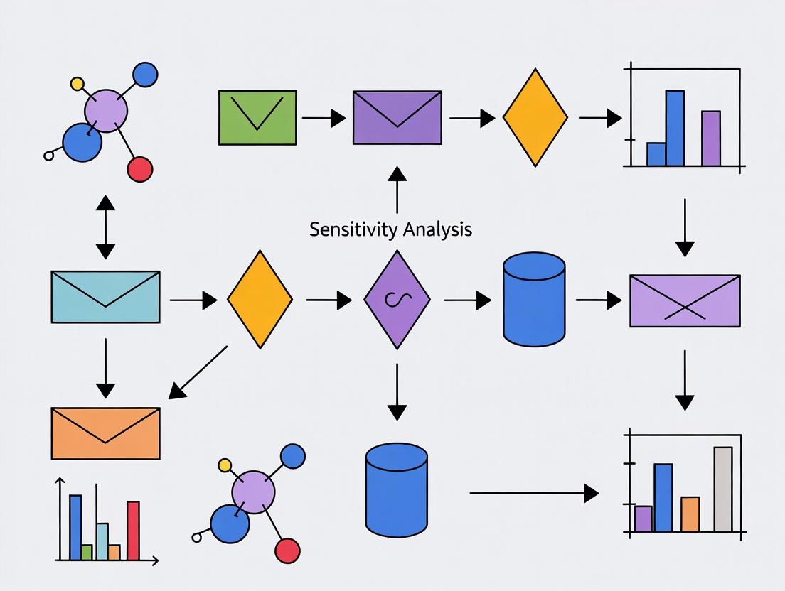
Visualizing SA Workflow and Biomechanical Pathways
Title: SA-Driven Model Calibration & Validation Workflow
Title: SA-Critical Parameters in Bone Mechanotransduction
The Scientist's Toolkit: Essential Research Reagents & Solutions
| Item/Category | Function in SA-Informed Biomechanics Research |
|---|---|
| High-Fidelity 3D Scanners (e.g., µCT, Laser) | Provides precise geometric input parameters and their population variance for model construction. Critical for geometry SA. |
| Biaxial/Triaxial Material Testing System | Characterizes anisotropic, nonlinear material properties (e.g., tendon, artery, bone) to define accurate parameter ranges for SA. |
| Digital Image Correlation (DIC) System | Provides full-field experimental strain data for validating model outputs identified as sensitive in SA. |
| Parameter Sampling Software (e.g., SALib, Dakota) | Implements algorithms (Sobol', Morris, FAST) to generate efficient input parameter sets for global SA. |
| Statistical Computing Environment (R, Python) | For calculating sensitivity indices (Sobol'), visualizing results, and performing uncertainty quantification. |
| Validated Finite Element Software (e.g., FEBio, Abaqus) | The core simulation platform. Must allow for batch/scripted runs to automate thousands of SA simulations. |
| Synthetic Biomimetic Phantoms | Provides a controlled, repeatable experimental platform for isolating and testing the impact of specific sensitive parameters. |
Sensitivity Analysis (SA) is a fundamental methodology in biomechanical modeling, used to quantify how the uncertainty in a model's output can be apportioned to different sources of uncertainty in its inputs. Within this thesis overview, we differentiate between two primary paradigms: Local Sensitivity Analysis (LSA) and Global Sensitivity Analysis (GSA). LSA assesses the impact of small perturbations around a nominal point in the input parameter space, often using derivatives. In contrast, GSA explores the entire input space, evaluating the effect of large variations and interactions among parameters. The choice between LSA and GSA has profound implications for the reliability, interpretation, and predictive power of biomechanical models, which are critical for applications ranging from implant design to drug delivery system development.
Core Methodological Distinctions
Local Sensitivity Analysis (LSA)
LSA is a one-at-a-time (OAT) method that computes the partial derivative of the model output with respect to an input parameter at a specific baseline value. It is computationally inexpensive but provides information only for the immediate vicinity of the chosen point.
Primary Methods:
- Finite Difference Method: Approximates the derivative: S_i = (f(x_i + Δx_i) - f(x_i)) / Δx_i.
- Direct Differentiation: Used in algorithmic differentiation, often applied within finite element (FE) solvers for biomechanics.
Global Sensitivity Analysis (GSA)
GSA methods vary all inputs simultaneously over their entire feasible ranges to apportion output variance to individual inputs and their interactions. They are computationally demanding but provide a comprehensive view.
Primary Methods:
- Variance-Based Methods (Sobol' Indices): Decompose output variance into contributions from individual parameters and parameter interactions.
- Elementary Effects Method (Morris Method): A screening method that provides qualitative measures of influence and interaction at moderate computational cost.
- Regression-Based Methods: Use standardized regression coefficients (SRC) on samples from a Monte Carlo simulation.
Quantitative Comparison
Table 1: Key Distinctions Between LSA and GSA
| Feature | Local Sensitivity Analysis (LSA) | Global Sensitivity Analysis (GSA) |
|---|---|---|
| Scope of Analysis | Single, nominal point in input space | Entire input parameter space |
| Parameter Interactions | Cannot detect interactions | Explicitly quantifies interaction effects |
| Computational Cost | Low (n+1 model runs for n parameters) | High (100s to 1000s of model runs) |
| Output Metric | Local derivatives (e.g., ∂Y/∂X_i) | Global indices (e.g., Sobol' Si, STi) |
| Primary Use Case | System linearity verification, gradient-based optimization | Model reduction, factor prioritization, uncertainty quantification |
| Typical Biomechanical Application | Linear elastic material model near a reference load; Pharmacokinetic (PK) model at standard dose | Nonlinear, large-deformation tissue models; Population PK/PD models with wide covariate ranges |
Table 2: Common Sensitivity Indices and Their Interpretation
| Index | Name | Range | Interpretation |
|---|---|---|---|
| S_i | First-order Sobol' Index | [0, 1] | Fraction of output variance due to input X_i alone. |
| S_Ti | Total-order Sobol' Index | [0, 1] | Fraction of variance due to X_i including all interactions with other inputs. |
| μ* (Morris) | Elementary Effects Mean | - | Measures overall influence of the parameter. |
| σ (Morris) | Elementary Effects Std. Dev. | - | Indicates involvement in interactions or nonlinear effects. |
Experimental Protocols for Cited Key Studies
Protocol 1: Local SA of a Knee Joint Finite Element Model
- Model Setup: Develop a validated FE model of a tibiofemoral joint in a commercial solver (e.g., Abaqus, FEBio).
- Parameter Selection: Define baseline values and perturbation magnitude (±1%) for inputs: Young's modulus of cartilage, menisci, and ligaments.
- Execution: For each parameter i, run two simulations: one at baseline (f(x)) and one with the parameter perturbed (f(x_i + Δx_i)).
- Calculation: Compute the normalized local sensitivity coefficient: LSC_i = (ΔOutput / Output_baseline) / (Δx_i / x_i_baseline).
- Output: Rank parameters by absolute LSC value to identify the most locally influential material property.
Protocol 2: Global SA (Sobol' Method) of a Bone Remodeling Algorithm
- Model Definition: Implement a computational bone remodeling model (e.g., based on strain energy density).
- Input Distributions: Define plausible probability distributions (e.g., uniform, normal) for all uncertain inputs (e.g., remodeling rate coefficient, error threshold, initial density).
- Sampling: Generate a (quasi-)random sample matrix (e.g., using Saltelli's extension of Sobol' sequences) of size N(2k+2), where k is the number of parameters.
- Model Evaluation: Run the biomechanical model for each row in the sample matrix to produce the corresponding output (e.g., final bone density).
- Index Calculation: Use the model outputs to calculate first-order (S_i) and total-order (S_Ti) Sobol' indices via variance decomposition.
- Interpretation: Identify which parameters contribute most to output variance. A large difference between S_Ti and S_i indicates significant interaction effects.
Visualizations of Workflows and Relationships
Title: Local Sensitivity Analysis (LSA) Workflow
Title: Global Sensitivity Analysis (GSA) Workflow
Title: Decision Tree for Choosing LSA or GSA
The Scientist's Toolkit: Research Reagent Solutions
Table 3: Essential Tools and Software for Sensitivity Analysis in Biomechanics
| Item / Solution | Function in Sensitivity Analysis | Example Product/Software |
|---|---|---|
| Finite Element Software with SA Plugins | Provides built-in tools for direct local sensitivity computation, often via direct differentiation. | Abaqus (Isight), COMSOL, FEBio with SA plugin. |
| Global SA Software Libraries | Implements advanced sampling and index calculation algorithms for GSA. | SALib (Python), GNU R sensitivity package, Simlab (JRC). |
| Quasi-Random Sequence Generators | Creates efficient space-filling samples for GSA to improve convergence. | Sobol' sequence, Latin Hypercube Sampling (LHS) algorithms. |
| High-Performance Computing (HPC) Cluster | Enables the thousands of model runs required for rigorous GSA of complex biomechanical models. | Local SLURM clusters, cloud computing (AWS, Azure). |
| Statistical & Data Analysis Software | Used to post-process results, visualize sensitivity indices, and perform regression-based SA. | Python (Pandas, Matplotlib, SciPy), R, MATLAB. |
| Uncertainty Quantification (UQ) Frameworks | Integrated platforms that couple forward modeling with parameter sampling and SA. | DAKOTA (Sandia), OpenTURNS, UQLab. |
When to Use Each Method: Guidelines for Biomechanical Modeling
Use Local Sensitivity Analysis (LSA) when:
- The model is confirmed to be linear or monotonic over the expected range of parameter variation.
- The analysis goal is to understand system behavior at a specific operating point (e.g., normal gait loading).
- Computational resources are severely limited, and a first-order approximation is acceptable.
- The gradient information is required for deterministic optimization or inverse problem-solving.
Use Global Sensitivity Analysis (GSA) when:
- The model is inherently nonlinear or exhibits threshold behaviors (common in tissue damage or failure models).
- Input parameters are uncertain across wide ranges (e.g., patient-specific material properties in a population study).
- The goal is to identify non-influential parameters for model reduction.
- Understanding interactions between parameters is critical (e.g., between drug diffusion rate and tissue degradation in a delivery model).
- The analysis is a precursor to robust design or comprehensive uncertainty quantification.
The distinction between local and global sensitivity analysis is not merely technical but philosophical, reflecting a choice between a focused, efficient probe and an exhaustive, systems-level exploration. In biomechanical modeling research—where complexity, nonlinearity, and uncertainty are paramount—GSA is often necessary for credible and generalizable results, despite its cost. LSA remains valuable for well-defined sub-problems and gradient-based applications. A robust SA strategy, potentially employing GSA for factor screening followed by targeted LSA, is essential for strengthening the inferential chain from model prediction to scientific insight or clinical decision-making.
In biomechanical modeling research, sensitivity analysis (SA) is a fundamental methodology for understanding the influence of model assumptions and input variability on simulated outcomes. This guide establishes the core terminology—Input Parameters, Output Responses, and Quantifying Uncertainty—within this context. Biomechanical models, ranging from finite element models of bone stress to multiscale models of cartilage lubrication or drug delivery in tissues, are complex and inherently uncertain. A rigorous SA framework is essential to assess model credibility, identify critical biological or mechanical factors, and guide resource-efficient experimentation and drug development.
Core Terminology: Definitions and Relationships
Input Parameters: These are the model parameters whose values are not derived from the model itself but must be supplied from external sources (e.g., experimental data, literature, estimation). In biomechanics, these can be geometric (e.g., bone dimensions, tissue layer thicknesses), material (e.g., Young's modulus, permeability, viscoelastic coefficients), kinematic (e.g., joint angles, loading rates), or biological (e.g., cell proliferation rate, drug diffusion coefficient). Parameters can be deterministic (fixed values) or stochastic (described by probability distributions).
Output Responses: Also called Quantities of Interest (QoIs), these are the results computed by the model. They are the target of the analysis and should be clinically or biologically relevant. Examples include peak stress/strain in a bone implant, contact pressure in a joint, rate of drug release from a polymeric scaffold, or predicted tissue deformation during surgical simulation.
Quantifying Uncertainty: This is the process of characterizing the degree of confidence in model predictions. It stems from two primary sources:
- Parameter Uncertainty: Arises from incomplete knowledge about the true values of input parameters (e.g., natural biological variation, measurement error).
- Model Structure Uncertainty: Arises from simplifications, missing physics, or incorrect assumptions in the model formulation itself (e.g., assuming isotropic versus anisotropic material behavior).
SA provides the mathematical tools to propagate input uncertainties to the output responses, thereby quantifying the overall uncertainty in predictions.
Title: The Role of Core Terminology in Sensitivity Analysis
Methodological Protocols for Sensitivity Analysis
The following experimental/computational protocols are standard in modern biomechanical SA.
3.1 Global Variance-Based Sensitivity Analysis (Sobol' Method) This protocol quantifies how much of the output variance each input parameter (or interactions between parameters) is responsible for.
- Step 1: Probabilistic Input Design. Define a joint probability distribution for all N uncertain input parameters (e.g., uniform, normal, log-normal based on experimental data).
- Step 2: Generate Sample Matrices. Create two N x M sample matrices (A and B), where M is the sample size (typically thousands), using quasi-random sequences (e.g., Sobol' sequence).
- Step 3: Construct Hybrid Matrices. For each parameter i, create a matrix C_i where all columns are from A, except the i-th column, which is from B.
- Step 4: Model Execution. Run the biomechanical model for all samples in matrices A, B, and each C_i, collecting the output QoI for each run.
- Step 5: Index Calculation. Compute the first-order (Si) and total-order (STi) Sobol' indices using variance estimators:
- Si = Variance due to parameter i alone / Total variance.
- STi = (Variance due to parameter i and all its interactions) / Total variance.
- Step 6: Interpretation. S_i identifies the most influential standalone parameter. S_Ti identifies parameters that are influential through interactions. A large difference between S_Ti and S_i suggests significant interaction effects.
3.2 Local Derivative-Based Sensitivity Analysis This protocol assesses the local effect of a small parameter change around a nominal value (e.g., a baseline patient geometry).
- Step 1: Establish Baseline. Define the nominal set of input parameters, x₀, and compute the baseline output, y₀ = f(x₀).
- Step 2: Parameter Perturbation. Perturb each parameter x_i by a small amount Δx_i (e.g., ±1%).
- Step 3: Finite Difference Calculation. For each parameter, compute the normalized local sensitivity coefficient (LSC):
- LSCi = [ ( f(x₀ + Δxi) - f(x₀) ) / f(x₀) ] / [ Δxi / x₀i ].
- Step 4: Ranking. Rank parameters by the absolute magnitude of LSC_i. This is computationally cheap but only valid locally.
Quantitative Data from Contemporary Research
Table 1: Exemplar Sensitivity Indices from a Finite Element Model of Vertebral Strength
| Input Parameter (Distribution) | Nominal Value | First-Order Sobol' Index (S_i) | Total-Order Sobol' Index (S_Ti) | Key Insight |
|---|---|---|---|---|
| Trabecular Bone Modulus (Normal, μ=300 MPa, σ=45 MPa) | 300 MPa | 0.52 | 0.61 | Dominant standalone factor. |
| Cortical Bone Thickness (Uniform, 0.5-1.5 mm) | 1.0 mm | 0.18 | 0.45 | High interaction with geometry. |
| Endplate Strength (Log-normal) | 25 MPa | 0.10 | 0.12 | Minor, independent influence. |
| Disc Nucleus Pressure (Normal) | 0.8 MPa | 0.05 | 0.22 | Low standalone, high interaction. |
Table 2: Local Sensitivity of Knee Contact Mechanics to Material Properties
| Perturbed Parameter (Baseline) | Change | Peak Contact Pressure Change | Normalized LSC |
|---|---|---|---|
| Meniscus Compressive Modulus (5 MPa) | +10% | -3.2% | -0.32 |
| Articular Cartilage Permeability (1.5e-15 m⁴/Ns) | +10% | +1.8% | +0.18 |
| Ligament Stiffness (Linear, 250 N/mm) | +10% | < 0.5% | < 0.05 |
The Scientist's Toolkit: Research Reagent Solutions
Table 3: Essential Computational & Experimental Tools for SA in Biomechanics
| Item/Category | Function in SA Context | Example/Note |
|---|---|---|
| Quasi-Random Sequence Generators | Create efficient, space-filling input parameter samples for global SA. | Sobol' sequences, Latin Hypercube Sampling (LHS). |
| High-Performance Computing (HPC) Cluster | Enables the thousands of model runs required for Monte Carlo and global methods. | Cloud-based (AWS, Google Cloud) or local clusters. |
| Uncertainty Quantification Software Libraries | Provide pre-built algorithms for SA and statistical analysis. | SALib (Python), UQLab (MATLAB), Dakota (Sandia). |
| Micro-CT / MRI Imaging Data | Provides population-derived distributions for geometric input parameters. | Source for statistical shape models and density variation. |
| Biaxial/Triaxial Material Testers | Quantifies stochastic material properties for soft tissues (ligaments, cartilage). | Outputs mean and standard deviation for constitutive model parameters. |
| Digital Image Correlation (DIC) Systems | Provides full-field experimental strain data for validating uncertain model outputs. | Gold standard for comparing simulated vs. actual deformation. |
Title: Workflow for Uncertainty Quantification in Biomechanical Modeling
The Critical Role of SA in Model Development, Verification, and Credibility
Sensitivity Analysis (SA) is an indispensable mathematical and computational methodology within biomechanical modeling research. It systematically investigates how the uncertainty in the output of a model (numerical or otherwise) can be apportioned to different sources of uncertainty in its inputs. This guide details its critical role in model development, verification, and the establishment of credibility, forming a cornerstone for robust research in biomechanics, orthopedics, and drug development for musculoskeletal diseases.
Theoretical Foundations of SA in Biomechanics
Biomechanical models are complex, integrating anatomical geometry, material properties, boundary conditions, and loading scenarios. SA provides the framework to:
- Identify Influential Parameters: Distinguish critical model inputs (e.g., ligament stiffness, cartilage permeability) from non-influential ones, guiding focused experimental data collection.
- Assess Model Robustness: Determine if model predictions remain stable under plausible variations in inputs, a key aspect of verification.
- Simplify Models: Enable model reduction by fixing non-influential parameters, enhancing computational efficiency.
- Inform Decision-Making: Quantify confidence in model-based predictions, such as implant performance or tissue stress levels, essential for translational applications.
Core SA Methodologies: Protocols and Application
Two primary classes of SA are employed, each with specific experimental (computational) protocols.
Local Sensitivity Analysis (LSA)
LSA evaluates the effect of small perturbations of an input parameter around a nominal value, often computing partial derivatives.
Experimental Protocol (One-at-a-Time - OAT):
- Define a nominal set of input parameters ( P0 = {p1^0, p2^0, ..., pn^0} ).
- Run the baseline simulation to obtain output ( Y_0 ).
- For each parameter ( pi ): a. Perturb the parameter by a small ( \Delta pi ) (e.g., ±1%). b. Run a new simulation with the set ( {p1^0, ..., pi^0 + \Delta pi, ..., pn^0} ). c. Compute the local sensitivity measure (e.g., normalized derivative): ( Si = (\Delta Y / Y0) / (\Delta pi / pi^0) ).
- Rank parameters by the absolute magnitude of ( S_i ).
Global Sensitivity Analysis (GSA)
GSA apportions output variance to the full distribution of input parameters, exploring the entire input space and capturing interactions.
Experimental Protocol (Variance-Based using Sobol' Indices):
- Define probability distributions for all uncertain input parameters.
- Generate two independent sampling matrices (A and B) of size ( N \times n ) using quasi-random sequences (e.g., Sobol' sequence).
- Construct ( n ) additional matrices ( A_B^{(i)} ), where column ( i ) is from matrix B and all other columns are from A.
- Run the model for all ( N \times (n + 2) ) sample points.
- Compute First-order (main) Sobol' indices ( Si ) (effect of ( pi ) alone) and Total-order Sobol' indices ( S{Ti} ) (effect of ( pi ) including all interactions): [ Si = \frac{V{pi}(E{\sim pi}(Y|pi))}{V(Y)}, \quad S{Ti} = 1 - \frac{V{\sim pi}(E{pi}(Y|\sim pi))}{V(Y)} ]
- ( S{Ti} - Si ) quantifies interaction strength for parameter ( i ).
Table 1: Comparison of SA Methods in Biomechanical Modeling
| Feature | Local SA (OAT) | Global SA (Variance-Based) |
|---|---|---|
| Input Space | Local, around a point | Global, across full distributions |
| Interaction Effects | Cannot detect | Explicitly quantifies |
| Computational Cost | Low ((n+1) runs) | High ((N \times (n+2)) runs) |
| Typical Output Metric | Normalized derivative ( S_i ) | Sobol' indices (( Si ), ( S{Ti} )) |
| Best For | Simple models, gradient-based optimization | Credibility assessment, complex nonlinear models |
Table 2: Illustrative SA Results from a Finite Element Knee Model
| Parameter (Input ( p_i )) | Nominal Value | Local Sensitivity ( S_i ) | First-Order Sobol' Index ( S_i ) | Total-Order Sobol' Index ( S_{Ti} ) |
|---|---|---|---|---|
| Ligament Stiffness | 200 N/mm | 0.85 | 0.52 | 0.68 |
| Cartilage Elastic Modulus | 10 MPa | 0.15 | 0.08 | 0.35 |
| Meniscus Material Properties | Hyperelastic | 0.10 | 0.05 | 0.22 |
| Bone Geometry (Condyle Radius) | 22 mm | 0.45 | 0.30 | 0.31 |
| Output: Peak Contact Stress in Tibial Cartilage |
The SA-Enhanced Model Workflow
Diagram Title: SA-Integrated Model Development and Credibility Workflow
The Scientist's Toolkit: Research Reagent Solutions
Table 3: Essential Tools for Conducting SA in Biomechanics
| Item | Function in SA | Example/Note |
|---|---|---|
| High-Performance Computing (HPC) Cluster | Enables thousands of model runs required for GSA. | Cloud-based (AWS, Google Cloud) or local clusters. |
| SA-Specific Software Libraries | Implements sampling and index calculation algorithms. | SALib (Python), OpenTURNS (C++/Python), Dakota (Sandia Labs). |
| Quasi-Random Sequence Generators | Generates efficient, space-filling input samples. | Sobol', Halton, or Latin Hypercube Sampling (LHS) algorithms. |
| Finite Element Analysis Software | The core biomechanical simulator. | FEBio, Abaqus, ANSYS with scripting API for batch runs. |
| Parameter Distribution Fitting Tools | Defines statistical input distributions from experimental data. | SciPy (Python), R fitdistrplus package. |
| Visualization & Post-Processing Suites | Creates sensitivity indices plots, tornado charts, interaction diagrams. | Matplotlib/Seaborn (Python), ParaView for spatial sensitivity. |
Signaling Pathway of Model Credibility Attainment
Diagram Title: SA as the Central Signaling Pathway to Model Credibility
Within the thesis of biomechanical modeling research, SA is not merely an optional step but a critical, integrating methodology that transforms a model from a complex hypothesis into a credible tool for scientific insight and decision-making. It rigorously connects model development with verification and validation, providing the quantitative evidence necessary to trust model predictions in drug development, surgical planning, and medical device evaluation.
Historical Perspective and Evolution of SA in Biomedical Engineering
Sensitivity Analysis (SA) is a critical methodological pillar in biomedical engineering, providing systematic techniques to quantify how uncertainty in a model's outputs can be apportioned to different sources of uncertainty in its inputs. This paper, framed within a broader thesis on the overview of sensitivity analysis in biomechanical modeling research, traces the historical development and evolution of SA, highlighting its transition from a simple parameter perturbation tool to a sophisticated framework essential for model credibility, regulatory compliance, and clinical translation.
Historical Trajectory: From Simple Methods to Complex Systems
The application of SA in biomedical engineering has paralleled the increasing complexity of computational models. The evolution can be segmented into distinct eras.
1. The Era of Local Methods (1970s-1990s): Early biomechanical models, often linear and low-dimensional, employed local SA, primarily using derivative-based approaches (e.g., one-at-a-time - OAT). The focus was on understanding the immediate neighborhood of a nominal parameter set.
2. The Shift to Global Methods (1990s-2010s): As models grew to incorporate nonlinearities, feedback, and stochastic elements (e.g., pharmacokinetic/pharmacodynamic - PK/PD, cardiovascular dynamics), local SA proved insufficient. Global SA (GSA) methods, which explore the entire input space, became the standard. Techniques like Sobol’ indices, Fourier Amplitude Sensitivity Testing (FAST), and Morris screening enabled the ranking of influential parameters and interaction effects.
3. The Modern Era of Integration and High-Dimensionality (2010s-Present): Contemporary challenges involve complex, multi-scale models (e.g., in-silico clinical trials, systems pharmacology), "black-box" machine learning models, and the need for integration with uncertainty quantification (UQ) and model verification/validation (V&V) workflows. SA is now a mandatory component for regulatory submission (e.g., FDA's ASME V&V 40 standard) and is applied to models with thousands of inputs.
Table 1: Evolution of Primary SA Methods in Biomedical Engineering
| Era | Primary Methods | Key Characteristics | Typical Biomechanical Application |
|---|---|---|---|
| Local (1970s-90s) | One-at-a-Time (OAT), Derivative-based | Computationally cheap; ignores interactions & global space. | Linear elastic bone/implant stress analysis. |
| Global (1990s-2010s) | Morris (Screening), Sobol’ (Variance-based), FAST | Explores full input space; ranks parameters, detects interactions. | PK/PD models, cardiac electrophysiology models, tissue growth models. |
| Modern (2010s-) | Sobol’ (via meta-models), DALI, Polynomial Chaos, ML-based SA | Handles high-dimensionality, integrates UQ & V&V, model-agnostic. | Multi-scale cancer models, population-based in-silico trials, AI/ML diagnostic classifiers. |
Table 2: Prevalence of SA Methods in Recent Biomedical Literature (Sample Analysis)
| SA Method | % of Reviewed Papers (2020-2024) | Primary Field of Application |
|---|---|---|
| Variance-based (Sobol’) | 38% | Systems biology, pharmacology, cardiovascular models. |
| Morris Screening | 25% | Initial screening for high-dimensional biomechanical & tissue models. |
| Regression-based | 18% | Clinical outcome prediction models, epidemiological models. |
| Derivative-based (Local) | 10% | Continuum-scale biomechanics (FEA of joints/implants). |
| Other/ML-based | 9% | Deep learning model interpretation, image-based diagnostics. |
Experimental Protocols for Key Sensitivity Analyses
Protocol 1: Global Variance-Based SA (Sobol’ Indices) for a PK/PD Model Objective: To quantify the contribution of individual PK parameters and their interactions to the variance in the predicted drug effect over time.
- Model Definition: Define the PK/PD model (e.g., two-compartment PK with an Emax PD model). Specify all input parameters (e.g., clearance, volumes, EC50).
- Parameter Distributions: Assign plausible probability distributions (e.g., log-normal) to each uncertain input parameter based on prior literature or experimental data.
- Sample Generation: Generate two independent random matrices (A and B) of size N x k (N~1000-5000, k=#parameters) using a quasi-random sequence (Sobol’ sequence).
- Model Evaluation: Create a set of hybrid matrices to compute first-order and total-effect indices. Run the model for each row in all matrices, generating the output of interest (e.g., AUC, Cmax, effect at time t).
- Index Calculation: Compute first-order (Si) and total-effect (STi) Sobol’ indices using the estimator of Saltelli (2010). Si measures the direct contribution of Xi, while STi includes all interaction effects.
- Interpretation: Rank parameters by STi. Parameters with STi > 0.1 are typically considered influential. The sum of all Si indicates the presence of interaction effects.
Protocol 2: Morris Screening for a High-Dimensional Bone Remodeling FEA Model Objective: To identify the most influential material properties and loading conditions in a complex finite element model of bone adaptation with >50 inputs.
- Factor Prioritization: List all uncertain input factors (e.g., elastic moduli of cortical/trabecular bone, muscle force magnitudes, remodeling rate constants).
- Discretization & Trajectory Design: Define a plausible range (p-levels) for each factor. Generate
rrandom trajectories (r=20-50) in the input space, where each trajectory changes one factor at a time. - Elementary Effect Calculation: For each trajectory, compute the Elementary Effect (EE) of factor Xi: EE_i = [Y(X1,..., Xi+Δ,..., Xk) - Y(X)] / Δ, where Δ is a predetermined step size.
- Statistical Analysis: For each factor Xi, calculate the mean (μ) and standard deviation (σ) of its absolute EE across all trajectories. Plot μ* (mean of |EE|) vs σ.
- Factor Screening: Factors with high μ* are considered to have a large overall influence. Factors with high σ indicate significant interactions or nonlinear effects. Select the top ~10-15 factors for subsequent, more detailed GSA.
Visualizations
Title: Evolution of Sensitivity Analysis Methods Over Time
Title: Workflow for Global Variance-Based Sensitivity Analysis
The Scientist's Toolkit: Key Research Reagent Solutions
Table 3: Essential Software and Computational Tools for Modern SA
| Tool/Reagent | Function/Description | Typical Use Case |
|---|---|---|
| SALib (Python Library) | Open-source library implementing Sobol’, Morris, FAST, and others. | Accessible GSA for custom models; integration into simulation pipelines. |
| Dakota (Sandia NL) | Advanced UQ/SA toolkit with optimization capabilities. | Large-scale, high-performance computing (HPC) SA for complex biomechanics. |
| Gaussian Process / Kriging Meta-models | Surrogate models to approximate complex simulations for efficient SA. | Enabling GSA for computationally expensive FEA or agent-based models. |
| SUMO Toolbox (Matlab) | Advanced SA, UQ, and meta-modeling with GUI and scripting. | SA for systems biology and pharmacological models developed in Matlab/Simulink. |
| Sensitivity Analysis Plugin (COMSOL) | Integrated local and global SA within a multiphysics FEA environment. | Direct SA of coupled physics problems (e.g., electro-thermal tissue ablation). |
| Global SA in PK/PD Software (e.g., Monolix, NONMEM) | Built-in SA workflows for population pharmacokinetic analysis. | Quantifying parameter influence on drug exposure and response variability. |
The historical perspective reveals that Sensitivity Analysis in biomedical engineering has evolved from a peripheral check to a central, indispensable component of the model-based research and development lifecycle. Its maturation, driven by increasing model complexity and regulatory expectations, has produced a robust toolkit of global methods. For today's researcher, effectively applying SA is no longer optional but a fundamental practice for ensuring the reliability, interpretability, and defensibility of biomechanical models in drug development and therapeutic innovation.
How to Perform Sensitivity Analysis: Methods, Tools, and Real-World Biomechanical Applications
Within the broader thesis on the overview of sensitivity analysis (SA) in biomechanical modeling research, this guide explores three foundational methodological approaches. Sensitivity analysis is critical in this domain for identifying which model input parameters—such as material properties, boundary conditions, or physiological forces—most influence outputs like stress, strain, or failure prediction. This process validates models, enhances efficiency by focusing on key parameters, and quantifies uncertainty, directly impacting applications in implant design, surgical planning, and drug delivery device development.
Core Methodologies
One-at-a-Time (OAT) Screening
A local SA method where one input parameter is varied while all others are held at baseline values.
Protocol:
- Define a baseline point ( x0 = (x1^0, x2^0, ..., xk^0) ) in the k-dimensional parameter space.
- For each parameter ( xi ), define a range of variation ( [xi^0 - \Deltai, xi^0 + \Delta_i] ).
- For ( i = 1 ) to ( k ):
- Vary ( x_i ) across its defined range.
- Hold all other parameters ( x{j \neq i} = xj^0 ).
- Compute the model output ( y ).
- Analyze the output variation, often via elementary effects: ( EEi = [y(xi^0+\Deltai) - y(xi^0)] / \Delta_i ).
Limitations: Cannot detect interactions between parameters; results are valid only locally around the chosen baseline.
Morris Screening (Elementary Effects Method)
A global screening method that improves upon OAT by efficiently sampling the input space to provide a measure of global sensitivity.
Protocol:
- Discretize the input space into a p-level grid for each of the k factors.
- Generate an initial random baseline vector ( x^* ).
- Construct a trajectory through the input space by randomly changing one factor at a time. The step size ( \Delta ) is a multiple of ( 1/(p-1) ).
- For a trajectory starting at ( x^{(0)} ), a series of points ( x^{(0)}, x^{(1)}, ..., x^{(k)} ) is generated, each differing from the previous in one component.
- The elementary effect for factor ( i ) is calculated as: ( EE_i(x) = \frac{[y(x^{(i)}) - y(x^{(i-1)})]}{\Delta} )
- Repeat for r random trajectories (typically 10-50).
- Compute sensitivity metrics:
- ( \mui^* = \frac{1}{r} \sum{j=1}^{r} |EE_i^j| ) (measures overall influence).
- ( \sigmai = \sqrt{\frac{1}{r-1} \sum{j=1}^{r} (EEi^j - \mui)^2 } ) (measures nonlinear/interaction effects).
Derivative-Based Methods (Local and Global)
These methods use partial derivatives to quantify sensitivity, formalized as Local Sensitivity Analysis (LSA) or extended to Global Sensitivity Analysis via the Derivative-based Global Sensitivity Measure (DGSM).
Protocol for LSA:
- Select a nominal point ( x_0 ).
- Compute the partial derivative ( \frac{\partial y}{\partial xi} ) at ( x0 ), analytically or via finite differences: ( \frac{\partial y}{\partial xi} \approx \frac{y(xi^0+\epsilon) - y(x_i^0)}{\epsilon} ).
- Normalize to obtain sensitivity coefficients (e.g., ( Si = (\partial y / \partial xi) \cdot (x_i^0 / y^0) ) ).
Protocol for DGSM:
- Assume inputs are independent random variables with probability density functions.
- Compute the partial derivative ( \frac{\partial y}{\partial x_i} ) at many points in the input space (via Monte Carlo).
- Calculate the DGSM index: ( \nui = \int{\Omega} (\frac{\partial y}{\partial x_i})^2 dx ), where ( \Omega ) is the input space.
- Often compared to total Sobol' indices, as ( \nu_i ) provides an upper bound.
Comparative Analysis and Data Presentation
Table 1: Methodological Comparison for Biomechanical Application
| Feature | OAT Screening | Morris Method | Derivative-Based (LSA) | Derivative-Based (DGSM) |
|---|---|---|---|---|
| Scope | Local | Global | Local | Global |
| Interaction Detection | No | Yes (via σ) | No | Indirect (via ν bounds) |
| Computational Cost | Very Low (k+1 runs) | Low (r*(k+1) runs) | Very Low (k+1 runs) | High (Monte Carlo based) |
| Primary Output | Elementary Effect | μ* (importance), σ (interactions) | Local Sensitivity Coefficients | DGSM indices (ν_i) |
| Key Advantage | Simplicity, intuitive | Efficient global screening | Precise local gradient | Theoretical link to variance |
| Main Disadvantage | Misses interactions/ non-linearities | Qualitative ranking | Not valid for large ranges | Higher cost than Morris |
Table 2: Illustrative Quantitative Results from a Tendon Biomechanics Model
| Parameter (Example) | OAT EE | Morris μ* | Morris σ | LSA Coefficient | DGSM ν_i (Normalized) |
|---|---|---|---|---|---|
| Elastic Modulus (E) | 12.5 | 11.8 | 1.2 | 0.95 | 0.61 |
| Fiber Diameter (d) | 8.1 | 7.9 | 3.5 | 0.62 | 0.52 |
| Load Frequency (f) | -4.2 | 4.5 | 0.8 | -0.31 | 0.08 |
| Damping Ratio (ζ) | 0.7 | 0.8 | 0.1 | 0.05 | 0.01 |
Visualized Workflows and Relationships
Title: OAT Screening Workflow for Biomechanical Models
Title: Morris Method Global Screening Procedure
Title: Relationship Between SA Methods in Research
The Scientist's Toolkit: Research Reagent Solutions
Table 3: Essential Computational Tools for Sensitivity Analysis in Biomechanics
| Item/Category | Function in SA | Example Solutions/Software |
|---|---|---|
| SA-Specific Libraries | Pre-implemented algorithms for OAT, Morris, DGSM. | SALib (Python), GSA (MATLAB), sensitivity (R). |
| Numerical Solver | Core engine to compute model outputs for perturbed inputs. | FEBio, ANSYS, COMSOL, Abaqus, OpenSim. |
| Scripting Interface | Automates parameter variation, batch job submission, and results collection. | Python, MATLAB, R. |
| High-Performance Computing (HPC) | Manages the hundreds to thousands of model runs required for global SA. | Slurm, PBS, cloud compute instances (AWS, GCP). |
| Data & Visualization Suite | Processes and visualizes sensitivity indices and rankings. | NumPy/Pandas (Python), ggplot2 (R), Paraview for spatial fields. |
| Uncertainty Quantification (UQ) Framework | Integrates SA with broader calibration, validation, and UQ workflows. | DAKOTA, UQLab, OpenTURNS. |
1. Introduction within Biomechanical Modeling Research Sensitivity Analysis (SA) is a cornerstone of robust biomechanical modeling, which seeks to understand the complex relationship between biological inputs (e.g., material properties, loading conditions, geometric parameters) and model outputs (e.g., stress, strain, displacement). Global SA techniques are essential for quantifying how uncertainty in model inputs contributes to uncertainty in the output, identifying non-influential parameters to reduce model complexity, and guiding experimental design. This guide provides an in-depth technical examination of three pivotal global SA methods: Sobol' indices, Fourier Amplitude Sensitivity Testing (FAST), and Polynomial Chaos Expansion (PCE), contextualized within modern biomechanical research.
2. Core Methodologies and Mathematical Foundations
2.1 Sobol' Indices (Variance-Based Method) Sobol' indices decompose the total variance of the model output into contributions from individual inputs and their interactions.
- First-Order Index (Si): Measures the contribution of input *Xi* alone to the output variance.
- Formula: ( Si = \frac{V{Xi}[E{\mathbf{X}{\sim i}}(Y|Xi)]}{V(Y)} )
- Total-Order Index (S{Ti}): Measures the total contribution of input *Xi*, including all its interactions with other inputs.
- Formula: ( S{Ti} = 1 - \frac{V{\mathbf{X}{\sim i}}[E{Xi}(Y|\mathbf{X}{\sim i})]}{V(Y)} )
2.2 Fourier Amplitude Sensitivity Testing (FAST) FAST transforms a multi-dimensional integral into a one-dimensional one by exploring the parameter space along a defined search curve. The variance contribution of each parameter is linked to the amplitude of its characteristic frequency in the Fourier-transformed output.
- Search Curve: ( Xi(s) = Gi(\sin(\omegai s)) ), where ( \omegai ) are integer frequencies assigned to each parameter.
- The output ( Y(s) ) becomes a periodic function, and its power spectrum at frequency ( \omegai ) is proportional to the variance from ( Xi ).
2.3 Polynomial Chaos Expansion (PCE) PCE represents the random model output as a spectral expansion in terms of orthogonal polynomial basis functions ( \Psi_{\boldsymbol{\alpha}} ) of the uncertain inputs.
- Expansion: ( Y = \sum{\boldsymbol{\alpha} \in \mathbb{N}^M} c{\boldsymbol{\alpha}} \Psi_{\boldsymbol{\alpha}}(\mathbf{X}) )
- Sobol' indices can be computed analytically and post-hoc from the PCE coefficients ( c_{\boldsymbol{\alpha}} ), as the variance decomposes by the orthogonality of the basis.
3. Quantitative Comparison of Methods Table 1: Comparative Summary of Advanced Global SA Techniques
| Feature | Sobol' Indices | FAST | Polynomial Chaos Expansion (PCE) |
|---|---|---|---|
| Core Principle | Variance decomposition via Monte Carlo | Spectral analysis via parameter space traversal | Surrogate modeling via orthogonal polynomial expansion |
| Computational Cost | High (requires ~N*(M+2) model runs) | Moderate | Low once surrogate is built; cost in training data |
| Interactions | Explicitly quantified (higher-order indices) | Can be estimated via extended FAST | Naturally captured in the expansion |
| Output | First & total-order indices | First-order indices primarily | Full surrogate model & analytical indices |
| Best For | Detailed variance attribution, small-to-medium M | Screening, moderate M, models with periodic response | Expensive models, derivative-based analysis, many queries |
4. Experimental Protocols for Implementation
4.1 Protocol for Computing Sobol' Indices via Saltelli's Algorithm
- Define Input Distributions: Assign probability distributions (e.g., uniform, normal) to all M uncertain biomechanical parameters.
- Generate Matrices: Create two (N x M) random sample matrices (A and B) using quasi-random sequences (e.g., Sobol' sequence).
- Construct Hybrid Matrices: For each parameter i, create matrix C_i, where column i is from B and all other columns are from A.
- Model Evaluation: Run the biomechanical model (e.g., finite element analysis) for all rows in A, B, and each C_i (Total runs = N*(M+2)).
- Calculate Indices: Use the model outputs to compute ( V(Y) ), ( E(Y) ), and variances of conditional expectations to compute ( Si ) and ( S{Ti} ).
4.2 Protocol for PCE-Based SA in a Bone Remodeling Model
- Parameter Selection: Identify M uncertain inputs (e.g., Young's modulus, permeability, applied load magnitude).
- Basis Construction: Choose orthogonal polynomial families matching input distributions (e.g., Legendre for uniform, Hermite for normal).
- Design of Experiments: Generate input samples using advanced schemes like LHS or optimal design from the polynomial basis.
- Surrogate Training: Execute the full biomechanical model for each sample. Solve for PCE coefficients via regression or spectral projection.
- Validation & SA: Validate the PCE surrogate against a hold-out test set. Compute Sobol' indices directly via summation of squared coefficients grouped by index.
5. Visualization of Workflows
Workflow for Sobol' Indices Computation
PCE-Based Sensitivity Analysis Workflow
6. The Scientist's Toolkit: Research Reagent Solutions
Table 2: Essential Software Tools & Libraries for Global SA
| Item (Software/Library) | Function in SA | Typical Use in Biomechanics |
|---|---|---|
| SALib (Python) | Implements Sobol', FAST, and other SA methods. | Direct integration with Python-based modeling pipelines for parameter screening and ranking. |
| UQLab (MATLAB) | A comprehensive uncertainty quantification framework featuring PCE and SA. | Building surrogates for complex finite element models and performing derivative-based SA. |
| Dakota (C++/API) | A versatile optimization/UQ toolkit from Sandia National Labs. | Coupling with commercial FEA software (Abaqus, FEBio) for large-scale, high-performance SA studies. |
| Chaospy (Python) | Advanced library for constructing polynomial chaos expansions. | Creating custom PCE surrogates for stochastic biomechanical simulations with non-standard input distributions. |
| OpenTURNS (C++/Python) | An industrial library for treatement of uncertainties in numerical simulations. | Robust sensitivity and reliability analysis of implant designs under uncertain physiological loads. |
Sensitivity Analysis (SA) is a critical methodological component in biomechanical modeling, used to quantify how uncertainty in a model's input parameters contributes to uncertainty in its outputs. Within the broader thesis of "Overview of sensitivity analysis in biomechanical modeling research," this guide provides a practical implementation framework. It enables researchers to enhance model credibility, identify key biological drivers, and optimize experimental design in areas like orthopedics, cardiovascular mechanics, and drug delivery systems.
Foundational SA Methods and Selection Criteria
Selection of an SA method depends on model linearity, computational cost, and the desired analysis (screening or quantitative). The core methodologies are summarized below.
Table 1: Core Sensitivity Analysis Methods and Applications
| Method | Type | Key Metric | Ideal Model Characteristics | Biomechanical Application Example |
|---|---|---|---|---|
| Morris Method | Global, Screening | Elementary Effects (μ*, σ) | High-dimensional, computationally expensive (10-50 inputs) | Screening material properties in a finite element (FE) bone model. |
| Sobol' Indices | Global, Variance-based | First-order (Si), Total-order (STi) | Nonlinear, non-monotonic, moderate computational cost (≤50 inputs) | Quantifying influence of muscle activation parameters on joint contact forces. |
| Fourier Amplitude Sensitivity Test (FAST) | Global, Variance-based | First-order indices | Moderate dimensions, periodic sampling | Analyzing soft tissue constitutive model parameter sensitivity. |
| Local (One-at-a-Time - OAT) | Local | Partial derivatives | Linear, additive, rapid execution | Preliminary check of a new pharmacokinetic-pharmacodynamic (PK-PD) model. |
Software Toolkit Implementation Guide
SALib (Python Ecosystem)
SALib is an open-source Python library for performing global SA.
Experimental Protocol: Implementing Sobol' Analysis with SALib
- Problem Definition: Define the model's input parameters and their distributions (e.g., uniform, normal).
- Sample Generation: Use
SALib.sample.saltellito generate the model input sample matrix (N*(2D+2) samples). - Model Evaluation: Run the biomechanical model (e.g., an OpenSim simulation or custom PDE solver) for each input sample to compute output(s) of interest (e.g., peak stress, diffusion rate).
- Analysis: Compute first and total-order Sobol' indices using
SALib.analyze.sobol.
Example Code Snippet:
Dakota (Sandia National Labs)
Dakota is a comprehensive toolkit for optimization and uncertainty quantification, suitable for high-performance computing (HPC) environments.
Experimental Protocol: Morris Screening with Dakota
- Configuration: Create an input file (
dakota.in) specifying methodmorris, parameter ranges, number of trajectories, and output variables. - Interface Setup: Configure Dakota to interface with your simulation code (e.g., Abaqus, FEBio) via system calls or file I/O.
- Execution: Run Dakota from the command line:
dakota -i dakota.in -o dakota.out. - Post-processing: Dakota outputs measures of μ (mean) and σ (standard deviation) of elementary effects for ranking parameter influence.
Custom Implementation in MATLAB/Python
Custom scripts offer maximum flexibility for integrating SA into proprietary or specialized modeling pipelines.
Protocol: Custom Local Sensitivity Analysis
- Baseline: Define a set of nominal parameter values (p0).
- Perturbation: For each parameter i, create a perturbed set pi = p0, but with pi[i] = p0[i] * (1 + Δ), where Δ is a small fraction (e.g., 0.01).
- Evaluation: Compute the model output for p0 and each p_i.
- Calculation: Compute normalized sensitivity coefficients: Si = (Output(pi) - Output(p0)) / (Δ * Output(p0)).
Visualization of SA Workflow in Biomechanics
Diagram Title: SA Workflow in Biomechanical Modeling
The Scientist's Toolkit: Essential Research Reagents & Software
Table 2: Key Software and Computational Reagents for SA
| Item Name | Category/Type | Primary Function in SA |
|---|---|---|
| SALib | Python Library | Provides turnkey functions for sampling (Saltelli, Morris) and analysis (Sobol', FAST) for global SA. |
| Dakota | HPC Toolkit | Enables large-scale parametric studies, optimization, and SA tightly coupled with simulation codes on clusters. |
| OpenSim | Biomech. Simulation | Provides musculoskeletal models; SA is used to identify critical muscle-tendon or kinematic parameters. |
| FEBio | FE Biomechanics | Solves nonlinear biomechanics FE problems; SA determines sensitive material properties or boundary conditions. |
| NumPy/SciPy | Python Libraries | Core numerical backends for custom SA implementations and data processing. |
| MATLAB Global Optimization Toolbox | Commercial Library | Includes functions for conducting SA, particularly useful for models already built in MATLAB/Simulink. |
| Jupyter Notebook | Development Environment | Ideal for interactive exploration, visualization, and documentation of SA results. |
| ParaView/Matplotlib | Visualization Tools | Critical for creating publication-quality plots and charts of sensitivity indices and parameter interactions. |
Advanced Considerations & Future Directions
- High-Dimensional & Emulator-Based SA: For models with runtimes of hours/days, replace the full model with a Gaussian Process or Polynomial Chaos Expansion emulator to make global SA feasible.
- Time-Varying SA: Compute sensitivity indices at each time point for dynamic outputs (e.g., joint angle over gait cycle) to understand parameter influence evolution.
- Integration with Uncertainty Quantification (UQ): SA should be part of a larger UQ workflow, where it directly informs which parameters require precise calibration or probabilistic representation.
- Experimental Design: Results from SA can prioritize which physical experiments (e.g., mechanical tissue testing) will most effectively reduce model output uncertainty.
Implementing robust SA using these toolkits moves biomechanical modeling from a purely descriptive endeavor to a predictive, hypothesis-testing framework, directly supporting research credibility and drug/device development.
This case study is situated within a broader thesis investigating sensitivity analysis (SA) in biomechanical modeling research. SA is a critical methodology for quantifying how the uncertainty in the output of a model can be apportioned to different sources of uncertainty in the model inputs. In the context of finite element (FE) modeling of bone and orthopedic implants, SA provides a systematic framework for identifying the most influential material properties, geometric parameters, and boundary conditions. This identification is paramount for model simplification, validation, and ensuring that research and development efforts focus on parameters that materially affect predictive outcomes.
Core Concepts: Global vs. Local Sensitivity Analysis
Local Sensitivity Analysis (One-at-a-Time - OAT): Assesses the effect of varying one parameter at a time around a nominal value (e.g., central difference derivative). It is computationally efficient but cannot explore the full input space or capture interactions between parameters.
Global Sensitivity Analysis (Variance-Based Methods): Quantifies the contribution of each input parameter, and its interactions with others, to the output variance over the entire multi-dimensional parameter space. The most common indices are the first-order Sobol' index (Si), measuring the main effect, and the total-effect Sobol' index (STi), which includes interaction effects.
Key Parameters in Bone and Implant FE Models
The table below summarizes common parameters subjected to sensitivity analysis in this domain.
Table 1: Key Input Parameters for Sensitivity Analysis in Bone-Implant FE Models
| Parameter Category | Specific Parameter | Typical Range/Variation | Primary Output Metrics of Interest |
|---|---|---|---|
| Bone Material Properties | Elastic Modulus (Cortical) | 10-20 GPa | Bone strain, implant micromotion, interface stress |
| Elastic Modulus (Trabecular) | 0.1-1.5 GPa | Periprosthetic strain, stress shielding | |
| Yield Strength / Failure Criteria | Variable by density | Risk of fracture, fatigue failure | |
| Poisson's Ratio | 0.1 - 0.3 | Strain distribution | |
| Implant Material Properties | Elastic Modulus (e.g., Ti, CoCr, PEEK) | 1-210 GPa | Stress transfer, interfacial stress |
| Coef. of Friction (Bone-Implant) | 0.2 - 0.6 | Micromotion, initial stability | |
| Geometric Parameters | Cortical Bone Thickness | 1 - 5 mm | Strain concentration, stiffness |
| Implant Taper Angle / Stem Geometry | Design-dependent | Primary stability, stress peaks | |
| Bone-Implant Interface Gap Size | 0 - 500 µm | Micromotion, load transfer pathway | |
| Loading & Boundary Conditions | Gait Cycle Magnitude & Direction | ISO 7206 standards | Cyclic stress, fatigue safety factor |
| Muscle Force Magnitude & Line of Action | Subject-specific variation | Joint contact force, bending moments | |
| Bone Boundary Conditions (Fixity) | Fully fixed vs. elastic support | Model stiffness, stress distribution |
Experimental Protocols for Cited Sensitivity Analyses
Protocol 1: Global SA for a Cemented Tibial Component
- Objective: Rank the importance of material and interface parameters on cement mantle stress.
- Model: 3D FE model of a proximal tibia with a cemented implant under walking load.
- Input Parameters (8): Bone modulus, cement modulus, bone-cement friction, cement-implant friction, load magnitude, load angle, cement thickness, presence of defects.
- SA Method: Sobol' indices using a quasi-random (Sobol') sequence sample of 10,000 model evaluations.
- Workflow: 1. Define probability distribution for each input (e.g., uniform ±20%). 2. Generate input sample matrix. 3. Run FE analysis for each sample. 4. Extract output (max cement von Mises stress). 5. Calculate first-order (Si) and total-effect (STi) indices via post-processing.
- Key Finding: Bone modulus and load magnitude had the highest main effects (Si > 0.5), while friction parameters showed minimal influence but notable interactions (STi > S_i).
Protocol 2: Local SA for a Dental Implant's Primary Stability
- Objective: Determine the relative effect of bone quality and surgical technique on implant stability.
- Model: Axisymmetric FE model of a mandible with a threaded dental implant under oblique load.
- Input Parameters (5): Trabecular bone density (D1-D4), cortical bone thickness (1-2mm), implant insertion torque, bone-implant contact ratio (%).
- SA Method: OAT local sensitivity using a central difference approach (±10% change).
- Workflow: 1. Establish a baseline model (D2 bone, 1.5mm cortex). 2. For each parameter, create a 'low' and 'high' model while holding others constant. 3. Run FE simulations. 4. Calculate normalized sensitivity coefficient:
S = (ΔOutput / Output_baseline) / (ΔInput / Input_baseline). 5. Rank parameters by absoluteSvalue. - Key Finding: Trabecular bone density was the most sensitive parameter (
S= 1.2), followed by cortical thickness (S= 0.8). Insertion torque had a non-linear, less sensitive effect.
Visualization of Sensitivity Analysis Workflow
Diagram Title: Global Sensitivity Analysis Workflow for FE Models
The Scientist's Toolkit: Research Reagent Solutions
Table 2: Essential Computational & Experimental Tools for Parameter Studies
| Item / Solution | Function / Rationale |
|---|---|
| FE Software with Scripting API (e.g., Abaqus/Python, ANSYS/APDL, FEBio) | Enables automated batch creation, parameter modification, simulation execution, and result extraction, which is essential for running the 1000s of models required for global SA. |
| Sensitivity Analysis Libraries (e.g., SALib for Python, UQLab for MATLAB) | Provides standardized, peer-reviewed implementations of SA methods (Sobol', Morris, FAST) to ensure correct calculation of sensitivity indices from input/output data. |
| High-Performance Computing (HPC) Cluster | Drastically reduces the wall-clock time for computationally expensive FE models, making global SA studies feasible within research timelines. |
| Micro-CT Imaging System | Provides subject-specific 3D geometry and bone mineral density distribution, which can be directly converted to heterogeneous material properties in the FE model, reducing geometric uncertainty. |
| Mechanical Testing System (Biaxial/Instron) | Used for material property characterization (e.g., of bone specimens or implant coatings) to define accurate and population-variable input ranges for the SA. |
| Digital Image Correlation (DIC) | Provides full-field experimental strain measurements on bone-implant constructs during bench testing. This data is the gold standard for validating the strain predictions of the FE model across the parameter space. |
This case study is framed within the broader thesis "Overview of Sensitivity Analysis in Biomechanical Modeling Research." Sensitivity Analysis (SA) is a critical methodology for quantifying how uncertainty in the input parameters of a computational model propagates to uncertainty in the model outputs. In biomechanics, where models of physiological systems are inherently complex and subject to parameter variability, SA provides a rigorous framework for identifying key drivers of behavior, validating models, and informing experimental design. This guide focuses on the application of SA to advanced Fluid-Structure Interaction (FSI) models in musculoskeletal and cardiovascular systems, two domains where the interplay between fluid flow and soft tissue deformation is paramount.
Core Methodologies for Sensitivity Analysis in FSI Models
Local vs. Global SA Approaches
- Local SA (One-at-a-Time - OAT): Perturbs one input parameter at a time around a nominal value (e.g., central finite differences). Efficient but fails to capture interactions.
- Variance-Based Global SA (Sobol' Indices): Decomposes the output variance into contributions from individual parameters and their interactions. Computationally expensive but comprehensive.
- Morris Screening: A global screening method that calculates elementary effects through efficient sampling, ranking parameters by their influence.
- Surrogate-Assisted SA: Employs a computationally cheap meta-model (e.g., Gaussian Process, Polynomial Chaos Expansion) to approximate the full FSI model, enabling rapid SA.
Experimental Protocol for a Typical SA Workflow in Cardiovascular FSI
- Model Definition: Develop a 3D patient-specific FSI model of, for example, an aortic aneurysm. Key components include arterial wall (hyperelastic material), blood (Navier-Stokes equations), and coupled solver.
- Parameter Selection & Ranges: Identify uncertain input parameters (X) and define their plausible physiological ranges based on literature or patient cohort data.
- Sampling Design: Generate input sample matrix using Latin Hypercube Sampling (LHS) or Sobol sequences to ensure space-filling properties.
- Model Execution: Run the high-fidelity FSI model (or its surrogate) for each input sample set.
- Output Quantification: Record quantities of interest (Y) such as maximum wall stress, flow displacement, or oscillatory shear index.
- SA Computation: Apply the chosen SA method (e.g., calculate Sobol' indices) to quantify the contribution of each input to the variance of each output.
- Interpretation: Identify the most influential ("sensitive") parameters guiding further research or clinical decision-making.
Summarized Data from Recent Studies
Table 1: SA Results in Cardiovascular FSI Models (Aortic Applications)
| Study Focus | SA Method | Key Input Parameters | Output Metric | Most Sensitive Parameters (Top 2) |
|---|---|---|---|---|
| Abdominal Aortic Aneurysm (AAA) Wall Stress | Sobol' Indices | Wall Stiffness, Peak Systolic Pressure, Thrombus Properties | Maximum Wall Stress | Peak Systolic Pressure (S~0.65), Wall Stiffness (S~0.25) |
| Thoracic Aortic Dissection | Morris Screening | Initial Tear Size, Blood Pressure, Tissue Strength | False Lumen Flow Rate | Initial Tear Size (μ* ~ 0.42), Diastolic Pressure (μ* ~ 0.31) |
| Aortic Valve Leaflet Dynamics | Polynomial Chaos | Leaflet Elastic Modulus, Annulus Diameter, Cardiac Output | Coaptation Area | Leaflet Elastic Modulus (Total SI > 0.7), Cardiac Output |
Table 2: SA Results in Musculoskeletal FSI Models (Synovial Joint Applications)
| Study Focus | SA Method | Key Input Parameters | Output Metric | Most Sensitive Parameters |
|---|---|---|---|---|
| Knee Joint Lubrication | Local OAT | Cartilage Permeability, Synovial Fluid Viscosity, Load Rate | Minimum Film Thickness | Cartilage Permeability (ΔY ~ 40%), Load Rate (ΔY ~ 25%) |
| Hip Joint Capsule Pressure | Global Variance-Based | Capsule Laxity, Fluid Injection Volume, Muscle Force | Intra-Articular Pressure | Fluid Injection Volume (S1 ~ 0.55), Capsule Stiffness (S1 ~ 0.30) |
Visualization of Workflows and Relationships
Cardiovascular FSI-SA Workflow
SA Method Selection Logic
The Scientist's Toolkit: Research Reagent Solutions
Table 3: Essential Computational Tools & Materials for FSI-SA
| Item Name | Category | Function in FSI-SA Research |
|---|---|---|
| OpenFOAM | CFD/FSI Solver | Open-source library for solving continuum mechanics problems; provides flexible FSI solvers (e.g., solidFoam, pimpleFoam with coupling). |
| FEBio | Biomechanics Solver | Specialized finite element software for biomechanics and biophysics, with growing FSI capabilities and integrated SA plugins. |
| SALib (Python) | SA Library | A comprehensive Python library for performing Sobol', Morris, and other global SA methods; facilitates workflow integration. |
| Dakota | Optimization/SA Toolkit | Sandia National Labs' software providing a wide range of SA algorithms, designed for integration with high-performance computing models. |
| Simvascular | Cardiovascular Modeling Pipeline | Open-source platform for patient-specific cardiovascular modeling, simulation, and analysis, incorporating SA frameworks. |
| LHS Design Scripts | Sampling Tool | Custom or library scripts (e.g., in scipy) to generate statistically robust input parameter samples for global SA. |
| Hyperelastic Constitutive Models | Material Definition | Mathematical models (e.g., Mooney-Rivlin, Ogden) to define non-linear, anisotropic soft tissue behavior in the solid solver. |
| Patient-Specific Geometric Meshes | Model Geometry | High-quality volumetric meshes derived from medical imaging, representing the anatomic region of interest for FSI simulation. |
Leveraging SA for Drug Delivery System Design and Medical Device Optimization
Sensitivity Analysis (SA) is a foundational methodological pillar in computational biomechanics, enabling researchers to quantify how uncertainty in a model's input parameters contributes to uncertainty in its outputs. Within the context of drug delivery system (DDS) design and medical device optimization, SA transitions from an abstract statistical exercise to a critical engineering tool. It systematically identifies which material properties, physiological conditions, and design tolerances most significantly impact performance metrics like drug release kinetics, stent deformation, or catheter flow profiles. This guide details the technical implementation of SA, providing protocols and data frameworks to anchor these methods within contemporary research.
Core Methodologies in Sensitivity Analysis
SA techniques are broadly categorized into local and global methods. The selection of methodology is dictated by the model's linearity, parameter interactions, and computational cost.
Local Sensitivity Analysis (One-at-a-Time - OAT)
Local SA evaluates the effect of a small perturbation of a single parameter around a nominal value, holding all others constant. It is computationally efficient but fails to capture interactions or effects across the entire parameter space.
- Protocol: For a model Y = f(P₁, P₂,..., Pₙ), the local sensitivity index Sᵢ for parameter Pᵢ is often computed as the normalized partial derivative: Sᵢ = (∂Y/∂Pᵢ) × (Pᵢ / Y), evaluated at the baseline point.
Global Sensitivity Analysis (GSA)
GSA apportions output uncertainty to input uncertainty across their entire feasible ranges, capturing interaction effects. The two predominant methods are:
- Sobol' Indices: A variance-based method that decomposes the output variance into contributions from individual parameters and their interactions.
- Protocol:
- Define probability distributions for all k input parameters.
- Generate N samples using a Sobol' sequence (quasi-random sampling) to create two N × k matrices, A and B.
- Construct k additional matrices Cᵢ, where column i is from B and all other columns are from A.
- Run the model for all sample sets (A, B, and all Cᵢ).
- Compute first-order (Sᵢ) and total-order (Sₜᵢ) indices using estimators based on the resulting model outputs.
- Protocol:
- Morris Method (Elementary Effects): A screening method that provides qualitative rankings of parameter importance with moderate computational cost.
- Protocol:
- Define a p-level grid for each of k parameters.
- Generate r random "trajectories" through the parameter space. Each trajectory starts at a random grid point, and each parameter is varied once, in a random order, by a fixed Δ.
- For each parameter, compute the Elementary Effect: EEᵢ = [f(..., Pᵢ+Δ, ...) - f(..., Pᵢ, ...)] / Δ.
- Compute the mean (μ) and standard deviation (σ) of the absolute EEᵢ across r trajectories. High μ indicates strong influence; high σ indicates nonlinearity or interactions.
- Protocol:
Application to Drug Delivery System Design
SA is instrumental in optimizing complex, multi-parameter DDS like polymeric nanoparticles and implantable scaffolds.
Case Study: PLGA Nanoparticle Drug Release
A mechanistic model of drug release from Poly(lactic-co-glycolic acid) (PLGA) nanoparticles may include parameters for polymer degradation rate, drug diffusivity, initial drug loading, and nanoparticle radius. A global SA reveals which factors dominate release profile (e.g., burst vs. sustained release).
Table 1: Sobol' Indices for a PLGA Nanoparticle Release Model
| Parameter (Nominal Value ± Range) | First-Order Index (Sᵢ) | Total-Order Index (Sₜᵢ) | Key Insight |
|---|---|---|---|
| Polymer Degradation Rate (0.1 ± 0.05 day⁻¹) | 0.68 | 0.75 | Primary driver of long-term release. |
| Drug Diffusivity in Polymer (1e-16 ± 5e-17 m²/s) | 0.15 | 0.31 | Moderate main effect, high interaction. |
| Nanoparticle Radius (100 ± 20 nm) | 0.08 | 0.12 | Minor influence within tested range. |
| Initial Drug Loading (10 ± 2 wt%) | 0.05 | 0.10 | Least influential parameter. |
Experimental Workflow for Model Calibration & SA:
Title: SA-Driven Drug Delivery System Optimization Workflow
The Scientist's Toolkit: Research Reagent Solutions for DDS Modeling & Validation
Table 2: Essential Materials for DDS Development & SA Validation
| Item | Function in SA Context |
|---|---|
| Poly(D,L-lactide-co-glycolide) (PLGA) | Model biodegradable polymer; its degradation rate (Mw change) is a key SA parameter. |
| Fluorescent Dye (e.g., Coumarin-6) | Drug surrogate for non-invasive, real-time tracking of release kinetics in validation experiments. |
| Dialysis Membranes (MWCO) | Enable sink condition maintenance for in vitro release studies to validate computational models. |
| Dynamic Light Scattering (DLS) Instrument | Characterizes nanoparticle size and polydispersity—critical input parameters for release models. |
| Phosphate Buffered Saline (PBS) / Simulated Body Fluids | Provides physiologically relevant medium for in vitro degradation and release testing. |
Application to Medical Device Optimization
In medical devices, SA predicts performance under anatomical and material variability, ensuring robustness and safety.
Case Study: Coronary Stent Expansion
A Finite Element Analysis (FEA) model of stent expansion incorporates material plasticity, balloon pressure, vessel wall properties, and plaque composition. SA identifies which uncertainties most affect critical outputs like stent malapposition and tissue stress.
Table 3: Morris Method Results for a Stent Expansion FEA Model
| Parameter | μ* (Mean of | EE | ) | σ (Std. Dev. of EE) | Importance Ranking |
|---|---|---|---|---|---|
| Balloon Inflation Pressure (1.2 ± 0.2 MPa) | 0.85 | 0.10 | 1 (Most Influential) | ||
| Plaque Tensile Strength (1.5 ± 0.5 MPa) | 0.62 | 0.25 | 2 | ||
| Stent Strut Thickness (80 ± 10 μm) | 0.41 | 0.15 | 3 | ||
| Vessel Wall Elastic Modulus (5.0 ± 1.5 MPa) | 0.30 | 0.08 | 4 |
μ computed from absolute Elementary Effects.
SA in Stent Design and Risk Assessment Pathway:
Title: SA-Integrated Medical Device FEA and Risk Assessment
The synergistic application of SA in DDS and device development creates a rigorous, predictive framework. By identifying non-influential parameters, SA reduces experimental dimensionality, focusing resources on critical factors. This accelerates the transition from empirical prototyping to computationally-guided, robust design, directly contributing to the reliability and efficacy of final biomedical products. Integrating SA as a mandatory step in the modeling workflow is paramount for advancing predictive biomechanics and translation to clinical applications.
Overcoming Challenges: Best Practices for Efficient and Robust Sensitivity Analysis
Within the thesis "Overview of sensitivity analysis in biomechanical modeling research," a central computational challenge is the curse of dimensionality. As biomechanical models incorporate increasingly detailed representations of tissues, implants, and drug interactions, the parameter space expands exponentially. This whitepaper provides an in-depth technical guide to strategies for navigating high-dimensional parameter spaces, enabling robust sensitivity analysis and model calibration in biomechanical and related biomedical research.
Understanding the Curse in Biomechanical Context
High-dimensionality arises from multiple model inputs: material properties (Young's modulus, viscosity), geometric parameters, boundary conditions, and drug-specific coefficients (e.g., diffusion rates, binding affinities). Traditional sampling and analysis methods become computationally intractable.
Table 1: Dimensionality Challenges in Exemplary Biomechanical Models
| Model Type | Typical Parameters | Parameter Count | Key Dimensionality Source |
|---|---|---|---|
| Whole-Bone Implant Stress | Bone density, implant stiffness, interface healing rate | 15-25 | Spatial heterogeneity of tissue properties |
| Intervertebral Disc Degeneration | Proteoglycan content, collagen fiber angles, osmotic pressure | 20-30 | Multi-scale biochemical & mechanical factors |
| Drug-Eluting Stent Deployment | Polymer coating thickness, drug diffusivity, arterial wall elasticity | 30-50 | Coupled pharmacokinetic-pharmacodynamic (PK/PD) & structural mechanics |
| Tumor Biophysics under Therapy | Cell proliferation rate, drug uptake, tissue permeability, mechanical stress | 50-100+ | Spatially-varying cell phenotypes & treatment parameters |
Core Dimensionality Reduction Strategies
Sensitivity Analysis for Parameter Screening
Global Sensitivity Analysis (GSA) identifies non-influential parameters to be fixed, reducing effective dimensionality.
Experimental Protocol: Morris Method Screening
- Define Parameter Ranges: Set physiologically/pharmacologically plausible min/max for each of k parameters.
- Generate Trajectories: Construct r random trajectories in parameter space. Each parameter is varied across p discrete levels.
- Model Execution: Run the biomechanical model for each sample point (total runs = r * (k+1)).
- Compute Elementary Effects (EE): For parameter i, EE_i = [f(x1,..., xi+Δ,..., xk) - f(x)] / Δ.
- Aggregate Statistics: Calculate mean (μ) and standard deviation (σ) of absolute EE_i across trajectories. High μ indicates strong influence; high σ indicates nonlinearity/interaction.
- Screen: Fix parameters with μ and σ below a defined threshold (e.g., 5% of max μ).
Table 2: Comparison of GSA Methods for High-Dimensional Screening
| Method | Computational Cost (Runs) | Handles Interactions? | Best For |
|---|---|---|---|
| Morris (Screening) | ~ O(100 * k) | Limited | Initial filtering in very high-D spaces (>50 params) |
| Sobol' (Variance-Based) | ~ O(1000 * k) | Yes | Detailed analysis post-screening (<30 params) |
| Fourier Amplitude Sensitivity Test (FAST) | ~ O(100 * k) | No | Monotonic models, moderate dimensionality |
| Active Subspaces | ~ O(10 * k) to identify subspace | Yes | Models with dominant low-dimensional structure |
Diagram Title: Global Sensitivity Analysis (GSA) Dimensionality Reduction Workflow
Surrogate Modeling (Metamodeling)
Replace the computationally expensive biomechanical model with a fast, approximate function.
Experimental Protocol: Gaussian Process (GP) Surrogate Construction
- Design of Experiments (DoE): Generate a space-filling sample of n points in the d-dimensional parameter space (e.g., using Latin Hypercube Sampling).
- High-Fidelity Model Runs: Execute the full biomechanical model at each of the n sample points. Record outputs of interest (e.g., peak stress, diffusion profile).
- GP Training: Assume the model response, f(x), is a realization of a GP: f(x) ~ GP(m(x), k(x,x')).
- Choose mean function m(x) (often zero or constant).
- Select kernel/covariance function k (e.g., Matérn 5/2 for flexibility).
- Hyperparameter Optimization: Optimize kernel parameters (length scales, variance) by maximizing the log marginal likelihood of the observed data.
- Validation: Test surrogate predictions on a held-out test set. Use metrics like R² or Root Mean Square Error (RMSE). Iterate with more samples if accuracy is insufficient.
- Deployment: Use the trained GP for massive Monte Carlo simulations, sensitivity analysis, or optimization at negligible cost.
Active Subspaces and Projection Methods
Identify low-dimensional linear combinations of input parameters that dominate output variation.
Experimental Protocol: Active Subspace Identification
- Gradient Sampling: Compute the gradient ∇_x f(x) at M random points in parameter space. This may require adjoint methods or finite differences.
- Covariance Matrix Construction: Form the matrix C = (1/M) Σ (∇f)(∇f)ᵀ.
- Spectral Decomposition: Perform eigenvalue decomposition: C = W Λ Wᵀ.
- Identify Active Subspace: Partition eigenvectors: W = [W1, W2], where W1 contains eigenvectors corresponding to the n largest eigenvalues (the "active" subspace, n << d).
- Projection: Create active variables y = W1ᵀ x. The model response is approximated as f(x) ≈ g(y), a function of only n variables.
- Verify: Plot sufficient summary statistics (e.g., f(x) vs. y) to confirm the relationship.
The Scientist's Toolkit: Research Reagent Solutions
Table 3: Essential Toolkit for High-Dimensional Analysis in Biomechanics
| Tool/Reagent | Category | Function in Analysis |
|---|---|---|
| SALib (Sensitivity Analysis Library) | Software (Python) | Implements Morris, Sobol', FAST, and other GSA methods directly. |
| GPy / GPflow | Software (Python) | Libraries for constructing and training Gaussian Process surrogate models. |
| Dakota | Software (C++/Python) | Comprehensive toolkit from Sandia National Labs for optimization, uncertainty quantification, and sensitivity analysis. |
| Latin Hypercube Sampler | Algorithm | Generates efficient, space-filling initial designs for exploring high-D spaces. |
| Adjoint Solver | Computational Method | Enables efficient computation of model gradients for gradient-based GSA and active subspaces (often built into FEA software like COMSOL or FEniCS). |
| High-Performance Computing (HPC) Cluster | Infrastructure | Provides parallel processing for "embarrassingly parallel" model runs during sampling. |
| Tensor Decomposition Libraries (e.g., TensorLy) | Software | For models where parameters naturally form tensors (e.g., spatially-varying fields), enabling advanced dimensionality reduction. |
Diagram Title: Strategy Decision Tree for Curse of Dimensionality
Application to Drug Development Biomechanics
In drug development, biomechanical models predict device efficacy (e.g., stent scaffolding) or tissue response (e.g., bone anabolism). Key steps include:
- Parameter Prioritization: Use GSA on a coupled PK/PD-biomechanical model to identify which drug release parameters most influence therapeutic strain levels.
- Uncertainty Quantification: Use a validated surrogate model to propagate uncertainty from in vitro measurements to clinical-scale predictions.
- Optimal Design: Perform robust optimization in the reduced active subspace to design a drug-eluting implant coating that performs well across patient variability.
Table 4: Example Results from a Synthetic Bone Healing Model Dimensionality Study
| Strategy | Original Params | Effective Params | Computational Cost | Variance Explained |
|---|---|---|---|---|
| Baseline (Brute-Force) | 42 | 42 | 10,000 runs (infeasible) | 100% (reference) |
| Morris → Sobol' | 42 | 9 | 500 (Morris) + 20,000 (Sobol') = 25,500 | 98.5% |
| GP Surrogate | 42 | 42 | 500 training runs + negligible prediction cost | 99.2% (on test set) |
| Active Subspaces | 42 | 3 | 300 gradient runs + exploration in 3D | 96.0% |
Navigating high-dimensional parameter spaces is not a singular task but a strategic process. By sequentially applying screening methods like Morris, building efficient surrogate models (e.g., GPs), and exploiting low-dimensional structure (e.g., active subspaces), researchers can tame the curse of dimensionality. This enables comprehensive sensitivity analysis, robust calibration, and reliable prediction within complex biomechanical models, directly advancing the thesis goal of rigorous sensitivity analysis in biomechanical modeling for research and drug development.
Within a comprehensive thesis on sensitivity analysis (SA) in biomechanical modeling—a field crucial for understanding implant performance, tissue mechanics, and drug delivery systems—a central challenge emerges. High-fidelity, finite element (FE) or multibody dynamics models, essential for capturing physiological realism, are often prohibitively expensive to evaluate thousands of times, as required by robust global SA methods like Sobol’ indices. This computational bottleneck directly limits the depth and scope of analysis. This guide details two synergistic strategies to overcome this: building fast statistical approximations (surrogate models) and employing algorithms that intelligently select simulation points (smart sampling), thereby making comprehensive SA feasible in biomechanical research and pharmaceutical development.
Surrogate Modeling (Emulators) for Biomechanical Systems
Surrogate models, or emulators, are mathematical approximations constructed from a limited set of carefully chosen runs of the high-fidelity "true" model.
Core Methodologies
Gaussian Process (GP) Regression / Kriging: A Bayesian non-parametric approach that not only predicts an output at a new input point but also provides an estimate of its own uncertainty. This is particularly valuable for SA and uncertainty quantification.
- Experimental Protocol for Implementation:
- Design of Experiment (DoE): Select an initial set of
ninput parameter combinations (e.g., material properties, loading conditions) using a space-filling design (e.g., Latin Hypercube Sampling, LHS). - High-Fidelity Simulation: Execute the biomechanical FE model for each of the
ninput sets to collect output data (e.g., peak stress, strain energy). - Model Training: Assume the relationship between inputs
xand outputyis:y = f(x) + ε. Modelf(x)as a GP defined by a mean function (often zero) and a covariance kernelk(x, x')(e.g., Matérn 5/2). Estimate kernel hyperparameters (length scales, variance) via maximum likelihood estimation. - Prediction: For a new input
x*, the GP provides a predictive distribution (meanμ*and varianceσ*²).
- Design of Experiment (DoE): Select an initial set of
- Experimental Protocol for Implementation:
Polynomial Chaos Expansion (PCE): Represents the model output as a sum of orthogonal polynomial basis functions of the random input parameters. Highly efficient for propagating uncertainty when the model is smooth.
- Experimental Protocol for Implementation:
- Input Parameter Specification: Define the probabilistic distribution (e.g., uniform, normal) for each uncertain biomechanical input parameter.
- Basis Selection: Choose a polynomial family orthogonal with respect to the input distributions (e.g., Legendre for uniform, Hermite for normal).
- Coefficient Calculation: Compute the expansion coefficients using spectral projection (quadrature) or linear regression. A common method is using quadrature points from a sparse grid as the training simulations.
- Surrogate Evaluation: The PCE surrogate is a simple polynomial, allowing instantaneous evaluation and direct computation of Sobol’ indices from the coefficients.
- Experimental Protocol for Implementation:
Artificial Neural Networks (ANNs): Flexible, deep learning-based function approximators capable of capturing highly non-linear and high-dimensional relationships.
- Experimental Protocol for Implementation:
- Data Preparation: Split the simulation data (from a large DoE) into training, validation, and test sets (e.g., 70/15/15). Normalize both input and output data.
- Network Architecture Definition: Design a feed-forward network with input layer (size = number of parameters), multiple hidden layers with activation functions (e.g., ReLU), and an output layer.
- Training: Minimize the loss function (Mean Squared Error) using an optimizer (e.g., Adam). Use the validation set for early stopping to prevent overfitting.
- Validation: Assess the trained ANN on the unseen test set.
- Experimental Protocol for Implementation:
Quantitative Comparison of Surrogate Model Performance
Table 1: Comparison of Key Surrogate Modeling Techniques for Biomechanical SA.
| Technique | Theoretical Strength | Computational Cost to Build | Best For | Provides Intrinsic Uncertainty Estimate? | Typical Training Sample Size (for ~10 parameters) |
|---|---|---|---|---|---|
| Gaussian Process | Interpolation, Uncertainty Quantification | High (O(n³) inversion) | Expensive, deterministic models; < 1000 simulations | Yes | 50 - 500 |
| Polynomial Chaos | Fast evaluation, Direct SA indices | Medium (depends on quadrature/regression) | Smooth models with well-defined input distributions | Via bootstrap, not intrinsic | 100 - 1000 (sparse grid) |
| Neural Network | High-dimensional, non-linear problems | Very High (training time) | Very large datasets (>1000s simulations) | No (requires ensembles) | 1000+ |
| Radial Basis Functions | Simplicity, adaptability | Low to Medium | Irregularly spaced data, moderate dimensionality | No | 50 - 300 |
Smart Sampling Techniques
Smart sampling strategies aim to maximize information gain while minimizing the number of costly simulations.
Core Methodologies
Sequential Design (Active Learning): Iteratively selects the next simulation point where the surrogate model is most uncertain or where an acquisition function is optimized.
- Experimental Protocol (Using GP with Expected Improvement):
- Build an initial GP surrogate with an LHS of
n=20points. - Define an acquisition function,
a(x), e.g., Expected Improvement (EI):EI(x) = E[max(0, f(x) - f(x^+))], wheref(x^+)is the current best output. - Find the input
x_newthat maximizesa(x). - Run the high-fidelity simulation at
x_new, add the result to the training set, and update the GP surrogate. - Repeat steps 2-4 until a computational budget or convergence criterion is met.
- Build an initial GP surrogate with an LHS of
- Experimental Protocol (Using GP with Expected Improvement):
Latin Hypercube Sampling (LHS): A space-filling, one-shot DoE that ensures each input parameter is stratified across its range.
- Sobol’ Sequence: A quasi-random, low-discrepancy sequence that provides more uniform coverage of the input space than random sampling, accelerating surrogate model convergence.
Integrated Workflow for Sensitivity Analysis
The combination of smart sampling and surrogate modeling creates a powerful, adaptive workflow for SA.
Integrated SA Workflow Using Emulators & Smart Sampling
The Scientist's Toolkit: Research Reagent Solutions
Table 2: Essential Computational Tools and Libraries for Implementation.
| Item / Software Library | Primary Function | Key Application in Workflow |
|---|---|---|
| Dakota (Sandia Labs) | Optimization & UQ toolkit | Integrated surrogate modeling (GP, PCE) and sampling algorithms. |
| GPy / GPflow (Python) | Gaussian Process modeling | Building and training custom GP surrogates with various kernels. |
| Chaospy / UQLab (Python/MATLAB) | Uncertainty Quantification | Polynomial Chaos Expansion construction, sensitivity index computation. |
| PyTorch / TensorFlow | Deep Learning frameworks | Designing and training ANN-based surrogate models. |
| SALib (Python) | Sensitivity Analysis | Easy interface to compute Sobol' indices from model outputs or surrogates. |
| LHS & Sobol' Seq. Codes | Design of Experiment | Generating initial and sequential sample points in hyperparameter space. |
| Abaqus / FEBio / OpenSim | High-Fidelity Biomech. Solver | The "ground truth" simulator for generating training data. |
| ParaView/Matplotlib | Visualization | Analyzing and presenting spatial simulation results and SA findings. |
Identifying and Handling Correlated Input Parameters in Complex Biological Systems
Within the broader thesis on sensitivity analysis in biomechanical modeling research, a critical and often underappreciated challenge is the presence of correlated input parameters. These dependencies, whether structural or functional, violate the standard independence assumptions of many global sensitivity analysis (GSA) methods, leading to biased importance rankings and misleading model interpretations. This guide provides a technical framework for identifying, quantifying, and correctly handling parameter correlations in complex biological systems, such as signaling networks, pharmacokinetic-pharmacodynamic (PK/PD) models, and multiscale biomechanical simulations.
Correlations arise from multiple sources:
- Structural Model Constraints: Conservation laws (mass, energy) and thermodynamic relationships create built-in dependencies.
- Biological Coupling: Parameters representing linked biological processes (e.g., enzyme synthesis and degradation rates) are often correlated in vivo.
- Experimental & Identification Error: Limited or noisy data during model calibration can induce artificial correlations between estimated parameters.
- Prior Knowledge & Distributions: Informative priors in Bayesian frameworks may specify dependency structures.
Ignoring these correlations can cause variance-based sensitivity indices (e.g., Sobol indices) to be inaccurate, as they apportion influence incorrectly between correlated factors, potentially obscuring true key drivers or inflating the importance of spurious ones.
Methodologies for Detection and Quantification
Pre-Calibration Analysis
Method: Prior to model simulation, analyze the defined joint probability distribution of input parameters. Protocol:
- Define the multivariate distribution of all uncertain inputs (e.g., via expert elicitation or literature meta-analysis).
- Calculate the variance-covariance matrix or correlation matrix (for linear relationships).
- For non-linear dependencies, use rank correlation coefficients (Spearman’s ρ) or mutual information to quantify associations.
- Perform a Principal Component Analysis (PCA) on the parameter space to identify orthogonal directions of combined variation.
Post-Calibration Analysis
Method: Analyze the posterior distribution of parameters after fitting the model to experimental data. Protocol:
- Employ Markov Chain Monte Carlo (MCMC) sampling or a similar Bayesian inference technique to obtain the posterior parameter distribution.
- From the MCMC chain samples, compute the posterior correlation matrix.
- Visualize pairwise dependencies using scatter plot matrices or kernel density estimates.
- Calculate the posterior covariance matrix’s eigenvalues to assess identifiability; near-zero eigenvalues indicate strong correlation and potential non-identifiability.
Table 1: Quantitative Metrics for Correlation Assessment
| Metric | Formula (Conceptual) | Scope | Interpretation |
|---|---|---|---|
| Pearson's r | ( r = \frac{\text{cov}(X,Y)}{\sigmaX \sigmaY} ) | Linear Dependence | -1 (perfect negative) to +1 (perfect positive). 0 implies no linear correlation. |
| Spearman's ρ | ( \rho = 1 - \frac{6 \sum d_i^2}{n(n^2-1)} ) | Monotonic Dependence | Assesses rank-order relationship, robust to outliers. |
| Mutual Information (I) | ( I(X;Y) = \iint p(x,y) \log \frac{p(x,y)}{p(x)p(y)} dx dy ) | General Dependence | ≥ 0. 0 indicates independence. Captures non-linear relationships. |
| Condition Number (of Cov. Matrix) | ( \kappa = \frac{\lambda{max}}{\lambda{min}} ) | Overall Identifiability | High κ (> 10^3) indicates strong multicollinearity and ill-posed calibration. |
Handling Strategies and Sensitivity Analysis Methods
Methods Robust to Correlation
a) Sobol’ Indices with Dependent Inputs (Kucherenko Approach)
- Protocol: Decompose total variance considering the full input distribution, not assuming independence. Use numerical integration or Monte Carlo over conditional distributions. Compute main and total effect indices that account for dependencies.
- Application: Suitable when correlation structure is known and can be sampled from.
b) Moment-Independent Sensitivity Indices (δ-indices)
- Protocol: Measure the effect of an input on the entire output distribution (e.g., using the Kullback-Leibler divergence). Computed via Monte Carlo sampling and kernel density estimation.
- Application: Provides a comprehensive measure of influence, naturally incorporating correlation effects.
c) Regression-Based Methods on Structured Designs
- Protocol: Use Latin Hypercube Sampling (LHS) with a prescribed correlation structure. Perform Partial Least Squares Regression (PLSR) or Ridge Regression on model outputs to derive sensitivity metrics, as these methods handle multicollinearity.
- Application: Effective for high-dimensional models with many correlated factors.
Model Reparameterization
Protocol: Transform the original correlated parameters θ into a set of independent (orthogonal) parameters φ.
- Perform PCA on the covariance matrix of θ.
- Define new parameters φ as the principal component scores.
- Perform sensitivity analysis on φ.
- Back-transform the results to the original space θ using the eigenvector matrix for interpretation. Benefit: Allows the use of standard GSA methods on the orthogonalized set.
Diagram 1: Workflow for PCA-based Reparameterization
Variance Decomposition in the Presence of Correlation
The total variance V(Y) can be decomposed to account for correlation, separating the independent contribution of a parameter from its correlative contribution with others. This helps distinguish between a parameter's intrinsic effect and its effect due to linkage with others.
Case Study: PK/PD Model of a Tyrosine Kinase Inhibitor
System: A physiologically-based PK model linked to a PD model of tumor cell inhibition via the MAPK/ERK pathway. Key correlated parameters include drug clearance (CL) and volume of distribution (Vd), and the phosphorylation rates of MEK and ERK.
Experimental Protocol for Correlation Handling:
- Define Input Distribution: Assign log-normal distributions to all kinetic parameters. Define a multivariate distribution where CL and Vd have a prior correlation (ρ ≈ -0.7) based on population PK studies.
- Sampling: Generate 10,000 parameter sets using LHS with a Cholesky decomposition to impose the CL-Vd correlation.
- Model Execution: Run the coupled PK/PD model for each parameter set.
- Sensitivity Analysis: Apply the Kucherenko method for dependent inputs to compute Sobol' total-order indices for the output AUC of tumor volume reduction.
- Comparison: Compute standard Sobol' indices (erroneously assuming independence) for comparison.
Table 2: Comparison of Sensitivity Indices (Total-Order)
| Parameter | Standard Sobol' (Assumes Independence) | Kucherenko Method (Accounts for CL-Vd Correlation) | Key Insight |
|---|---|---|---|
| Drug Clearance (CL) | 0.45 | 0.38 | Influence is overestimated when ignoring correlation. |
| Volume (Vd) | 0.15 | 0.22 | Influence is underestimated; its link to CL amplifies its role. |
| MEK Phosphorylation Rate | 0.72 | 0.75 | Uncorrelated to PK parameters, so index is stable. |
| ERK Phosphorylation Rate | 0.68 | 0.70 | Uncorrelated to PK parameters, so index is stable. |
The Scientist's Toolkit: Research Reagent Solutions
Table 3: Essential Tools for Parameter Correlation Analysis
| Item / Solution | Function in Context |
|---|---|
| Global Sensitivity Analysis Library (GSLib) | Open-source software (e.g., SALib in Python) containing implementations of Sobol', Morris, and Fourier Amplitude Sensitivity Test (FAST) methods, extendable for correlation. |
| UQLab (Uncertainty Quantification) | A comprehensive MATLAB framework offering advanced tools for dependent sensitivity analysis, Bayesian inversion, and PCA-based techniques. |
| Bayesian Inference Software (Stan/PyMC3) | Probabilistic programming languages used to sample from complex posterior distributions, directly revealing parameter correlations via MCMC diagnostics. |
| Correlated Parameter Samplers | Algorithms for generating samples from multivariate distributions (e.g., copula-based sampling, Cholesky decomposition of covariance matrices) for robust experimental design. |
| High-Performance Computing (HPC) Cluster Credits | Essential for running thousands of complex biomechanical model simulations required for Monte Carlo-based GSA with many correlated inputs. |
Best Practices and Recommendations
- Always Assess First: Begin any GSA with an assessment of potential parameter dependencies using prior knowledge and post-calibration diagnostics.
- Choose Method by Context: Use moment-independent indices (δ) for a holistic view or variance-based methods on orthogonalized parameters for a detailed breakdown.
- Communicate Dependencies: Clearly report the correlation structure assumed in any analysis, as it fundamentally alters the interpretation of results.
- Iterate with Model Development: Strong, unidentifiable correlations may indicate over-parameterization; consider model reduction or seeking novel data to break the correlation.
Effectively managing correlated parameters transforms a potential source of error into a deeper understanding of a biological system's structure, leading to more reliable, interpretable, and useful biomechanical models for research and drug development.
Interpreting Non-Linear and Interaction Effects in Model Outputs
Within the thesis on Overview of sensitivity analysis in biomechanical modeling research, a critical challenge is the accurate interpretation of complex model behaviors. Biomechanical models, particularly those in orthopedics, cardiovascular research, and drug development for musculoskeletal disorders, often incorporate non-linear terms (e.g., quadratic, exponential) and interaction effects between input parameters. Sensitivity analysis (SA) is the primary methodology for apportioning output uncertainty to these complex model structures. This guide provides a technical framework for interpreting these effects from model outputs, ensuring robust conclusions in research and development.
Fundamental Concepts
Non-Linear Effects: Occur when the relationship between an input parameter and the model output is not proportional. A small change in input can lead to a disproportionately large or small change in output. Common forms include saturation, thresholds, and exponential growth/decay.
Interaction Effects: Occur when the effect of one input parameter on the output depends on the value of another input parameter. This indicates that parameters are not independent in their influence.
Sensitivity Analysis Methods:
- Local SA: Examines output variation around a specific point in the input space (e.g., using partial derivatives). Limited for interpreting global non-linear and interaction effects.
- Global SA: Examines output variation across the entire input space. Variance-based methods (e.g., Sobol’ indices) are essential for quantifying interaction effects.
Key Quantitative Metrics for Interpretation
The following metrics, derived from global variance-based SA, are fundamental for interpretation.
Table 1: Key Sensitivity Indices for Non-Linear and Interaction Effects
| Index | Mathematical Symbol | Interpretation | Value Range | Indicates |
|---|---|---|---|---|
| First-Order (Main) Effect | ( S_i ) | Fraction of output variance explained by input ( X_i ) alone. | [0, 1] | Direct, linear/main effect. |
| Total-Order Effect | ( S_{Ti} ) | Fraction of output variance explained by ( X_i ) and all its interactions with other inputs. | [0, 1] | Total importance, including all interactions. |
| Interaction Effect | ( S{Ti} - Si ) | Fraction of output variance due only to interactions involving ( X_i ). | ≥ 0 | Presence and strength of interactions. |
| Second-Order Effect | ( S_{ij} ) | Fraction of variance due specifically to the interaction between ( Xi ) and ( Xj ). | ≥ 0 | Strength of a specific pairwise interaction. |
Experimental Protocols for Quantifying Effects
Protocol 1: Variance-Based Global Sensitivity Analysis (Sobol' Method)
Objective: To compute first-order ((Si)) and total-order ((S{Ti})) Sobol' indices for a biomechanical model. Methodology:
- Parameter Sampling: Generate two independent sampling matrices (A and B) of size (N \times k), where (N) is the sample size (e.g., 10,000) and (k) is the number of uncertain input parameters. Use quasi-random sequences (Sobol' sequences).
- Model Evaluation: Run the model for all (N) rows in A and B to create output vectors (yA) and (yB).
- Construct Hybrid Matrices: For each parameter (i), create matrix A(_B^{(i)}) where column (i) is taken from B and all other columns from A.
- Variance Computation: Compute total variance (V(Y)) from the outputs. Estimate partial variances:
- (Vi = V[E(Y|Xi)]) is estimated using outputs from A and A(_B^{(i)}).
- Index Calculation:
- (Si = Vi / V(Y))
- (S{Ti} = 1 - \frac{V[E(Y|\mathbf{X}{\sim i})]}{V(Y)}), where (\mathbf{X}_{\sim i}) denotes all parameters except (i).
Protocol 2: Visualizing Interactions via Finite Difference Ratios
Objective: To visually confirm and explore the nature of a detected interaction between two parameters. Methodology:
- Fix a Baseline: Hold all other input parameters at their nominal values (e.g., mean).
- Create a 2D Grid: Define a range for parameter (Xi) and parameter (Xj).
- Compute the Output: Evaluate the model across the full 2D grid.
- Plot and Interpret: Create a 3D surface or 2D contour plot. Non-parallel contours or a twisted surface indicate an interaction. Compute the cross-derivative (\partial^2 Y / \partial Xi \partial Xj); a non-zero value quantifies the interaction at a point.
The Scientist's Toolkit: Research Reagent Solutions
Table 2: Essential Computational Tools for Sensitivity Analysis
| Item | Function in Analysis | Example Software/Package |
|---|---|---|
| Quasi-Random Sequence Generator | Efficiently samples high-dimensional input space for global SA. | SobolSeq, SALib (Python), randtoolbox (R) |
| Variance-Based SA Library | Computes Sobol' indices from model output data. | SALib (Python), sensobol (R), SIMLAB |
| Non-Linear Regression Tool | Fits emulators (surrogate models) to complex model outputs. | Gaussian Process Regression (GPy, scikit-learn), Polynomial Chaos Expansion (Chaospy) |
| High-Performance Computing (HPC) Scheduler | Manages thousands of complex biomechanical model runs. | SLURM, PBS Pro, AWS Batch |
| Visualization Suite | Creates interaction plots, Pareto charts, and spider plots. | Matplotlib/Seaborn (Python), ggplot2 (R), ParaView (3D) |
Visualizing Interpretation Workflows
Title: Workflow for Interpreting Effects via SA
Title: Decomposition of Variance for Effect Interpretation
Sensitivity Analysis (SA) is a cornerstone of robust biomechanical modeling, enabling researchers to quantify how uncertainty in model inputs propagates to variation in outputs. Within drug development, this is critical for validating musculoskeletal, cardiovascular, and orthopedic models used in device testing, surgical planning, and therapeutic intervention studies. An optimized SA workflow ensures efficiency, reproducibility, and clarity, from initial design to final visualization.
Core SA Methods in Biomechanics: A Comparative Framework
Selecting the appropriate SA method depends on the model's computational cost, linearity, and the desired insight (local vs. global).
Table 1: Key Sensitivity Analysis Methods for Biomechanical Models
| Method | Scope | Key Metric | Computational Cost | Ideal Use Case in Biomechanics |
|---|---|---|---|---|
| One-at-a-Time (OAT) | Local | Partial Derivative | Very Low | Screening; initial parameter ranking for complex models. |
| Morris Method | Global | Elementary Effects (μ*, σ) | Moderate | Factor screening for models with many (10-50) inputs. |
| Sobol’ Indices | Global | Variance-Based (Si, STi) | High (1,000s-10,000s runs) | Definitive quantification of main & interaction effects. |
| Fourier Amplitude Sensitivity Test (FAST) | Global | Variance-Based (Si) | Moderate-High | Models with periodic output; efficient main effect calculation. |
| Polynomial Chaos Expansion (PCE) | Global | Coefficients & Sobol' Indices | Moderate (after surrogate built) | Expensive finite element or multibody dynamics models. |
An Optimized End-to-End SA Workflow
The following diagram outlines a systematic, four-stage workflow for conducting SA in biomechanical studies.
Diagram Title: Four-Stage SA Workflow for Biomechanical Models
Detailed Protocol: Global Variance-Based SA Using Sobol' Indices
This protocol is the gold standard for nonlinear biomechanical models where interaction effects are suspected.
Materials & Software: Biomechanical simulation software (e.g., OpenSim, FEBio, AnyBody), Python 3.9+ with libraries (SALib, NumPy, Pandas, Matplotlib), high-performance computing cluster or local workstation.
Procedure:
- Parameter Definition: For a musculoskeletal model, define
nuncertain input parameters (e.g., tendon stiffnesses, muscle maximum forces, ligament attachment points). Assign a plausible probability distribution to each (e.g., Uniform ±20% of nominal value). - Sample Generation: Using the
SALib.sample.saltellifunction, generateN * (2n + 2)model input samples, whereNis a base sample size (e.g., 512-2048). - Automated Model Execution: Write a wrapper script that:
- Reads one row of the sample matrix.
- Modifies the biomechanical model file (e.g., OpenSim
.osim, FEBio.feb) accordingly. - Executes the simulation (e.g., inverse kinematics, static optimization, finite element analysis).
- Parses the output file to extract the quantity of interest (QoI).
- Appends the QoI to a results array.
- Index Calculation: Use
SALib.analyze.sobolto compute first-order (S1), total-order (ST), and second-order indices from the input-output data. - Uncertainty Quantification: Perform bootstrapping (1000 resamples) on the results to estimate 95% confidence intervals for each sensitivity index.
- Visualization: Create a horizontal bar chart of
STindices with confidence intervals. Parameters withSTsignificantly greater than zero are deemed influential.
Essential Visualization Techniques for SA Results
Effective visualization communicates the SA results at a glance.
Table 2: Standard SA Visualization Charts and Their Application
| Chart Type | Purpose | Best For | Interpretation Guide |
|---|---|---|---|
| Tornado Chart | Display local sensitivity of output to ±Δ inputs. | OAT, Presentation to non-experts. | Bar length = effect size. Compare relative influence. |
| Scatter Plot Matrix | Reveal relationships & non-linearities between inputs & output. | Global SA, Initial data exploration. | Look for patterns (linear, parabolic, complex) in each panel. |
| Heatmap of Sobol' Indices | Compare S1 and ST across many parameters & outputs. |
Multi-output models, Reporting. | Large gap between S1 and ST indicates strong interactions. |
| Radial (Spider) Plot | Show sensitivity of multiple outputs to one parameter's variation. | Comparing model responses. | Shape distortion indicates which outputs are most affected. |
Diagram Title: SA Visualization Selection Guide
The Scientist's Toolkit: Key Reagents & Software
Table 3: Essential Tools for Advanced SA in Biomechanics
| Item | Category | Function/Benefit | Example (Not Exhaustive) |
|---|---|---|---|
| SALib | Software Library (Python) | Open-source library for implementing Morris, Sobol', FAST, and other SA methods. Simplifies sampling & analysis. | https://salib.readthedocs.io |
| UQLab | Software Framework (MATLAB) | Comprehensive framework for uncertainty quantification, including advanced SA, surrogate modeling, and reliability analysis. | https://www.uqlab.com |
| OpenSim | Biomechanical Modeling | Open-source platform for modeling, simulating, and analyzing musculoskeletal systems. Primary simulation engine for many SA studies. | https://opensim.stanford.edu |
| Dakota | Software Toolkit | Extensive toolkit from Sandia National Labs for optimization and UQ, interfacing with many simulation codes. | https://dakota.sandia.gov |
| High-Performance Computing (HPC) Cluster | Hardware | Enables execution of 10,000s of simulation runs required for global SA of complex models in a feasible timeframe. | University/Institutional clusters, Cloud computing (AWS, Azure). |
| Jupyter Notebook / R Markdown | Documentation Tool | Creates reproducible, narrative-driven workflows that integrate code, results, and visualizations, ensuring SA transparency. | https://jupyter.org |
Benchmarking and Validation: Ensuring Credibility and Compliance in Biomechanical SA
Within the broader thesis on the overview of sensitivity analysis (SA) in biomechanical modeling research, a critical gap exists between performing SA and formally validating the predictive accuracy of the resultant model. This guide establishes a rigorous framework to explicitly link SA methodologies to quantifiable improvements in model predictive performance. The paradigm shift advocated here moves SA from a peripheral diagnostic tool to a core, iterative component of the model development and validation cycle, particularly for applications in drug development and translational biomechanics.
Foundational SA Methods and Their Validation Outputs
Sensitivity Analysis quantifies how uncertainty in a model's output can be apportioned to different sources of uncertainty in its inputs. The choice of SA method dictates the type of validation metric it can inform.
Table 1: SA Methods and Corresponding Validation Metrics
| SA Method Category | Specific Technique | Key Outputs | Linked Validation Metric |
|---|---|---|---|
| Local (Derivative-based) | Finite Differences, Direct Differentiation | Local sensitivity coefficients (∂Y/∂Xᵢ) |
Parameter identifiability, Confidence intervals for predictions |
| Global (Variance-based) | Sobol’ Indices, FAST, Morris Screening | First-order (Sᵢ) & total-order (Sₜᵢ) indices | Uncertainty quantification, Factor prioritization for experimental design |
| Sampling-based | Latin Hypercube, Monte Carlo | Scatter plots, Correlation coefficients (Pearson, Spearman) | Prediction interval calibration, Robustness assessment |
| Emulator-based | Gaussian Process, Polynomial Chaos Expansion | Metamodel predictions, Global sensitivity from emulator | Surrogate model error, Cross-validation error linkage |
Core Validation Framework Protocol
The proposed framework is iterative, involving three linked phases.
Phase 1: SA-Driven Model Reduction & Identifiability
Protocol:
- Construct a prior model with all hypothesized mechanisms.
- Perform global SA (e.g., Sobol’ indices) using a clinically plausible parameter space.
- Rank parameters by total-order sensitivity index (Sₜᵢ).
- Fix or remove parameters with Sₜᵢ below a defined threshold (e.g., < 0.01).
- On the reduced model, perform identifiability analysis (profile likelihood) using synthetic data. Output: A simplified, identifiable core model ready for calibration.
Phase 2: Calibration with SA-Informed Priors
Protocol:
- Use SA results (Sᵢ) to inform Bayesian prior distributions. High-sensitivity parameters receive weakly informative priors; low-sensitivity parameters can be tightly constrained.
- Calibrate the model against a primary dataset (e.g., in-vitro biomechanical test, Phase I PK/PD data) using Markov Chain Monte Carlo (MCMC).
- Validate the calibrated model against a secondary, unseen dataset (e.g., different loading condition, different patient cohort). Validation Metric: Calculate the Normalized Root Mean Square Error (NRMSE) and Bayesian R² for the prediction.
Phase 3: Predictive Accuracy Assessment via Uncertainty Propagation
Protocol:
- Propagate the full posterior parameter distribution (from Phase 2) through the model to generate prediction intervals (PIs) for a novel experimental scenario.
- Conduct the novel physical experiment (e.g., new drug dosage, new kinematic condition).
- Quantify the percentage of experimental data points falling within the 95% prediction interval (PI coverage).
- Measure the sharpness (average width) of the prediction intervals. Validation Metric: A model has high predictive accuracy if it achieves ~95% PI coverage with the sharpest possible intervals.
Validation Framework Linking SA to Predictive Accuracy
Case Application: Osteoporosis Drug Effect on Bone Fracture Risk
Model: Finite Element model of proximal femur under stance loading. SA Goal: Identify parameters governing fracture load prediction for SA-driven validation.
Table 2: SA Results for Bone Fracture Model
| Input Parameter | Range | First-Order Sobol' Index (Sᵢ) | Total-Order Sobol' Index (Sₜᵢ) | Action for Validation |
|---|---|---|---|---|
| Trabecular Bone Modulus | 50-500 MPa | 0.52 | 0.58 | Calibrate with high priority |
| Cortical Bone Thickness | 1.0-2.0 mm | 0.28 | 0.31 | Calibrate |
| Drug Efficacy (on turnover) | 0.5-1.5 | 0.15 | 0.18 | Inform prior for clinical extrapolation |
| Cartilage Material Property | 5-15 MPa | 0.02 | 0.03 | Fixed to literature value |
Validation Experiment:
- Calibration: Use micro-CT and mechanical test data from ex-vivo osteoporotic bone samples (n=10) to calibrate modulus and thickness.
- Prediction: Propagate uncertainties to predict fracture load for a separate hold-out set of samples (n=5).
- Outcome: The model achieved 100% PI coverage (5/5 experimental fracture loads within the 95% PI) with an average PI width of ± 450N, demonstrating validated predictive accuracy for this context.
The Scientist's Toolkit: Research Reagent Solutions
Table 3: Essential Materials & Computational Tools
| Item / Reagent | Function in SA-Validation Framework | Example Vendor / Software |
|---|---|---|
| Global SA Software | Computes variance-based sensitivity indices from complex models. | SALib (Python), UQLab (MATLAB) |
| Bayesian Calibration Tool | Infers parameter posteriors using MCMC sampling. | PyMC (Python), Stan |
| Uncertainty Quantification Suite | Propagates parameter distributions to model outputs. | Chaospy (Python), Dakota (Sandia) |
| Biomechanical Simulation Core | Solves the underlying boundary-value problem. | FEBio, ABAQUS, OpenSim |
| High-Performance Computing Cluster | Enables computationally intensive SA and MCMC runs. | Local University HPC, AWS/Azure Cloud |
| Standardized Biomaterial Test Data | Provides calibration and validation datasets. | Open Science Framework Repositories |
Drug Effect Pathway Linked to SA Parameter
This framework establishes a closed-loop process where Sensitivity Analysis is not an endpoint but a directive for model refinement, calibration, and ultimately, rigorous validation of predictive accuracy. By explicitly linking SA indices to validation metrics like prediction interval coverage and sharpness, researchers can deliver biomechanical models with quantified reliability, directly supporting robust decision-making in drug development and personalized medicine.
Sensitivity Analysis (SA) is a fundamental component in the validation and refinement of biomechanical models, which are critical for musculoskeletal research, implant design, and drug development for musculoskeletal diseases. This analysis quantifies how uncertainty in model inputs (e.g., material properties, boundary conditions, physiological parameters) influences the uncertainty in model outputs (e.g., stress, strain, displacement). Within the broader thesis on the overview of SA in biomechanical modeling research, this guide provides a technical, comparative analysis of prevailing SA methodologies, offering domain-specific recommendations for researchers and drug development professionals.
Local Sensitivity Analysis (LSA)
Experimental Protocol: LSA, often using the One-At-a-Time (OAT) method, involves varying one input parameter by a small amount (typically ±1-10% from its nominal value) while keeping all others fixed. The partial derivative of the output with respect to that input is computed, often via finite difference methods in computational models (e.g., Finite Element Analysis in biomechanics). Strengths: Computationally inexpensive; provides clear gradient information at a specific point in the input space. Weaknesses: Explores only a localized region of the input space; cannot capture interactions between parameters; results are dependent on the chosen nominal point.
Global Sensitivity Analysis (GSA)
GSA assesses the effects of input variations across their entire possible ranges, capturing interactions and providing a more comprehensive view. 2.2.1 Variance-Based Methods (Sobol' Indices) Experimental Protocol: Inputs are sampled from their joint probability distribution using sequences like Sobol' or Halton. The model is evaluated for each sample set. The total output variance is decomposed into contributions from individual inputs and their interactions. First-order (main effect) and total-order Sobol' indices are calculated via Monte Carlo integration. Strengths: Quantifies interaction effects; provides robust, distribution-based insights; model-independent. Weaknesses: Computationally demanding (requires thousands of model runs); interpretation can be complex with many inputs.
2.2.2 Morris Method (Elementary Effects) Experimental Protocol: A computationally efficient screening method. Parameters are varied across "p" levels in a discretized grid. The elementary effect for each parameter is calculated as the difference in output divided by the parameter change. The mean (μ) and standard deviation (σ) of these effects across multiple trajectories are used to rank parameter importance and identify nonlinear/interactive effects. Strengths: More efficient than full variance-based methods; good for screening many parameters. Weaknesses: Provides qualitative (ranked) rather than quantitative variance contributions; trajectory design can influence results.
2.2.3 Fourier Amplitude Sensitivity Testing (FAST) Experimental Protocol: Each input parameter is oscillated at a unique integer frequency. The model output is then decomposed using a Fourier transform. The portion of the output variance attributable to a specific input is identified by the amplitude at its assigned frequency and its harmonics. Strengths: Efficient calculation of first-order indices. Weaknesses: Historically, computing total-order indices was difficult (extended by RBD-FAST); can be less intuitive to implement.
Table 1: Quantitative Comparison of SA Method Characteristics
| Method | Computational Cost (Typical # Runs) | Output Metric | Captures Interactions? | Primary Use Case |
|---|---|---|---|---|
| Local (OAT) | ~n+1 | Partial Derivatives | No | Local gradient checking, model calibration |
| Morris Screening | ~r*(n+1) (r=10-50) | Mean (μ) & Std Dev (σ) of EEs | Indicated by high σ | Parameter screening for large models (n>20) |
| FAST | ~500-1000 | First-Order Sensitivity Indices | No | Efficient main effect analysis |
| Sobol' (Variance) | ~N*(n+2) (N=1000+) | 1st & Total-Order Indices | Yes (explicitly) | Final, comprehensive analysis of critical parameters |
Table 2: Domain-Specific Recommendations for Biomechanical Modeling
| Research Domain | Recommended SA Method(s) | Rationale |
|---|---|---|
| Orthopedic Implant Design | Sobol' / Morris + Local Refinement | Identify critical material/interface properties; refine manufacturing tolerances. |
| Soft Tissue Mechanics | Variance-Based (Sobol') | Capture complex, nonlinear material model interactions (e.g., hyperelastic parameters). |
| Drug Efficacy on Bone Density | Morris Screening -> FAST | Screen many physiological parameters efficiently; then quantify main effects of drug targets. |
| Multiscale Modeling | Hierarchical SA (Morris at macro, Sobol' at micro) | Manage computational cost while probing cross-scale sensitivity. |
Experimental Workflow for Global SA in Biomechanics
Detailed Protocol: Implementing a Variance-Based SA for a Knee Joint FEM
- Model Definition: Develop a finite element model of the tibiofemoral joint, incorporating bone, cartilage, and menisci.
- Input Parameter Selection: Define uncertain inputs (e.g., Young's modulus of cartilage (Ecart), Poisson's ratio (ν), ligament stiffness (klig), load magnitude (F)).
- Assign Distributions: Define plausible probability distributions for each input (e.g., E_cart ~ Uniform(5, 15) MPa).
- Sampling: Generate a Sobol' sequence sample matrix of size N x (2n+2), where n is the number of parameters and N is the base sample count (e.g., 1024).
- Model Execution: Run the FEM for each parameter set in the sample matrix to compute output(s) of interest (e.g., maximum cartilage contact stress).
- Index Calculation: Use the model outputs to compute first-order (Si) and total-order (STi) Sobol' indices via established estimators (e.g., Saltelli 2010).
- Interpretation: Identify parameters with high STi as most influential. A large difference between STi and S_i indicates significant interaction effects.
GSA Workflow for Biomechanical Models
The Scientist's Toolkit: Essential Research Reagent Solutions
Table 3: Key Computational & Experimental Tools for SA in Biomechanics
| Item / Reagent | Function / Description | Example in Biomechanical SA |
|---|---|---|
| Finite Element Software (FEA) | Solves partial differential equations governing mechanics. | Platform for model execution (e.g., Abaqus, FEBio). Integrated or coupled with SA scripts. |
| SA Software Library | Provides algorithms for sampling and index calculation. | Python libraries (SALib, UQpy), R (sensitivity), or MATLAB toolboxes automate SA workflows. |
| High-Performance Computing (HPC) Cluster | Enables parallel processing of thousands of model runs. | Essential for running GSA (Sobol') on complex, high-fidelity 3D models. |
| Parameterized CAD/Geometry | Allows automatic variation of geometric inputs. | For SA on anatomical dimensions (e.g., bone curvature, implant size). |
| Stochastic Material Testing Data | Provides empirical distributions for input parameters. | Used to define realistic ranges/distributions for material properties (e.g., tendon modulus). |
| Visualization & Post-Processing Suite | Analyzes and presents multi-dimensional SA results. | Tools like Paraview or custom Python/Matlab scripts for plotting indices and interaction graphs. |
Signaling Pathway Sensitivity in Musculoskeletal Drug Development
In drug development for diseases like osteoarthritis, compounds target specific signaling pathways (e.g., Wnt/β-catenin, TGF-β) that influence chondrocyte behavior and cartilage homeostasis. SA can be applied to computational pharmacokinetic-pharmacodynamic (PK-PD) models of these pathways to identify which reaction rates or binding affinities most critically affect the desired therapeutic output.
TGF-β Pathway for Cartilage Drug Targets
SA Protocol for PK-PD Pathway Model:
- Develop a system of ordinary differential equations (ODEs) representing the pathway.
- Define uncertain kinetic parameters (e.g., kon, koff, k_phos, degradation rates) as inputs.
- Define model output as the concentration of a key product (e.g., type II collagen).
- Apply the Morris method to screen all parameters across physiological ranges.
- Perform a variance-based SA (Sobol') on the top 10 parameters identified by Morris.
- Result: Identifies which specific biochemical step is most sensitive to perturbation, guiding targeted drug design and revealing potential off-target effects.
The selection of an SA method is contingent upon the specific phase of the biomechanical modeling research, the computational cost of the model, and the questions posed. Local SA serves for initial checks, Morris for efficient screening, and variance-based methods for definitive, quantitative analysis of critical models. Integrating robust SA protocols, as outlined in this guide, strengthens model credibility, directs experimental resource allocation, and ultimately enhances the translational impact of biomechanical research in clinical and drug development settings.
The Role of SA in Regulatory Submissions (FDA, EMA) and the ASME V&V 40 Framework
Within the thesis Overview of sensitivity analysis in biomechanical modeling research, this document examines the critical role of Sensitivity Analysis (SA) in validating computational models for regulatory submission. Agencies like the U.S. Food and Drug Administration (FDA) and the European Medicines Agency (EMA) increasingly require rigorous verification, validation, and uncertainty quantification (VVUQ) for in silico evidence. The ASME V&V 40 framework provides a standardized risk-informed methodology to establish model credibility, with SA serving as a cornerstone for assessing uncertainty and building regulatory confidence.
Regulatory Context: FDA and EMA Expectations
Regulatory authorities recognize the value of computational modeling and simulation (CM&S) but mandate evidence of model reliability. SA is explicitly referenced in key guidance documents as a component of uncertainty analysis.
FDA Perspectives
The FDA's guidance, "Reporting of Computational Modeling Studies in Medical Device Submissions," emphasizes the need for a comprehensive uncertainty analysis, of which SA is a key part. For drug development, the FDA's "Physiologically Based Pharmacokinetic (PBPK) Analyses" guidance recommends conducting sensitivity analyses to identify critical model parameters.
EMA Perspectives
The EMA's "Qualification of novel methodologies for medicine development" provides a pathway for assessing model credibility. The EMA's Scientific Advice procedures encourage discussing SA plans early to agree on acceptable uncertainty bounds for predictive models.
Table 1: Regulatory References to Sensitivity Analysis
| Agency | Document/Program | Key SA-Related Requirement |
|---|---|---|
| FDA (CDRH) | Reporting of Computational Modeling Studies | Requires uncertainty analysis, including global SA to assess input variability impact. |
| FDA (CDER) | PBPK Analyses — Format and Content | Recommends local/global SA to identify and rank influential parameters. |
| EMA | Qualification of Novel Methodologies | Expects analysis of uncertainty sources and their impact on model output. |
| Both (FDA/EMA) | ASME V&V 40 Standard | Adopted as a consensus framework; SA is integral to Uncertainty Quantification. |
The ASME V&V 40 Framework and SA
ASME V&Q Q 40-2018, "Assessing Credibility of Computational Modeling through Verification and Validation: Application to Medical Devices," establishes a risk-informed framework for model credibility. SA is embedded within the "Uncertainty Quantification" step of the credibility assessment process.
Key Components of V&V 40 Relevant to SA
- Context of Use (COU): Defines the specific role and requirements of the model. The rigor of SA is scaled to the risk associated with the COU.
- Credibility Factors: SA directly supports the factors of Model Input Accuracy and Uncertainty Quantification.
- Risk-Informed Credibility: The required completeness of SA (e.g., local vs. global, number of parameters tested) is determined by the decision consequence of the model.
Table 2: SA Rigor as a Function of Model Risk (Per V&V 40)
| Decision Consequence (Risk) | Recommended SA Scope | Typical Regulatory Expectation |
|---|---|---|
| High (e.g., Primary evidence for safety) | Comprehensive Global SA (e.g., Sobol, Morris). Quantification of output uncertainty distributions. | Mandatory. Must be pre-specified in a Validation Plan. |
| Medium (e.g., Supporting evidence, dose selection) | Combination of local (OAT) and global methods on a subset of high-risk parameters. | Expected. Focus on parameters with highest uncertainty. |
| Low (e.g., Exploratory research) | Basic local SA (One-at-a-Time) may be sufficient. | May be recommended but not rigorously assessed. |
Methodologies for Regulatory-Grade Sensitivity Analysis
To meet regulatory standards, SA protocols must be robust, documented, and aligned with the COU.
Protocol for Local (One-at-a-Time) SA
Purpose: To preliminarily identify influential parameters and support global SA design.
- Define Baseline: Establish nominal values for all model inputs (n parameters).
- Define Perturbation Range: For each parameter p_i, define a physiologically/physically plausible range (e.g., ± 10%, 25%, or based on population SD).
- Perturb: Vary p_i across its range while holding all other parameters at nominal values.
- Compute Sensitivity Metric: Calculate the normalized sensitivity coefficient: S_i = (ΔQ/Q) / (Δp_i/p_i), where Q is the model output Quantity of Interest (QoI).
- Rank: Rank parameters by the magnitude of |S_i|.
Protocol for Global Variance-Based SA (e.g., Sobol Method)
Purpose: To quantify the contribution of each input parameter and its interactions to the total output variance. This is the gold standard for high-consequence models.
- Define Probability Distributions: Assign a probability distribution (e.g., uniform, normal, log-normal) to each uncertain input parameter.
- Generate Sample Matrices: Create two independent N x n sample matrices (A and B) using quasi-random sequences (e.g., Sobol sequence), where N is the sample size (typically 1,000-10,000).
- Construct Hybrid Matrices: For each parameter i, create a matrix A_B^(i) where column i is taken from B and all other columns from A.
- Model Evaluation: Run the computational model for all rows in A, B, and each A_B^(i), recording the QoI each time. Total model runs = N * (n + 2).
- Calculate Indices: Use the estimator formulas to compute:
- First-Order Sobol Index (Si): Measures the main effect of pi on the output variance.
- Total-Order Sobol Index (STi): Measures the total contribution of pi, including all interaction effects with other parameters.
- Interpretation: S_i indicates the variance reduction achievable by fixing p_i. S_Ti indicates the variance remaining if all parameters except p_i are fixed.
Title: V&V 40 and SA Workflow for Regulatory Submission
The Scientist's Toolkit: Essential Research Reagent Solutions
Table 3: Key Tools for Conducting Regulatory-Focused Sensitivity Analysis
| Item / Solution | Function in SA | Example/Note |
|---|---|---|
| Quasi-Random Sequence Generators | Creates efficient, space-filling samples for global SA (e.g., Sobol, Halton sequences). Reduces required model runs. | Libraries: SALib (Python), randtoolbox (R). |
| Variance-Based SA Software | Automates calculation of Sobol indices from model input/output data. | SALib (Python), Dakota (Sandia), UNICORN. |
| High-Performance Computing (HPC) Cluster | Enables thousands of complex biomechanical model runs required for global SA within feasible time. | Cloud (AWS, Azure) or on-premise clusters. |
| Uncertainty Quantification (UQ) Platform | Integrated suite for linking parameter distributions, running ensembles, and visualizing uncertainty. | ANSYS Minerva, Dassault SIMULIA Isight, Ubermag. |
| Parameter Distribution Database | Provides literature-derived statistical distributions for biological/physiological parameters (mean, SD, bounds). | FDA's MIDD+ Database, PhysioBench, literature meta-analyses. |
| Model Coupling Software | Manages data flow between different model components (e.g., FEA solver + circulatory model) during SA. | OpenCOR, MUSCLE3, preCICE. |
| Statistical Visualization Tools | Creates regulatory-ready plots: tornado diagrams (local SA), Sobol index bar charts, scatter plots. | Python (Matplotlib, Seaborn), R (ggplot2), JMP. |
Title: SA Method Selection Based on Risk and Goals
For biomechanical models intended to support regulatory submissions to the FDA or EMA, sensitivity analysis transitions from a research best practice to a regulatory necessity. The ASME V&V 40 framework provides a critical structure for justifying the scope and rigor of SA based on the model's risk-informed Context of Use. Implementing robust, well-documented global SA protocols, such as the Sobol method, generates the quantitative evidence required to satisfy regulatory expectations for uncertainty quantification and ultimately establish model credibility. This aligns with the broader thesis by demonstrating that SA is the linchpin connecting sophisticated biomechanical modeling to actionable, trusted results in the drug and device development pipeline.
1. Introduction & Context within Biomechanical Modeling Research
Sensitivity Analysis (SA) is a foundational methodology within biomechanical modeling research, serving as the primary means to quantify how uncertainty in a model's input parameters propagates to uncertainty in its outputs. This process is critical for establishing model credibility—the evidence and degree of belief that a computational model is reliable for its intended purpose. Within the thesis "Overview of sensitivity analysis in biomechanical modeling research," this case study exemplifies the application of SA to a high-stakes, clinically relevant domain. Patient-specific surgical planning models, particularly finite element (FE) models of bone structures or soft tissues, are inherently subject to uncertainties stemming from medical image segmentation, material property assignment, and boundary condition definition. SA provides the rigorous framework to test these models against the question: "Which uncertain inputs most influence the surgical plan's predicted outcome?"
2. Core Methodologies for Sensitivity Analysis
The selection of an SA method depends on the model's computational cost and the goal of the analysis. The following protocols are central to the field.
Protocol 2.1: Local (One-at-a-Time - OAT) Sensitivity Analysis
- Objective: To assess the local effect of a small perturbation in a single input parameter around a nominal value.
- Methodology:
- Define a baseline (nominal) set of input parameters, x₀.
- For each parameter xᵢ, vary it by a small amount (e.g., ±5%, ±1 standard deviation) while holding all other parameters at their nominal values.
- Run the computational model for each variation and record the output quantity of interest (QoI), y (e.g., peak von Mises stress, implant displacement).
- Calculate the local sensitivity measure, often as a normalized derivative: Sᵢ = (Δy / y₀) / (Δxᵢ / xᵢ₀).
Protocol 2.2: Global Variance-Based Sensitivity Analysis (Sobol' Indices)
- Objective: To apportion the total output variance to individual input parameters and their interactions, exploring the entire input space.
- Methodology:
- Define probability distributions for all uncertain input parameters (e.g., Young's modulus ~ Uniform(Min, Max), mesh density ~ Discrete).
- Generate a sequence of input samples using quasi-random sequences (e.g., Sobol' sequences). Two independent sample matrices A and B of size N x k (where k is the number of parameters) are created.
- Construct a set of hybrid matrices Aᴮ⁽ⁱ⁾, where column i is taken from B and all other columns from A.
- Run the model for all samples in A, B, and each Aᴮ⁽ⁱ⁾.
- Compute the first-order (main effect) Sobol' index: Sᵢ = Var[E(Y|Xᵢ)] / Var(Y). The total-effect index STᵢ (including interactions) is computed as: STᵢ = 1 - Var[E(Y|X₋ᵢ)] / Var(Y).
3. Applied Case Study: SA for Femoral Osteotomy Planning
Consider a patient-specific FE model to plan a corrective femoral osteotomy. The goal is to predict post-operative bone strain to assess risk of fracture or non-union.
- Key Uncertain Parameters: Cortical bone Young's Modulus (E_cort), trabecular bone density-property relationship slope (m_trab), magnitude of surgical compression force (F_comp), and mesh element size (h).
- Quantity of Interest: Peak von Mises stress in the peri-implant region.
- SA Application: A global variance-based SA (Protocol 2.2) is performed. The resulting Sobol' indices identify F_comp and E_cort as the dominant sources of output variance, guiding targeted data refinement (e.g., intra-operative force measurement, CT-based density calibration).
4. Data Presentation
Table 1: Sobol' Indices for Osteotomy Model QoIs
| Input Parameter | First-Order Index (Sᵢ) | Total-Effect Index (STᵢ) |
|---|---|---|
| Cortical Bone Young's Modulus (E_cort) | 0.45 | 0.52 |
| Surgical Compression Force (F_comp) | 0.38 | 0.41 |
| Trabecular Property Slope (m_trab) | 0.05 | 0.15 |
| Mesh Element Size (h) | 0.02 | 0.03 |
Table 2: Comparison of SA Methodologies
| Method | Scope | Computational Cost | Output Metric | Key Advantage |
|---|---|---|---|---|
| Local (OAT) | Local, nominal point | Low (k+1 runs) | Normalized derivative | Simple, intuitive, fast. |
| Global (Sobol') | Global, full space | High (N(k+2) runs) | Variance ratio (Sᵢ, STᵢ) | Captures interactions, robust. |
| Morris Screening | Global, screening | Medium (~ r(k+1) runs) | Elementary effect mean (μ*) & std (σ) | Efficient for ranking many inputs. |
| Fourier Amplitude (FAST) | Global | Medium (~ N_sk* runs) | Partial variance | Historically efficient for monotonic models. |
5. Visualizations
Workflow for Patient-Specific FE Modeling & SA
Logical Rationale for SA in Planning
6. The Scientist's Toolkit: Research Reagent Solutions
Table 3: Essential Resources for SA in Surgical Planning Models
| Item / Solution | Function / Purpose |
|---|---|
| FE Software (FEBio, Abaqus, ANSYS) | Core platform for building and solving the biomechanical boundary value problem. |
| SA Library (SALib, Dakota, UQLab) | Provides pre-implemented algorithms for sampling (Sobol', Morris) and index calculation. |
| Medical Image Toolkit (3D Slicer, SimpleITK) | Enables segmentation of patient anatomy from DICOM data to create 3D geometry. |
| Mesh Generation Tool (Gmsh, MeshLab) | Converts 3D geometry into a finite element mesh; controls element type and size (h). |
| CT Hounsfield Unit Calibration Phantom | Enables conversion of CT grayscale values to bone mineral density, reducing uncertainty in E_cort and m_trab. |
| Python/R MATLAB | Scripting environment to automate the coupling between FE software, SA library, and data analysis. |
| High-Performance Computing (HPC) Cluster | Facilitates the hundreds to thousands of model evaluations required for global SA in a feasible timeframe. |
Within the context of sensitivity analysis in biomechanical modeling research, the integration of Machine Learning (ML), Digital Twins, and Uncertainty Quantification (UQ) represents a paradigm shift. This integration enables the creation of high-fidelity, predictive, and robust in silico representations of biological systems. Sensitivity analysis remains the critical tool for elucidating which model inputs and parameters drive output variability, guiding model reduction, experiment design, and hypothesis generation. The convergence of these technologies enhances the scope, efficiency, and reliability of biomechanical models, accelerating translational research in areas like orthopedics, cardiovascular health, and drug delivery systems.
Foundational Concepts
Sensitivity Analysis in Biomechanics
Sensitivity Analysis (SA) quantifies how uncertainties in a model's outputs can be apportioned to different sources of uncertainty in its inputs. In biomechanics, inputs include material properties (e.g., Young's modulus, permeability), boundary conditions (loads, constraints), and geometric parameters.
- Local SA: Assesses output sensitivity to small perturbations around a nominal input value (e.g., using derivatives). Useful for linear, stable systems.
- Global SA: Evaluates effects across the entire input space, capturing interactions between parameters (e.g., using Sobol' indices, Morris method). Essential for non-linear, complex biomechanical models.
Machine Learning Integration
ML algorithms learn patterns from data to augment or replace components of traditional biomechanical workflows.
- Surrogate Modeling: ML models (e.g., Gaussian Processes, Neural Networks) are trained on simulation data to create fast-to-evaluate emulators of complex finite element models, enabling rapid SA and uncertainty propagation.
- Direct Feature Discovery: ML can identify non-intuitive relationships between inputs and outputs, complementing traditional SA.
Digital Twins
A biomechanical Digital Twin is a dynamic, virtual replica of a physical entity (e.g., a patient's heart, a knee joint) that is continuously updated with patient-specific data from imaging, sensors, and genomics. It moves beyond traditional "one-off" modeling to a living, adaptive system for prediction and personalization.
Uncertainty Quantification
UQ systematically characterizes and reduces uncertainties in model predictions. It involves:
- Forward UQ: Propagating input uncertainties through the model to determine output confidence intervals.
- Inverse UQ/Calibration: Inferring model inputs from observed data while accounting for measurement and model error.
Integrated Framework: ML-Enhanced Digital Twins with SA/UQ
The synergistic integration forms a closed-loop, adaptive system for biomechanical research.
Diagram Title: Closed-loop integration framework for biomechanical digital twins.
Workflow & Protocol
Protocol 1: Constructing an ML-Augmented Digital Twin for Patient-Specific Bone Mechanics
Data Acquisition & Geometry Generation:
- Acquire quantitative CT (qCT) scans of the target bone (e.g., femur).
- Segment the 3D geometry using validated software (Mimics, Simpleware).
- Assign spatially varying bone material properties (elastic modulus, density) based on calibrated CT Hounsfield Units.
High-Fidelity Model Construction (Digital Twin Core):
- Mesh the geometry with finite elements (Abaqus, FEBio).
- Apply physiological loading and boundary conditions based on motion capture data or standardized activities.
- Execute the simulation to solve for stress, strain, and displacement fields.
Surrogate Model Development (ML Integration):
- Design of Experiments: Define the input parameter space (e.g., key material property ranges, load magnitudes) using Latin Hypercube Sampling.
- High-Fidelity Model Execution: Run the finite element model for each sampled input set.
- Training Data Curation: Collect input-output pairs (parameters -> quantities of interest, e.g., peak von Mises stress).
- Model Training: Train a Gaussian Process Regressor or a Deep Neural Network on the dataset. Validate using k-fold cross-validation.
Global Sensitivity Analysis & UQ:
- Using the trained surrogate, perform variance-based global SA (e.g., compute Sobol' indices) to rank input parameters by their contribution to output variance.
- Propagate characterized input uncertainties (from population data) through the surrogate to generate probability distributions for key outputs.
Calibration & Update (Digital Twin Lifecycle):
- If new patient-specific data is available (e.g., post-implant strain measurements), use inverse UQ (Bayesian calibration) with the surrogate to update model parameters.
- The refined model becomes a more accurate digital twin for future predictions.
Table 1: Comparison of SA Methods in Integrated Biomechanics Workflows
| Method | Type | Computationally Efficient with Surrogate? | Captures Interactions? | Primary Use Case in Integrated Framework |
|---|---|---|---|---|
| Morris Screening | Global | Yes | Limited | Initial factor screening for high-dimensional models pre-surrogate training. |
| Sobol' Indices | Global | Yes (Requires ~1000s runs) | Yes | Definitive ranking of influential parameters for model reduction and UQ. |
| ANOVA/PRCC | Global | Yes | Varies | Sensitivity in linear/logistic regression models linking ML-predicted outputs to inputs. |
| Local Derivatives | Local | Extremely | No | Real-time parameter adjustment in calibrated digital twins for scenario exploration. |
Table 2: Impact of ML Surrogates on Computational Workflow Efficiency
| Metric | Traditional FEM Workflow (10^3 runs) | ML-Surrogate Enhanced Workflow | Efficiency Gain |
|---|---|---|---|
| Time for SA (Sobol') | ~1000 CPU hours | ~1 CPU hour (surrogate eval) + 50 CPU hours (FEM training data) | ~20x faster |
| Feasibility of Real-Time UQ | Not feasible | Feasible for clinical decision support | Enabled |
| Inverse Problem (Calibration) Cost | Prohibitive | Tractably iterative | >100x cost reduction |
The Scientist's Toolkit: Research Reagent Solutions
Table 3: Essential Tools for Integrated ML/Digital Twin Biomechanics Research
| Item/Software | Category | Function in Research |
|---|---|---|
| FEBio / Abaqus | Biomechanical Solver | Provides the high-fidelity simulation engine at the core of the digital twin. |
| PyTorch / TensorFlow | Machine Learning Framework | Enables the development and training of neural network-based surrogate models. |
| SALib (Python Library) | Sensitivity Analysis | Implements key global SA methods (Sobol', Morris) compatible with ML surrogate outputs. |
| UQLab / Dakota | UQ Framework | Comprehensive toolkits for forward/inverse UQ, Bayesian calibration, and reliability analysis. |
| 3D Slicer / MITK | Medical Image Analysis | Open-source platforms for segmenting patient-specific anatomy from clinical scans. |
| Gaussian Process Regression | ML Model Type | A preferred surrogate model that naturally provides uncertainty estimates on its predictions. |
| Docker/Singularity | Containerization | Ensures reproducibility of complex software stacks across research and clinical environments. |
Advanced Application: Drug Delivery & Tissue Engineering
Protocol 2: UQ in a Digital Twin for Scaffold-Based Bone Regeneration
Objective: Predict the probability of bone ingrowth failure in a biodegradable scaffold under mechanobiological stimuli.
Model Components:
- Biomechanical: Poroelastic finite element model of scaffold under load.
- Biological: Agent-based or PDE model of osteoblast migration and proliferation, driven by mechanical stimuli (e.g., fluid shear stress).
- Uncertain Parameters: Scaffold permeability, degradation rate, growth factor dose, initial cell seeding density.
Integrated Analysis Workflow:
Diagram Title: UQ workflow for scaffold bone regeneration digital twin.
- Key Output: A probabilistic map of bone formation versus time, with confidence bounds. SA identifies that scaffold permeability and growth factor release kinetics are the two most sensitive parameters, guiding experimental refinement.
The integration of Machine Learning, Digital Twins, and rigorous Uncertainty Quantification, underpinned by global sensitivity analysis, is transforming biomechanical modeling from a descriptive to a predictive, prescriptive science. This framework allows researchers to move from "what if" scenarios to quantified, actionable predictions of in vivo performance and treatment outcomes. It directly addresses the core challenge of variability in biological systems, making computational models more credible and impactful for personalized medicine and drug/device development. The future lies in the continuous, automated application of this loop, where digital twins learn and evolve in tandem with the biological entities they mirror.
Conclusion
Sensitivity analysis is not merely a technical step but a fundamental pillar of credible biomechanical modeling. This guide has traversed from foundational concepts to advanced applications, demonstrating that SA is critical for understanding model behavior, prioritizing research efforts, and managing inherent uncertainties in biological systems. By adopting robust methodological frameworks—from global techniques like Sobol indices to efficient surrogate models—researchers can transform opaque computational models into trustworthy tools. The integration of SA with validation protocols and regulatory frameworks paves the way for clinically impactful models, from optimized implant designs to personalized treatment plans. Future advancements lie in the seamless coupling of SA with AI-driven models and digital twin technologies, promising a new era of predictive, reliable, and translatable biomechanical simulations that accelerate innovation in drug development and patient care.