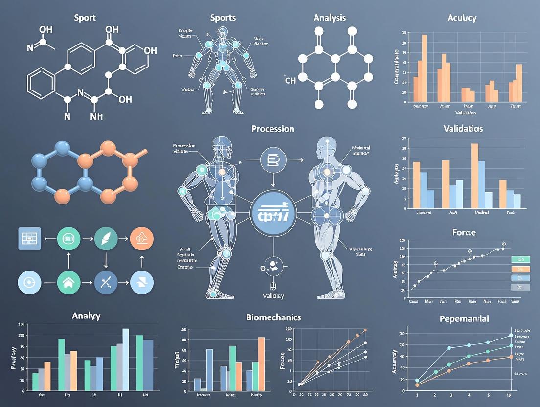
Revolutionizing Sports Biomechanics: How AI and Computer Vision Validate Human Movement for Research and Drug Development
This article explores the transformative integration of computer vision (CV) and artificial intelligence (AI) in sports biomechanics validation.
Revolutionizing Sports Biomechanics: How AI and Computer Vision Validate Human Movement for Research and Drug Development
Abstract
This article explores the transformative integration of computer vision (CV) and artificial intelligence (AI) in sports biomechanics validation. Aimed at researchers, scientists, and drug development professionals, it details the shift from traditional lab-based motion capture to markerless, data-rich analysis. We cover foundational principles, current methodological applications for quantifying athlete kinematics and kinetics, strategies for optimizing and troubleshooting AI models in real-world scenarios, and rigorous validation protocols comparing CV/AI to gold-standard systems. The synthesis provides a critical framework for adopting these technologies to enhance objectivity, scalability, and precision in human movement analysis for biomedical research and therapeutic development.
From Lab to Field: The Foundational Shift to AI-Powered Biomechanics
Application Notes
Computer vision (CV) for biomechanics validation represents a paradigm shift from traditional marker-based motion capture (MOCAP). It is defined as the use of algorithmic techniques to automatically extract, analyze, and validate quantitative measures of human movement from digital imagery, without the need for physical markers or sensors attached to the subject. Within sports biomechanics and drug development research, its core function is to provide a scalable, ecologically valid ground truth or comparative dataset for validating both AI-driven movement models and the biomechanical efficacy of therapeutic interventions.
The validation operates on two primary axes:
- Method Validation: CV systems (e.g., markerless pose estimation, 3D reconstruction from video) are validated against gold-standard laboratory systems (e.g., Vicon, Qualisys). Metrics include joint angle error (degrees), positional error (mm), and temporal parameters (s).
- Biomechanical Outcome Validation: CV-derived biomechanical variables (e.g., knee adduction moment, stride variability) are used as sensitive, objective endpoints to validate the physiological impact of sports training regimens or pharmacological treatments in clinical trials.
Table 1: Quantitative Performance Summary of Markerless CV vs. Marker-Based MOCAP
| Biomechanical Variable | Typical CV Error (vs. MOCAP) | Key Influencing Factors | Suitability for Drug Trial Endpoint |
|---|---|---|---|
| Joint Angles (Sagittal) | 2° - 5° RMSE | Camera view, motion speed | High (for gross motor tasks) |
| Joint Angles (Frontal/Transverse) | 5° - 10° RMSE | Camera number, occlusion | Moderate (requires multi-view setup) |
| Spatiotemporal (Stride Time) | < 2% error | Frame rate, algorithm | Very High |
| Keypoint Position (2D) | < 5 pixels error | Resolution, lighting | Low (primarily for validation) |
| Keypoint Position (3D) | 20 - 40 mm RMSE | Camera calibration, setup | Moderate to High (for movement volumes) |
| Peak Vertical Ground Reaction Force | 10% - 20% error (via ML models) | Model training data quality | High (emerging, needs trial-specific validation) |
Experimental Protocols
Protocol 1: Validation of Markerless 3D Pose Estimation for Gait Analysis in a Clinical Trial Setting
Objective: To establish the concurrent validity of a multi-camera markerless CV system for measuring knee flexion-extension range of motion during walking, for use as a primary endpoint in a knee osteoarthritis drug trial.
Materials & Reagents:
- The Scientist's Toolkit: Key Research Reagent Solutions
Item Function in Protocol Gold-Standard MOCAP System (e.g., Vicon) Provides reference 3D biomechanical data for validation. Synchronized Multi-Camera CV Array (≥4 cameras) Records video data for markerless 3D reconstruction. Calibration Object (e.g., Wand, L-Frame) Enables geometric calibration of all cameras to a common 3D coordinate system. Pose Estimation Algorithm (e.g., OpenPose, HRNet, DeepLabCut) Software to extract 2D human keypoints from video frames. 3D Triangulation & Biomechanics Software (e.g., Anipose, Theia3D, custom pipeline) Converts 2D keypoints from multiple views into 3D coordinates and computes joint angles. Standardized Walkway Controls for environment and ensures consistent task execution. Statistical Analysis Package (e.g., R, Python with SciPy) For calculating RMSE, Pearson's r, and Bland-Altman limits of agreement.
Methodology:
- Setup & Calibration: Position the gold-standard MOCAP system per manufacturer guidelines. Within the same capture volume, position the CV cameras (≥4, 100+ Hz) to maximize multi-view coverage. Perform a static calibration of both systems. Synchronize systems via a hardware trigger or post-hoc timestamp alignment.
- Participant Preparation: Fit participants with both retro-reflective markers for MOCAP and neutral-colored, form-fitting clothing for CV.
- Data Acquisition: Record participants performing 10 walking trials at a self-selected speed. Capture data simultaneously from both systems.
- Data Processing: a. Process MOCAP data using standard biomechanical modeling (e.g., Plug-in-Gait) to compute 3D knee angles. b. Process synchronized videos through the 2D pose estimation algorithm. c. Triangulate 2D keypoints to 3D using calibrated camera parameters. d. Apply the same biomechanical model to the CV-derived 3D keypoints to compute knee angles.
- Validation Analysis: For the primary outcome (peak knee flexion during stance), calculate Root Mean Square Error (RMSE), Pearson correlation coefficient (r), and 95% Bland-Altman limits of agreement between the CV and MOCAP systems across all trials and participants.
Visualization 1: CV Biomechanics Validation Workflow
Protocol 2: Validation of a Single-View CV Algorithm for Assessing Movement Quality in Sport
Objective: To validate a single-view, AI-based CV algorithm's ability to classify "proper" vs. "improper" squat technique against expert human rater consensus.
Methodology:
- Dataset Creation: Record frontal and sagittal video of athletes performing squats under varied conditions. A panel of 3 expert biomechanists provides a consensus label for each repetition (Proper/Improper).
- Algorithm Training: Train a deep learning model (e.g., a convolutional neural network with a temporal component) on the sagittal view videos and expert labels. Reserve a separate, participant-independent set for testing.
- Validation Protocol: Apply the trained model to the held-out test set videos.
- Analysis: Compute confusion matrices, accuracy, precision, recall, F1-score, and Cohen's Kappa between the algorithm's classification and the expert consensus.
Visualization 2: Movement Quality Validation Logic
This document details the application of core AI architectures for validating human movement in sports biomechanics research. The quantitative analysis of athletic motion—from gait and form to injury risk and intervention efficacy—requires precise, non-invasive, and scalable measurement tools. Convolutional Neural Networks (CNNs), 2D/3D pose estimation models, and 3D reconstruction techniques form an integrated pipeline to transform standard video data into clinically relevant, three-dimensional biomechanical parameters. This validation is critical for developing and assessing therapeutic interventions, including pharmacological agents aimed at enhancing recovery or performance.
Core Architectures: Technical Specifications & Comparative Performance
Convolutional Neural Networks (CNNs) for Feature Extraction
CNNs serve as the foundational feature extractors from raw image data. Architectures have evolved for efficiency and accuracy.
Table 1: Performance Comparison of Key CNN Backbones for Pose Estimation
| Model | Input Size | Params (M) | GFLOPs | Top-1 Acc (%) (ImageNet) | Common Use in Pose |
|---|---|---|---|---|---|
| ResNet-50 | 224x224 | 25.6 | 4.1 | 76.2 | Backbone for HRNet, SimpleBaseline |
| ResNet-152 | 224x224 | 60.2 | 11.6 | 78.3 | High-accuracy backbone |
| HRNet-W32 | 256x192 | 28.5 | 7.1 | 75.9 | Maintains high-res feature maps |
| EfficientNet-B0 | 224x224 | 5.3 | 0.39 | 77.1 | Mobile/edge deployment |
| MobileNetV3 | 224x224 | 5.4 | 0.22 | 75.2 | Real-time applications |
Protocol 1.1: CNN Feature Map Visualization for Biomechanical Cues Objective: To verify that the CNN backbone is activating on relevant biomechanical landmarks. Procedure:
- Input Preparation: Extract video frames from a high-speed camera capturing a sprint start.
- Forward Pass & Activation: Pass a frame through the chosen CNN (e.g., ResNet-50). Use a forward hook to extract the activation maps from the final convolutional layer.
- Grad-CAM Application: Compute gradients of the target output (e.g., classification score for "sprinting") relative to the activation maps. Generate a heatmap highlighting important regions.
- Validation: Overlay heatmap on original image. Confirm high activation aligns with anatomically critical zones (knee, ankle, hip joints, major muscle groups). Materials: Pre-trained CNN model, high-speed video dataset, PyTorch/TensorFlow with Grad-CAM library.
2D & 3D Human Pose Estimation Models
These models detect skeletal keypoints from monocular or multi-view video.
Table 2: Quantitative Performance of Pose Estimation Models on Standard Benchmarks
| Model (2D) | MPJPE (mm) | PCK@0.2 | Inference Time (ms) | Notes |
|---|---|---|---|---|
| HRNet | ~89.0 (Human3.6M) | ~92.7 (COCO) | 30 (GPU) | State-of-the-art accuracy |
| HigherHRNet | N/A | 90.6 (COCO) | 40 (GPU) | Excels at scale variation |
| Model (3D) | MPJPE (mm) | Protocol #1 | Input | Notes |
| VideoPose3D | 46.8 | 37.2 | 2D sequence | Temporal convolutions |
| PoseFormer | 44.3 | 35.2 | 2D sequence | Transformer-based |
| METRO (Mesh) | 54.0 (PA-MPJPE) | 50.9 | Image | Direct mesh regression |
Protocol 1.2: Multi-Camera 3D Pose Reconstruction for Biomechanics Objective: To reconstruct accurate 3D joint kinematics for inverse dynamics analysis. Procedure:
- System Setup: Synchronize ≥4 genlocked high-speed cameras (≥200 Hz) around a calibration volume encompassing the movement (e.g., a force plate).
- Calibration: Perform a dynamic wand calibration using Direct Linear Transform (DLT) to obtain intrinsic and extrinsic camera parameters.
- 2D Pose Estimation: Run a robust 2D pose estimator (e.g., HRNet) on synchronized frames from all cameras.
- Triangulation: Use the calibrated camera parameters and the 2D keypoints from multiple views to triangulate the 3D position of each joint per time frame.
- Filtering & Smoothing: Apply a low-pass Butterworth filter (cutoff frequency 10-15 Hz) to the 3D trajectory data to remove high-frequency noise. Materials: Synchronized multi-camera system, calibration wand, force plates, pose estimation software (e.g., Anipose, DeepLabCut).
3D Reconstruction & Human Mesh Recovery (HMR)
These models generate a full 3D mesh of the human body, providing surface geometry for detailed analysis.
Table 3: 3D Human Shape & Pose Estimation Models
| Model | Output | MPJPE (mm) | PVE (mm) | Key Feature |
|---|---|---|---|---|
| HMR (2018) | SMPL parameters | 88.0 | 139.3 | End-to-end regression |
| SPIN | SMPL parameters | 62.5 | 116.4 | Optimization loop within network |
| PARE | SMPL parameters | 57.9 | 103.2 | Part attention to occlusions |
| SPEC | SMPL parameters | 52.5 | 94.4 | Multi-spectral representation |
Protocol 1.3: Volumetric & Surface Analysis from Reconstructed Mesh Objective: To estimate segmental volumes and surface strain for muscle dynamics analysis. Procedure:
- Mesh Generation: Input multi-view video of an athlete into an HMR pipeline (e.g., SPIN or multi-view optimization) to generate a temporally coherent SMPL mesh sequence.
- Segmentation: Map the SMPL mesh vertices to predefined body segments (thigh, shank, torso).
- Volume Calculation: For each segment and time point, compute the volume enclosed by its mesh vertices using a convex hull or tetrahedralization algorithm.
- Surface Strain Analysis: Track the displacement of a grid of virtual markers on the mesh surface. Calculate Lagrangian strain between markers to estimate skin-surface deformation patterns during movement. Materials: Multi-view video data, SMPL model, 3D processing library (Trimesh, PyTorch3D).
Integrated Experimental Workflow for Drug Trial Validation
Diagram 1: AI Biomechanics Pipeline for Intervention Studies
The Scientist's Toolkit: Research Reagent Solutions
Table 4: Essential Materials & Computational Tools
| Item/Category | Example/Product | Function in Biomechanics AI Research |
|---|---|---|
| Motion Capture (Gold Standard) | Vicon, OptiTrack | Provides ground-truth 3D kinematics for training and validating AI models. |
| Force Measurement | AMTI, Kistler Force Plates | Measures ground reaction forces for inverse dynamics, validating AI-derived kinetics. |
| High-Speed Cameras | Phantom, GoPro (High Frame Rate) | Captures input video with minimal motion blur for accurate pose estimation. |
| Pose Estimation Framework | OpenPose, MMPose, DeepLabCut | Open-source libraries for training and deploying 2D/3D pose models. |
| 3D Human Model | SMPL, SMPL-X | Parametric body models enabling 3D mesh reconstruction from images. |
| Biomechanics Software | OpenSim, Visual3D | Used to compute joint kinetics (moments, powers) from AI-derived kinematics and force data. |
| Deep Learning Framework | PyTorch, TensorFlow | Core platforms for developing, training, and testing custom CNN and HMR architectures. |
| Synthetic Data Engine | NVIDIA Omniverse, Blender | Generates photorealistic, annotated training data for rare movements or pathology. |
Detailed Experimental Protocol: Gait Analysis for Intervention Assessment
Protocol 4.1: Quantifying Changes in Knee Kinematics Post-Therapeutic Intervention Objective: To detect statistically significant changes in knee adduction moment (KAM) and flexion angle following administration of a hypothetical osteoarthritis therapeutic.
Materials:
- Patients/athletes from clinical trial cohort.
- 8+ synchronized infrared cameras (Vicon) with retroreflective markers (gold standard).
- 4+ synchronized high-speed RGB cameras (validation & AI input).
- Force plates embedded in walkway.
- AI Pipeline: Custom HRNet -> Triangulation -> OpenSim pipeline.
Procedure:
- Baseline & Follow-up Data Acquisition: Record each subject walking at a self-selected speed. Collect synchronized marker data (Vicon), multi-view RGB video, and force plate data pre-intervention (Day 0) and post-intervention (Week 12).
- Gold Standard Processing: Process marker and force data in Visual3D to compute 3D knee angles and inverse dynamics KAM.
- AI-Derived Processing: a. 2D Pose: Run HRNet on all RGB camera views. b. 3D Triangulation: Use calibrated camera parameters to reconstruct 3D keypoints. c. Kinematic Filtering: Apply a 6Hz low-pass Butterworth filter. d. Kinetic Computation: Input filtered AI kinematics and force plate data into a scaled OpenSim model to compute KAM via inverse dynamics.
- Validation & Analysis: a. Calculate Pearson correlation (r) and Root Mean Square Error (RMSE) between AI-derived and Vicon-derived knee flexion angle and KAM for all baseline trials. b. For the primary endpoint, perform a paired-sample t-test (or non-parametric equivalent) on the AI-derived peak KAM and stance-phase knee flexion ROM between Day 0 and Week 12. c. Report effect size (Cohen's d).
Statistical Endpoints:
- Primary: Change in AI-derived peak KAM.
- Secondary: Correlation (r > 0.85) between AI and Vicon kinematics/kinetics; Change in knee flexion ROM.
Diagram 2: Gait Analysis Validation Workflow
1. Application Notes
The integration of computer vision (CV) and artificial intelligence (AI) is fundamentally validating and expanding the scope of sports biomechanics research. This paradigm shift moves beyond constrained laboratory settings, enabling the quantification of human movement in real-world athletic environments. The core advantages driving this transformation are markerless motion capture, enhanced ecological validity, and high-throughput data generation, which together create a powerful framework for both performance optimization and injury prevention research with applications extending to movement-related drug and therapeutic development.
- Markerless Systems: Traditional optical motion capture relies on physical markers and calibrated camera systems, creating subject preparation burden and laboratory confinement. Modern deep learning-based pose estimation algorithms (e.g., OpenPose, MediaPipe, AlphaPose, and customized CNN architectures) extract 2D or 3D keypoints directly from video streams. This eliminates the need for markers, reduces data collection barriers, and allows retrospective analysis of existing video footage.
- Ecological Validity: By operating in natural training and competition environments (fields, courts, gyms), markerless CV systems capture "in-situ" biomechanics. This yields data that more accurately reflects the cognitive loads, environmental constraints, and tactical decisions inherent to sport, leading to more generalizable and actionable findings for coaches and athletes.
- High-Throughput Data: The automation of motion analysis allows for the processing of thousands of movement trials across hundreds of athletes over entire seasons. This big-data approach facilitates longitudinal monitoring, the identification of subtle movement signatures associated with performance or injury risk, and the development of robust, population-level biomechanical models.
Table 1: Quantitative Comparison of Motion Capture Methodologies
| Feature | Traditional Marker-Based Systems | AI-Driven Markerless Systems |
|---|---|---|
| Setup Time | 30-60 minutes per subject | Minimal (camera setup only) |
| Capture Volume | Limited to lab space (e.g., 10m x 10m) | Scalable (entire field/court) |
| Output Data Rate | ~100-500 Hz | ~30-240 Hz (standard video) |
| Typical Accuracy (RMSE) | <1-2 mm (3D joint center) | 20-40 mm (3D, in-the-wild)* |
| Throughput (Subjects/Day) | Low (5-10) | Very High (50+) |
| Ecological Validity | Low | High |
*Accuracy continuously improving with advanced models and multi-view setups.
2. Experimental Protocols
Protocol 1: Validation of Markerless 3D Pose Estimation Against Gold-Standard Vicon System Objective: To determine the concurrent validity of a multi-camera AI markerless system for measuring lower-extremity joint kinematics during dynamic sporting tasks. Materials:
- 8-camera Vicon MX motion capture system (gold standard).
- 6 synchronized high-definition (1080p, 60Hz) consumer video cameras.
- Calibration object (checkerboard or L-frame).
- Informed consent from participant athletes. Method:
- Setup & Calibration: Position Vicon reflective markers on participant per Plug-in-Gait model. Arrange 6 video cameras around a 10m x 5m capture volume, ensuring overlapping fields of view. Perform a static volume calibration for both systems.
- Task Performance: Participants perform 5 trials each of: a) treadmill running at 4.0 m/s, b) side-step cutting, c) vertical jump.
- Synchronization: Use an audible/visual trigger event recorded by all systems for temporal synchronization.
- Data Processing: Process Vicon data using Nexus software to obtain 3D joint angles. Process synchronized video through a trained deep learning model (e.g., MotionBERT or SPIN) for 3D pose estimation, followed by biomechanical modeling to compute joint angles.
- Analysis: Calculate Root Mean Square Error (RMSE), Pearson's correlation coefficient (r), and Bland-Altman limits of agreement for key joint angles (knee flexion/abduction, hip rotation) across the movement cycle.
Protocol 2: High-Throughput Screening for Movement Asymmetries in a Collegiate Athletics Program Objective: To longitudinally screen an entire team for lower-limb asymmetries using markerless video analysis to identify athletes at elevated risk of overuse injury. Materials:
- 2 stationary IP cameras (4K, 60Hz) placed at sagittal and frontal planes.
- Cloud-based or local computing server for video processing.
- Standardized testing station (designated area on field). Method:
- Baseline Testing: At pre-season, each athlete performs 5 single-leg squats per leg in front of the cameras following a standardized protocol (stance, pace).
- Automated Processing: Videos are automatically uploaded and processed via a pose estimation pipeline (e.g., using OpenPose for 2D keypoints, then 3D reconstruction). Key metrics: knee valgus angle, hip drop (pelvic obliquity), and depth of squat symmetry index are calculated.
- Longitudinal Monitoring: The test is repeated monthly. Data is aggregated in a team dashboard.
- Risk Stratification: Athletes are flagged if their asymmetry index exceeds 15% or shows a negative trend exceeding 10% from baseline. Flagged athletes are referred for detailed biomechanical assessment.
3. The Scientist's Toolkit: Research Reagent Solutions
Table 2: Essential Materials for Markerless Sports Biomechanics Research
| Item | Function & Specification |
|---|---|
| Multi-Camera Sync System | Synchronizes frames from multiple cameras (µs precision) for accurate 3D reconstruction. (e.g., genlock cameras or hardware sync boxes). |
| Calibration Object (Charuco Board) | Provides known 3D-2D point correspondences for calibrating intrinsic/extrinsic camera parameters, essential for 3D triangulation. |
| High-Performance GPU Workstation | Accelerates the training and inference of deep learning models for pose estimation (e.g., NVIDIA RTX A6000 or comparable). |
| Pose Estimation Software (OpenPose/MediaPipe) | Open-source libraries providing pre-trained models for real-time 2D human pose estimation from video. |
| 3D Pose Estimation Model (e.g., VideoPose3D) | A PyTorch-based model for lifting 2D keypoints sequences to accurate 3D poses using temporal convolutional networks. |
| Biomechanical Analysis Software (OpenSim) | Open-source platform for modeling musculoskeletal structures and analyzing dynamics from 3D motion data. |
| Dedicated Data Pipeline (e.g., APE)* | An open-source Automated Pose Estimation pipeline for batch processing, model integration, and feature extraction. |
| Secure Data Storage (RAID/Cloud) | High-capacity, secure storage for large volumes of raw video and processed time-series biomechanical data. |
*Acquisition, Processing, and Exploration pipeline.
4. Visualizations
Within the thesis on "Computer Vision and AI for Sports Biomechanics Validation Research," the validation of novel athlete monitoring and drug efficacy protocols demands precise, real-time motion capture and analysis. This necessitates a hardware ecosystem comprising advanced cameras, depth sensors, and edge computing devices. These components are critical for generating high-fidelity biomechanical data (kinematics, kinetics) in ecologically valid environments, moving beyond constrained lab settings to real-world training grounds.
Hardware Specifications and Quantitative Comparison
Table 1: Camera System Comparison for Biomechanics Capture
| Parameter | High-Speed Optical (e.g., Vicon Bonita) | Rolling Shutter CMOS (e.g., GoPro Hero 12) | Global Shutter CMOS (e.g., Intel RealSense D455) |
|---|---|---|---|
| Max Frame Rate (fps) | 1,000 (at reduced resolution) | 240 (1080p) | 90 (1280x720) |
| Resolution | Up to 4MP | 5.3K (5312x2988) | 2MP (1280x800 per imager) |
| Shutter Type | Global | Rolling | Global |
| Key Strength | Gold-standard accuracy for marker-based 3D | Portable, high-resolution for field use | Synchronized depth + RGB, minimal motion blur |
| Primary Application | Lab-based validation of other systems | On-field qualitative and 2D quantitative analysis | Integrated 3D point cloud generation in dynamic scenes |
Table 2: Depth Sensor Technologies
| Technology | Time-of-Flight (ToF) (e.g., Microsoft Azure Kinect) | Structured Light (e.g., older Intel RealSense) | Stereo Vision (e.g., Intel RealSense D455) |
|---|---|---|---|
| Operating Range | 0.5 - 5.5 m | 0.2 - 1.2 m | 0.4 - 6 m |
| Depth Accuracy | ±1% of distance (e.g., ±1cm at 1m) | <2% at 1m | <2% at 2m |
| FPS (Depth Stream) | 30 | 90 | 90 |
| Sensitivity to Light | Moderate (can be affected by sunlight) | High (requires controlled light) | Low (uses ambient light) |
| Best For | Indoor court volumetric capture | Short-range, high-precision static scans | Outdoor and dynamic scene reconstruction |
Table 3: Edge Computing Platforms for On-Site AI
| Platform | NVIDIA Jetson AGX Orin | Intel NUC with Movidius Myriad X | Google Coral Dev Board |
|---|---|---|---|
| AI Performance (TOPS) | 275 (INT8) | ~4 TOPS (INT8) | 4 TOPS (INT8) |
| Power Draw (Typical) | 15-60W | 10-25W | ~5W |
| Memory | 32GB LPDDR5 | Up to 64GB DDR4 | 4GB LPDDR4 |
| Key Feature | Full AI pipeline training/inference | x86 compatibility with VPU | Low power, TPU co-processor |
| Use Case | Real-time multi-camera 3D pose estimation | Portable lab-grade analysis station | Wearable sensor data fusion |
Application Notes
AN-01: Synchronized Multi-Modal Data Capture Protocol
Purpose: To acquire temporally aligned RGB video, depth maps, and inertial measurement unit (IMU) data from athletes performing a standardized movement (e.g., a vertical jump or pitch) for validating biomechanical models. Procedure:
- Hardware Setup:
- Deploy a calibrated array of 3-6 global shutter cameras (e.g., FLIR Blackfly S) around the performance volume.
- Position one depth sensor (e.g., Azure Kinect DK) centrally for volumetric reference.
- Attach a wireless IMU (e.g., Xsens MTw Awinda) to the athlete's pelvis.
- Connect all devices to a master trigger box or use network-based synchronization (PTP).
- Software Configuration:
- Use a platform like Robot Operating System (ROS) or NI LabVIEW to create a synchronized recording node.
- Set all cameras to record at 120 Hz, depth sensor at 30 Hz, and IMU at 400 Hz.
- Calibration:
- Record a static calibration frame with a checkerboard and L-frame visible to all sensors.
- Perform dynamic wand calibration for motion capture space.
- Data Acquisition:
- Record athlete performing 10 trials of the target movement.
- Save all streams with a common global timestamp.
AN-02: On-Edge Pose Estimation & Anomaly Detection Workflow
Purpose: To process video feeds in real-time on the edge to compute biomechanical joint angles and flag deviations from normative patterns, potentially indicating fatigue or intervention effects. Procedure:
- Model Deployment:
- Quantize a pre-trained 3D human pose model (e.g., MediaPipe BlazePose or a custom PyTorch model) to INT8 format for the target edge device (e.g., NVIDIA Jetson).
- Edge Pipeline:
- Ingest RTSP video stream from a fixed-angle camera.
- Run the pose estimation model on every fifth frame (20 Hz effective) to conserve compute.
- Calculate key kinematic parameters (e.g., knee valgus angle, shoulder external rotation).
- Anomaly Detection:
- Compare real-time angles against a stored normative profile (mean ± 2SD) for the movement phase.
- If values exceed threshold for three consecutive analyses, flag an anomaly and save a 10-second video clip with overlaid angles.
- Output:
- Transmit summary statistics (mean max angle, anomaly count) and flagged clips to a central research database via 5G/Wi-Fi.
Experimental Protocols
EP-01: Validation of Depth Sensor against Force Plates for Ground Contact Time (GCT)
Objective: To validate depth sensor-derived temporal gait metrics against the gold standard (force plates) in a controlled sprint start. Materials: Force plate (e.g., AMTI), depth sensor (Azure Kinect DK), synchronization unit, calibration tools. Protocol:
- Place the force plate flush with the ground surface. Position the depth sensor 3m ahead of the plate's center, elevated 1m, pointing at the plate.
- Synchronize force plate and depth sensor data streams via an analog trigger.
- Record 20 sprint starts from a standardized block position. Athlete's lead foot must strike the force plate.
- Force Plate GCT: Calculate GCT as the duration the vertical ground reaction force exceeds 10N.
- Depth Sensor GCT:
- Use background subtraction to isolate the athlete.
- Apply a skeleton tracking algorithm (e.g., Kinect SDK) to identify the foot.
- Calculate GCT as the duration the foot centroid's vertical velocity is near zero (<0.5 m/s) and the foot is within a defined volume above the plate.
- Analysis: Perform Bland-Altman analysis to assess agreement between the two methods for GCT.
EP-02: Protocol for Assessing Edge Computing Latency in Real-Time Biofeedback
Objective: To quantify the end-to-end latency of a real-time biomechanical feedback system using an edge computing device. Materials: High-speed camera (1000fps reference), edge device (Javier Orin), LED indicator, video display. Protocol:
- Set up a high-speed camera pointing at both the video display and the LED indicator connected to the edge device's GPIO pin.
- On the edge device, run a simple OpenCV program that:
- Captures video from its own USB camera.
- Detects a specific color blob (e.g., bright green on the athlete's shoe).
- Triggers the GPIO pin to light the LED when the blob's vertical position exceeds a predefined pixel row.
- Start the high-speed camera recording. Have an operator move the green marker through the trigger point.
- Latency Calculation: From the high-speed footage, count the frames between the actual crossing of the pixel row and the illumination of the LED. Latency = (Frame Count) / (1000 fps).
Diagrams
Title: Data Flow for Biomechanics Hardware Ecosystem
Title: Real-Time Edge AI Analysis Pipeline
The Scientist's Toolkit: Research Reagent Solutions
Table 4: Essential Hardware and Software for CV Biomechanics Research
| Item | Function / Purpose |
|---|---|
| Calibrated Checkerboard | For intrinsic (lens distortion) and extrinsic (position/orientation) calibration of 2D cameras. |
| L-Frame (Wand) | Provides known scale and 3D reference points for multi-camera motion capture system calibration. |
| Retroreflective Markers | Used as high-contrast tracking points for gold-standard optical motion capture (e.g., Vicon). |
| Synchronization Pulse Generator | Sends simultaneous triggers to all cameras, sensors, and data acquisition devices to ensure temporal alignment. |
| OpenPose / MediaPipe / DLPack | Open-source software libraries for 2D and 3D human pose estimation from video, used for prototyping. |
| ROS 2 (Robot Operating System) | Middleware framework for managing synchronized data streams from heterogeneous sensors. |
| Docker Containers | To package and deploy consistent AI inference environments across different edge computing devices. |
| Force Plates & Pressure Mats | Gold-standard ground truth for kinetic variables (force, pressure) used to validate vision-based estimates. |
Application Notes
Gait Analysis
Quantitative gait analysis using computer vision (CV) and AI provides a non-invasive, markerless method for assessing human locomotion. Current research focuses on extracting spatiotemporal parameters (e.g., stride length, cadence, stance/swing phase ratio) and kinematic variables (joint angles) from 2D or 3D video feeds. Deep learning models, particularly pose estimation algorithms (e.g., OpenPose, MediaPipe, HRNet), form the backbone of this analysis, enabling high-throughput processing in ecological settings (e.g., clinics, training fields). Validation against gold-standard motion capture (MoCap) systems remains a core research theme, with recent studies reporting high correlation coefficients (>0.9) for key parameters under constrained conditions.
Key Quantitative Data:
| Parameter | CV/AI Method (2023-24) | Mean Error vs. MoCap | Correlation (r) | Typical Sample Rate | Research Context |
|---|---|---|---|---|---|
| Stride Length (m) | 3D Pose Estimation (Multi-view) | 0.02 m | 0.97 | 30-100 Hz | Neurological disorder progression |
| Knee Flexion Angle (deg) | 2D-to-3D Lifting Models | 3.5 deg | 0.94 | 60 Hz | Osteoarthritis monitoring |
| Cadence (steps/min) | Single-view 2D Pose Estimation | 2.1 steps/min | 0.99 | 30 Hz | Remote patient assessment |
| Ground Contact Time (ms) | Temporal CNN on Foot Keypoints | 12 ms | 0.96 | 120+ Hz | Running economy studies |
Injury Risk Assessment
AI-driven injury risk assessment leverages biomechanical data to identify aberrant movement patterns predictive of future injury. Machine learning classifiers (e.g., Support Vector Machines, Random Forests, and more recently, temporal convolutional networks) are trained on kinematic and kinetic time-series data to flag high-risk profiles. Primary foci include anterior cruciate ligament (ACL) injury risk from jump-landing mechanics and hamstring strain risk during sprinting. Research validates these models against prospective injury datasets, with area-under-curve (AUC) metrics for prediction being a critical performance indicator.
Key Quantitative Data:
| Injury Target | Predictive Biomechanical Features | AI Model Type | Reported AUC | Sensitivity/Specificity | Validation Cohort Size |
|---|---|---|---|---|---|
| Non-contact ACL | Knee valgus angle, trunk lateral flexion | Ensemble Learning | 0.88 | 0.82 / 0.81 | 250 athletes |
| Hamstring Strain | Peak thigh angular velocity, stride imbalance | Temporal CNN | 0.79 | 0.75 / 0.72 | 180 sprinters |
| Patellofemoral Pain | Hip adduction, contralateral pelvic drop | SVM with RBF kernel | 0.84 | 0.78 / 0.77 | 300 recreational runners |
| Shoulder (Labral) | Humeral rotation velocity, scapular dyskinesia | LSTM Network | 0.81 | 0.76 / 0.79 | 150 overhead athletes |
Performance Profiling
Performance profiling utilizes CV to deconstruct sport-specific movements for talent identification and training optimization. This involves creating athlete-specific digital biomechanical models to analyze efficiency, power generation, and technique. Dimensionality reduction (PCA, t-SNE) and clustering algorithms segment athletes into distinct performance phenotypes. Research validates these profiles against competitive outcomes or objective performance metrics (e.g., velocity, power output).
Key Quantitative Data:
| Sport | Profiled Movement | Key Profiling Metrics | AI/CV Technique | Correlation w/ Performance | Use Case Example |
|---|---|---|---|---|---|
| Sprinting | Block Start & Acceleration | Horizontal velocity, shin angle, reaction time | Optical Flow + Pose Estimation | r=0.91 w/ 10m time | Talent ID in youth academies |
| Swimming | Starts & Turns | Wall push-off force (proxy), glide efficiency | Underwater 3D Reconstruction | r=0.89 w/ turn time | Training intervention focus |
| Weightlifting | Clean & Jerk | Barbell path, joint velocity synergy, bar stability | Barbell tracking + Multi-person pose | r=0.85 w/ 1RM | Technique flaw diagnosis |
| Basketball | Jump Shot | Release angle, knee/elbow sync, shot arc | Video sequence classification (CNN) | r=0.78 w/ shooting % | Personalized coaching feedback |
Experimental Protocols
Protocol 1: Validation of Markerless Gait Analysis Against Reference MoCap
Objective: To validate the accuracy of a multi-view CNN-based 3D pose estimation system for gait parameter extraction.
- Participant Setup: Recruit cohort (e.g., n=20). Affix reflective markers for MoCap (e.g., Vicon system) per Plug-in-Gait model.
- System Synchronization: Genlock and synchronize MoCap cameras (120 Hz) with 4+ synchronized RGB video cameras (≥60 Hz, 4K resolution).
- Calibration: Perform static calibration of both systems using an L-frame for spatial alignment.
- Data Collection: Participants walk at self-selected speed on a 10m walkway. Collect 10 valid trials per participant.
- CV Processing: Process RGB feeds through 3D human pose estimator (e.g., VoxelPose, multi-view MediaPipe).
- Data Extraction: Extract 3D keypoints. Calculate spatiotemporal (stride length, cadence) and kinematic (sagittal plane joint angles) parameters.
- Statistical Analysis: Compute Pearson's r, root mean square error (RMSE), and Bland-Altman limits of agreement between CV and MoCap-derived time-series data for each parameter.
Protocol 2: Prospective Injury Risk Assessment Study
Objective: To develop and validate an AI model for predicting lower-limb injury from preseason movement screening.
- Cohort & Baseline Testing: Encode preseason biomechanical assessment of athletes (e.g., n=500). Perform standardized movement tasks (drop-jump, single-leg squat) in a CV lab (3D pose estimation).
- Feature Engineering: Extract time-series features (peak angles, angular velocities, asymmetries, coordination metrics).
- Ground Truth Labeling: Track all athletes prospectively for one competitive season. Record medically diagnosed injuries (e.g., ACL rupture, hamstring strain). Create binary labels (injured/not injured).
- Model Development: Split data (70/30 training/test). Train multiple classifiers (e.g., XGBoost, Temporal CNN) using features to predict injury label.
- Validation: Evaluate model on held-out test set. Report AUC, sensitivity, specificity, precision. Perform SHAP analysis for feature importance.
- External Validation: Where possible, test model on an independent cohort from a different institution.
Protocol 3: High-Throughput Performance Phenotyping Protocol
Objective: To create performance clusters for a specific sport action using unsupervised learning on CV-derived data.
- Data Acquisition: Record a large sample of athletes (n>200) performing the target skill (e.g., basketball free throw) under standardized conditions using a single calibrated high-speed camera.
- 2D/3D Pose Estimation: Process all videos with a consistent pose estimation framework (e.g., MediaPipe Pose).
- Kinematic Time-Series Extraction: Derive relevant time-normalized kinematic curves (e.g., shoulder-elbow-wrist angles for shooting).
- Dimensionality Reduction: Apply Principal Component Analysis (PCA) to the high-dimensional kinematic data to reduce to 5-10 principal components (PCs) explaining >90% variance.
- Clustering: Apply clustering algorithm (e.g., k-means, DBSCAN) to the PCs to identify distinct technique phenotypes.
- Profile Interpretation & Validation: Statistically compare performance outcome (e.g., shot accuracy, ball velocity) across clusters using ANOVA. Validate profiles with expert coach qualitative assessment.
Visualization
Title: Markerless Gait Analysis Validation Workflow
Title: Injury Risk AI Model Development Pipeline
Title: Unsupervised Performance Phenotyping Process
The Scientist's Toolkit: Research Reagent Solutions
| Item | Function in CV/AI Biomechanics Research | Example Product/Software |
|---|---|---|
| Multi-camera Synchronization System | Precisely synchronizes multiple high-speed RGB or IR cameras for 3D reconstruction. | OptiTrack Sync, Arduino-based custom triggers, Genlock-enabled cameras. |
| Calibration Object (L-Frame/Checkerboard) | Enables spatial calibration and scaling for 3D coordinate system alignment between cameras and MoCap. | Custom L-frame with reflective markers, large printed checkerboard (e.g., Charuco board). |
| High-Performance Pose Estimation Model | Pre-trained deep learning model for accurate 2D or 3D human pose estimation from video. | MediaPipe Pose, OpenPose, HRNet, AlphaPose, DeepLabCut. |
| Motion Capture (MoCap) System (Gold Standard) | Provides ground-truth 3D kinematic data for validation of CV/AI methods. | Vicon Nexus, Qualisys, OptiTrack. |
| Biomechanical Analysis Software | For processing motion data, calculating derivatives (velocity, acceleration), and generating standard metrics. | Visual3D, OpenSim, custom Python scripts (SciPy, NumPy). |
| Machine Learning Framework | Environment for developing, training, and validating custom injury risk or profiling models. | Python with Scikit-learn, TensorFlow, PyTorch, XGBoost. |
| Statistical Analysis Suite | For comprehensive validation statistics, including correlation, error analysis, and Bland-Altman plots. | R, Python (Statsmodels, Pingouin), GraphPad Prism. |
| High-Throughput Computing Cluster/GPU | Enables processing of large video datasets and training complex neural networks. | NVIDIA GPUs (e.g., A100, V100), Google Colab Pro, AWS EC2 instances. |
Methodologies in Action: Applying AI to Quantify Kinematics and Kinetics
This document details the end-to-end experimental pipeline for deriving validated biomechanical metrics from video data, framed within a thesis on computer vision (CV) and artificial intelligence (AI) validation for sports biomechanics. The protocols are designed to support rigorous research, including applications in performance optimization, injury prevention, and the assessment of therapeutic interventions in drug development. The integration of AI necessitates stringent validation against gold-standard measurement systems.
Core Pipeline: Workflow & Protocols
Diagram Title: Main CV Biomechanics Pipeline with Validation Loop
Protocol: Synchronized Multi-Modal Data Acquisition
Objective: To capture high-quality, synchronized video and ground-truth sensor data for AI model training and validation.
Equipment Setup:
- Cameras: Arrange multiple high-speed cameras (e.g., 120+ Hz) around the capture volume. Calibrate the 3D system using a wand with markers of known length.
- Ground-Truth Systems: Position force plates flush with the floor. Apply skin-mounted inertial measurement units (IMUs) and/or retro-reflective markers for optoelectronic motion capture (MoCap) on anatomical landmarks.
- Synchronization: Connect all devices (cameras, force plates, IMU receiver, MoCap system) to a common digital sync pulse generator.
Procedure:
- Record a static calibration trial of the subject in a known posture.
- For each dynamic trial (e.g., running jump, cutting maneuver), simultaneously trigger all systems.
- Record a minimum of N=20 trials per movement condition from ≥15 subjects to ensure statistical power for validation studies.
- Include a calibration object (e.g., checkerboard) in the first frame of video for intrinsic camera parameter verification.
Protocol: AI-Based 2D/3D Human Pose Estimation
Objective: To extract accurate 2D and 3D anatomical keypoints from video sequences.
Model Selection & Training:
- Select a state-of-the-art model (e.g., HRNet, ViTPose for 2D; VideoPose3D, MHFormer for 3D).
- Fine-tuning: If using domain-specific data (e.g., unusual camera angles, sports gear), fine-tune the model on a manually annotated dataset from your calibrated cameras. Use transfer learning, freezing early layers.
Inference & Post-Processing:
- Process video through the model frame-by-frame to generate keypoint coordinates (x, y, [z]) and confidence scores.
- Apply temporal filters (e.g., a Butterworth low-pass filter with a 6-10 Hz cutoff, or a Savitzky-Golay filter) to smooth keypoint trajectories. The exact cutoff frequency should be determined by residual analysis.
Protocol: Biomechanical Model Computation & Validation
Objective: To compute clinical/biomechanical metrics and validate them against gold-standard systems.
Kinematic Computation:
- Define a biomechanical skeleton model (e.g., a 15-segment, 6-degree-of-freedom model) using the tracked keypoints.
- Calculate joint angles (flexion/extension, abduction/adduction, rotation) using Cardan/Euler angle sequences (e.g., Y-X-Z for knee).
- Compute center of mass (COM) trajectory using anthropometric tables and segmental analysis.
Kinetic Computation (Inverse Dynamics):
- Input: Kinematic data + ground reaction force (GRF) data from force plates.
- Process: Use Newton-Euler equations of motion within software (e.g., OpenSim, custom Python/R scripts) to calculate net joint moments and powers.
Validation Analysis:
- Temporal Alignment: Synchronize AI-derived time series with MoCap/IMU data using cross-correlation on a known event (e.g., foot strike).
- Statistical Comparison: For key metrics (e.g., peak knee flexion angle, internal rotation moment), calculate:
- Root Mean Square Error (RMSE)
- Pearson's Correlation Coefficient (r)
- Bland-Altman Limits of Agreement (LoA)
Table 1: Representative Validation Metrics for AI vs. Optoelectronic MoCap
| Biomechanical Metric | Typical RMSE (AI vs. MoCap) | Typical Correlation (r) | Key Influencing Factors |
|---|---|---|---|
| Knee Flexion Angle (deg) | 3.5 - 6.0° | 0.97 - 0.99 | Camera viewpoint, clothing, movement speed, AI model architecture |
| Hip Abduction Angle (deg) | 4.0 - 8.0° | 0.90 - 0.98 | Soft tissue artifact, sensor placement error for validation data |
| Ankle Joint Moment (Nm/kg) | 0.15 - 0.30 Nm/kg | 0.85 - 0.95 | Accuracy of GRF measurement, segment inertial parameter estimation, filter choices |
| Stride Time (s) | 0.01 - 0.03 s | >0.99 | Frame rate of video, accuracy of event detection algorithm (e.g., foot contact from keypoints) |
Table 2: Key Experimental Parameters for Pipeline Validation Studies
| Parameter | Recommended Specification | Rationale |
|---|---|---|
| Camera Sample Rate | ≥120 Hz (≥200 Hz for high-speed movements) | To avoid aliasing and accurately capture rapid joint motions. |
| Number of Camera Views | Minimum of 2 for 3D reconstruction; 4-8 recommended for occlusion resilience | Ensures robust 3D triangulation and handles self-occlusion. |
| Video Resolution | ≥1920 x 1080 pixels (Full HD) | Higher resolution improves keypoint detection accuracy. |
| Validation Cohort Size | N ≥ 15 participants, with diverse movement patterns | Ensures generalizability of validation results and statistical power. |
| Gold-Standard Reference | 3D Optoelectronic MoCap (e.g., Vicon, Qualisys) sampled at ≥200 Hz, synchronized with force plates. | Provides the most accurate kinematic and kinetic ground truth for laboratory validation. |
The Scientist's Toolkit
Table 3: Essential Research Reagent Solutions & Materials
| Item Category / Name | Function & Application in Pipeline |
|---|---|
| High-Speed Video Cameras (e.g., GoPro Hero, Sony RX) | Provide the raw 2D video data. Must support external triggering and high frame rates for dynamic sports movements. |
| Calibration Wand & Checkerboard | Essential for determining the intrinsic (focal length, lens distortion) and extrinsic (position, orientation) parameters of cameras for 3D reconstruction. |
| OpenPose, HRNet, MediaPipe, ViTPose | Open-source and state-of-the-art AI libraries for 2D human pose estimation from video frames. The foundation for keypoint extraction. |
| OpenSim, Pyomeca, Biomechanics ToolKit (BTK) | Software toolkits for implementing biomechanical models, calculating inverse dynamics, and analyzing kinematic/kinetic data. |
| Force Platforms (e.g., AMTI, Kistler) | Measure ground reaction forces and moments. Critical for calculating joint kinetics (moments, powers) and validating AI-predicted loading events. |
| Inertial Measurement Units (IMUs) (e.g., Xsens, Noraxon) | Provide wearable ground-truth orientation data for segments. Used for field-based validation of AI-derived kinematics where MoCap is impossible. |
| Digital Sync Pulse Generator | Ensures sample-accurate temporal synchronization between all independent data streams (video, MoCap, force plates, IMUs). |
| Python/R with SciPy, NumPy, pandas | Core programming environments and libraries for data processing, filtering, statistical analysis (Bland-Altman, RMSE), and custom metric computation. |
Diagram Title: Statistical Validation Pathway for Biomechanical Metrics
The quantitative analysis of human movement is fundamental to sports biomechanics validation research. The extraction of 2D and 3D anatomical keypoints from video data provides a non-invasive, scalable method for kinematic assessment, enabling the evaluation of athletic performance, injury risk, and rehabilitation efficacy. Within the broader thesis on computer vision and AI for sports biomechanics, this document details application notes and protocols for prevalent keypoint extraction solutions: the open-source libraries OpenPose and MediaPipe, and select proprietary systems. Accurate validation of these tools against gold-standard motion capture is critical for their adoption in rigorous scientific and drug development research, where they may be used to quantify the biomechanical impact of therapeutic interventions.
OpenPose: A bottom-up, multi-person 2D pose estimation system. It first detects all body parts in an image (Part Affinity Fields), then associates them to form individual skeletons. It is renowned for its robustness in multi-person scenarios and its detailed model (25 body keypoints, plus hand and face models).
MediaPipe Pose: A BlazePose-based, top-down, lightweight solution optimized for real-time performance. It employs a two-step detector-tracker approach and is designed to run efficiently on a wide range of hardware. It offers 33 body keypoints, including specific torso and face landmarks for enhanced stability.
Proprietary Solutions (e.g., Theia3D, DARI Motion, Vicon Vue): These are often commercial, turn-key systems. They may use multi-view triangulation for 3D reconstruction, advanced deep learning models, or a fusion of sensor data. They typically provide direct 3D output, comprehensive support, and validation documentation, but at a significant cost and with less algorithmic transparency.
Quantitative Performance Comparison
Table 1: Comparative Summary of Keypoint Extraction Solutions (Data from recent benchmarks and published validations, 2023-2024).
| Feature / Metric | OpenPose | MediaPipe Pose | Proprietary (e.g., Theia3D) |
|---|---|---|---|
| Primary Output | 2D (3D via triangulation) | 2D (3D via lifter models) | Direct 3D |
| Inference Speed (FPS on CPU) | ~5-10 FPS | ~30-50+ FPS | Varies (often GPU-accelerated) |
| Keypoint Count (Body) | 25 (BODY_25 model) | 33 | Often 20-30+ |
| Multi-Person | Native, bottom-up | Requires separate detection | Typically native |
| Validation in Biomechanics | Moderate (extensive research use, variable accuracy) | Growing (good for gross motion) | High (often validated against optoelectronic systems) |
| Typical Accuracy (PCK@0.2 on COCO) | ~85% | ~88% | N/A (uses proprietary datasets) |
| 3D Root Mean Square Error (vs. Vicon) | ~30-50mm (from triangulated 2D) | ~25-40mm (from 3D lifter) | ~10-25mm |
| Cost | Free, Open-Source | Free, Open-Source | High (Licensing + Hardware) |
| Key Strength | Robust multi-person, customizable | Speed, accessibility, ease of use | Accuracy, direct 3D, support, validation |
Experimental Protocols for Biomechanical Validation
Protocol A: Single-Camera 2D Accuracy Validation
Objective: To validate the 2D keypoint accuracy of OpenPose and MediaPipe against manually annotated ground truth in a controlled lab setting.
Materials:
- High-speed camera (e.g., 120 Hz).
- Calibration board (checkerboard).
- A subject in athletic attire.
- A computer with OpenPose and MediaPipe installed.
Procedure:
- Camera Setup & Calibration: Mount the camera perpendicular to the plane of motion. Record a video of the checkerboard in various orientations to perform camera calibration and correct for lens distortion.
- Data Acquisition: Record the subject performing standardized movements (e.g., squats, gait cycles, overhead throws). Ensure the subject remains within the calibrated plane.
- Ground Truth Generation: Manually annotate keypoints (e.g., hip, knee, ankle) for a representative subset of frames (e.g., 100 frames) using a tool like LabelMe or COCO Annotator.
- Algorithm Processing: Process the same video sequence with both OpenPose and MediaPipe, extracting 2D keypoint coordinates.
- Analysis: Compute the Percentage of Correct Keypoints (PCK) at a threshold of 0.2 of the head segment length. Calculate the Mean Per Joint Position Error (MPJPE) in pixels between the algorithm output and manual annotations.
Protocol B: Multi-Camera 3D Reconstruction & Kinematic Error Assessment
Objective: To assess the error in 3D joint angles derived from triangulated OpenPose/MediaPipe keypoints compared to a synchronized gold-standard optoelectronic system (e.g., Vicon).
Materials:
- Test System: Minimum of 2 synchronized, calibrated high-definition video cameras (≥4 recommended).
- Gold Standard: Synchronized optoelectronic motion capture system (e.g., Vicon with ≥8 cameras).
- Protocol Subjects with reflective markers placed according to a biomechanical model (e.g., Plug-in Gait).
- A common synchronization trigger (e.g., LED light).
Procedure:
- System Synchronization & Calibration:
- Calibrate the Vicon system according to manufacturer specifications.
- Simultaneously calibrate the video camera network using a wand or checkerboard for 3D reconstruction. Establish a common global coordinate system with the Vicon system using a calibration frame.
- Set up a synchronization trigger visible to all systems.
- Data Collection: Record the subject performing dynamic sports tasks (e.g., cutting maneuver, jump landing). Ensure all reflective markers and body segments are visible.
- Data Processing:
- Vicon: Process marker trajectories, label markers, and compute 3D joint centers and angles using the defined biomechanical model.
- Video: Extract 2D keypoints from each camera view using OpenPose/MediaPipe. Use direct linear transform (DLT) or bundle adjustment to triangulate 3D keypoint coordinates.
- Temporal Alignment: Use synchronization signals to align data streams.
- Spatial Alignment: Perform a Procrustes analysis to align the two 3D coordinate systems.
- Outcome Measures: Calculate for key joints (knee, hip, ankle):
- Root Mean Square Error (RMSE) of 3D joint positions.
- RMSE and correlation (r) of joint angle time-series (e.g., knee flexion/extension).
- Bland-Altman plots to assess agreement and systematic bias.
(Title: 3D Keypoint Validation Protocol Workflow)
The Scientist's Toolkit: Essential Research Reagents & Materials
Table 2: Key "Research Reagent Solutions" for Keypoint Extraction Experiments.
| Item / Solution | Category | Function in Research |
|---|---|---|
| Vicon Motion Capture System | Gold-Standard Hardware | Provides ground truth 3D kinematic data for validation of vision-based methods. |
| Synchronization Trigger Box (e.g., LED) | Laboratory Equipment | Ensures temporal alignment between video cameras and other data acquisition systems. |
| Calibration Wand & Checkerboard | Calibration Tools | Enables geometric calibration of camera intrinsic/extrinsic parameters for accurate 2D/3D mapping. |
| OpenPose (BODY_25 Model) | Open-Source Software | Provides detailed, multi-person 2D pose estimation for custom analysis pipelines. |
| MediaPipe Pose (BlazePose) | Open-Source Software | Enables real-time, efficient 2D/3D pose estimation on commodity hardware for pilot studies. |
| Theia3D Markerless System | Proprietary Software | Offers a validated, commercial solution for direct 3D biomechanics from multi-view video. |
| Python (NumPy, SciPy, OpenCV) | Programming Environment | The core platform for data processing, triangulation, statistical analysis, and custom scripting. |
| COCO or MPII Annotator | Annotation Software | Creates manual 2D keypoint ground truth data for training or validating models. |
(Title: Solution Selection Logic for Biomechanics Research)
1. Introduction within Thesis Context This document serves as an application note within a doctoral thesis focused on validating computer vision (CV) and artificial intelligence (AI) models for sports biomechanics. The accurate derivation of foundational biomechanical variables—joint angles, angular velocities, and whole-body center of mass (COM)—is critical. The thesis posits that robust, markerless CV/AI systems must match or exceed the precision of traditional laboratory methods to be viable for real-world sports performance analysis and injury risk assessment, with downstream implications for pharmaceutical interventions targeting mobility and recovery.
2. Core Biomechanical Variables: Definitions and Quantitative Benchmarks The following variables are primary outputs for validation studies comparing CV/AI pipelines against gold-standard motion capture.
Table 1: Core Derived Biomechanical Variables
| Variable | Definition | Typical Gold-Standard Precision | Key Sports Application |
|---|---|---|---|
| Joint Angle | The relative orientation between two adjacent body segments in degrees (°). | < 1.0° RMSE for sagittal plane (e.g., knee flexion). | Gait analysis, technique optimization, identifying pathological movement. |
| Angular Velocity | The rate of change of a joint angle, in degrees per second (°/s). | < 10 °/s RMSE for dynamic tasks. | Quantifying explosive power (e.g., knee extension in sprinting), assessing joint control. |
| Whole-Body COM | The 3D spatial coordinate of the body's weighted average mass position. | < 15 mm RMSE for trajectory. | Balance assessment, calculation of derived measures (e.g., momentum, mechanical load). |
3. Experimental Protocols for Validation Research These protocols are designed to generate ground-truth data for validating CV/AI models.
Protocol 3.1: Concurrent Validation of Joint Kinematics Objective: To quantify the agreement between a markerless CV/AI system and synchronized marker-based motion capture during dynamic sports tasks. Materials:
- Optical motion capture system (e.g., Vicon, Qualisys) with >8 cameras.
- Synchronized high-speed video cameras (≥120 Hz) for CV system input.
- Calibrated force plates.
- Standardized athletic equipment (e.g., running treadmill, soccer ball, jump platform). Procedure:
- System Synchronization: Genlock or use analog/digital signals to synchronize motion capture and video cameras at a matched sampling frequency (≥120 Hz).
- Participant Preparation: Fit participants with retroreflective markers per a full-body model (e.g., Plug-in Gait, CAST). Record participant height, weight, and segment lengths.
- Static Calibration: Capture a 3-second static trial in a neutral T-pose to define participant-specific biomechanical models.
- Dynamic Task Execution: Participants perform a battery of sport-specific movements:
- Running: Steady-state running at 3.5 m/s, 5.0 m/s.
- Cutting: 45-degree and 90-degree side-step cuts at approach speed.
- Jumping: Countermovement jumps, drop jumps.
- Sport-Skill: Kicking, throwing, swinging.
- Data Processing:
- Gold Standard: Process marker trajectories, apply biomechanical model, filter data (low-pass Butterworth, 6-12 Hz cutoff), compute 3D joint angles and angular velocities.
- CV/AI System: Input video to the AI model (e.g., OpenPose, HRNet, DeepLabCut) to obtain 2D/3D keypoints. Apply same biomechanical model and filtering.
- Validation Analysis: Calculate Root Mean Square Error (RMSE), Pearson's correlation coefficient (r), and Bland-Altman limits of agreement for key joint angles (knee flexion, hip abduction) and angular velocities.
Protocol 3.2: Center of Mass Trajectory Estimation Objective: To validate the COM trajectory derived from a segmental method using CV/AI keypoints against the gold standard derived from force plate data (via the impulse-momentum theorem). Materials: As per Protocol 3.1, with critical emphasis on calibrated force plates. Procedure:
- Execute synchronized motion capture, video, and ground reaction force (GRF) collection during a task involving clear ground contact (e.g., running stride over force plate, landing from a jump).
- Gold Standard COM (Kinetic Method): Double-integrate the net GRF signal (minus body weight) to compute the acceleration, velocity, and displacement of the COM in the vertical and anteroposterior directions.
- Gold Standard COM (Kinematic Method): Calculate COM from the marker-based biomechanical model using segmental masses and positions.
- CV/AI COM Estimation: Apply a regression-based or geometric segmental model using the keypoints and participant mass to estimate segmental COM positions, then compute the whole-body COM.
- Validation Analysis: Compare the vertical displacement time series of the COM from the CV/AI method against both gold-standard methods using RMSE and cross-correlation.
4. Visualizing the Validation Workflow
Diagram Title: Validation Workflow for CV/AI Biomechanics Model
5. The Scientist's Toolkit: Research Reagent Solutions
Table 2: Essential Materials for Biomechanics Validation Studies
| Item / Solution | Function & Relevance to Validation |
|---|---|
| Retroreflective Markers & Sets | Create the optical "ground truth" for motion capture. Specific sets (e.g., Helen Hayes, IOR) define the biomechanical model. |
| Calibrated Force Plates | Provide kinetic gold standard for COM calculation and kinetic cross-validation (e.g., via inverse dynamics). |
| Synchronization Hub/Module | Critical for temporal alignment of multi-modal data streams (MoCap, video, force, EMG). |
| OpenPose, HRNet, DeepLabCut | Open-source AI pose estimation models serving as the experimental "reagent" under validation. |
| Biomechanical Modeling Software (OpenSim, Visual3D) | Software "reagents" used to apply segmental parameters and compute variables from both marker and keypoint data. |
| Standardized Athletic Turf & Equipment | Controlled environment ensures repeatability of sport-specific tasks across participants and sessions. |
| Statistical Package (R, Python SciPy) | For performing RMSE, correlation, and Bland-Altman analysis to generate validation metrics. |
Application Notes
Within the broader thesis on Computer Vision and AI for Sports Biomechanics Validation Research, the accurate prediction of Ground Reaction Forces (GRF) via inverse dynamics and AI models is a transformative capability. It enables the validation of biomechanical models against gold-standard force plates, providing a non-invasive, scalable, and ecologically valid method for human movement analysis. This is critical for sports performance optimization, injury mechanism elucidation, and quantitative assessment in clinical trials for musculoskeletal therapies.
Key Applications:
- Sports Science: In-field gait and jump analysis for load monitoring and technique refinement.
- Drug Development: Providing objective, quantitative biomechanical endpoints (e.g., gait symmetry, peak force) for Phase II/III trials of osteoarthritis or DMD therapeutics.
- Rehabilitation: Continuous assessment of functional recovery outside the lab.
- Prosthetics & Orthotics: Real-time evaluation of device efficacy and user adaptation.
Current AI Model Paradigms: The field has evolved from traditional biomechanical models to data-driven AI approaches. The following table summarizes key quantitative performance metrics from recent seminal studies.
Table 1: Performance Metrics of Selected AI Models for GRF/GRM Prediction
| Study (Year) | Input Data | Model Architecture | Output | Key Metric (Mean ± SD or RMSE) |
|---|---|---|---|---|
| Kidziński et al. (2020) | 2D Video (OpenPose keypoints) | Temporal Convolutional Network (TCN) | 3D GRF & Moment (GRM) | Correlation (ρ): 0.84-0.97 (GRF), 0.71-0.89 (GRM) |
| Johnson et al. (2021) | Markerless 3D Pose (Theia3D) | Feedforward Neural Network | Vertical GRF (vGRF) | Peak vGRF Error: 0.10 ± 0.07 N/BW |
| Zhang et al. (2022) | IMU (7 sensors, 52 features) | Bidirectional LSTM (BiLSTM) | 3D GRF | RMSE: 0.069-0.126 N/kg |
| Kanko et al. (2021) | 3D Kinematics (Motion Capture) | Ensemble of Dense Neural Networks | 3D GRF | Total GRF Error: 6.3% ± 2.1% (Walking), 7.9% ± 3.4% (Running) |
| This Thesis (Proposed) | Multi-view 2D Video → 3D Pose (CV Pipeline) | Attention-based Spatio-Temporal Graph ConvNet | 3D GRF & Center of Pressure | Target: RMSE < 0.1 N/kg, ρ > 0.95 |
Experimental Protocols
Protocol 1: Benchmarking AI-GRF against Force Plate Data
Objective: To validate the accuracy of a video-based AI model in predicting 3D GRF during dynamic tasks. Materials: Synchronized system: 4+ high-speed cameras (120Hz), calibrated force plates (1000Hz), standard marker set. Participants: N=20 athletes performing walking, running, and cutting maneuvers. Procedure:
- Calibration: Perform static and dynamic volume calibration for motion capture. Calibrate force plates to zero.
- Data Collection: Record synchronized 3D marker trajectories and raw GRF data for 10 successful trials per activity.
- Data Processing:
- Filter kinematic (low-pass 12Hz) and kinetic (low-pass 50Hz) data.
- Compute 3D joint centers and angles via inverse kinematics in OpenSim.
- Downsample and synchronize all data to 100Hz.
- AI Model Input Preparation:
- Path A (Gold Standard): Use processed 3D kinematics + anthropometrics as input to a biomechanically-informed neural network.
- Path B (Vision-Only): Render 2D video from 3D pose, run through a 2D-to-3D pose estimator (e.g., VideoPose3D), use the estimated 3D kinematics as model input.
- Training/Testing: Split data 70/15/15 (train/validation/test). Train model to minimize loss between predicted and measured GRF.
- Validation: Calculate RMSE, Pearson's r, and Bland-Altman limits of agreement on the test set.
Protocol 2: Pharmacological Intervention Assessment
Objective: To detect GRF-derived biomechanical changes pre- and post-administration of an analgesic in an OA population. Materials: As in Protocol 1. Add VAS pain scales and serum biomarker kits. Participants: N=15 patients with knee OA in a crossover design. Procedure:
- Baseline (Day 0): Collect GRF (force plate) and kinematic (video) data during level walking. Draw blood for biomarker (e.g., CTX-II) analysis.
- Intervention (Day 30): Post-treatment, repeat biomechanical and biomarker data collection.
- AI-Powered Analysis: Process all video through the validated AI-GRF model from Protocol 1.
- Endpoint Calculation: From both force plate and AI-predicted GRF, derive: Loading Rate (kN/s), Impulse (Ns), and Knee Adduction Moment Impulse (KAMi via inverse dynamics).
- Statistical Validation: Paired t-tests between force plate and AI-predicted changes in endpoints. Correlate biomechanical changes with biomarker and VAS changes.
Visualizations
Diagram 1: AI-GRF Model Development & Validation Workflow
Diagram 2: Spatio-Temporal Graph Neural Net for GRF
The Scientist's Toolkit: Research Reagent Solutions
Table 2: Essential Materials for AI-GRF Research
| Item / Solution | Function / Relevance | Example Vendor/Software |
|---|---|---|
| Multi-camera Motion Capture System | Provides synchronized, high-fidelity 2D video streams for 3D pose reconstruction. The input source for vision-based models. | Qualisys, Vicon, or custom multi-view RGB (e.g., Azure Kinect) |
| Force Platform | Provides the ground truth GRF data required for supervised training and validation of AI models. | AMTI, Kistler, Bertec |
| 3D Biomechanical Software | Performs inverse kinematics/dynamics to generate complementary features and validate model biomechanical plausibility. | OpenSim, Visual3D, Nexus |
| Deep Learning Framework | Enables the development, training, and deployment of custom spatio-temporal neural network architectures. | PyTorch, TensorFlow with Keras |
| Pose Estimation Library | Converts 2D video into 3D skeletal data; a critical pre-processing module. | OpenPose, MediaPipe, DeepLabCut, AlphaPose |
| Biomechanical Feature Extractor (Custom Code) | Calculates derived kinematic features (angles, angular velocities, etc.) from raw pose data to enhance model input. | Custom Python scripts (NumPy, SciPy) |
| Synchronization Hardware/Software | Ensures temporal alignment of video and force data (µs precision), critical for accurate model training. | TTL pulse systems, LabStreamingLayer (LSL) |
| Standardized Anthropometric Kit | For measuring subject mass, height, segment lengths; required for scaling generic models and inverse dynamics. | Stadiometer, scale, calipers |
Within the broader thesis on computer vision (CV) and AI for sports biomechanics validation, the application of these technologies to drug development represents a critical translational frontier. The validation of movement algorithms in athletic performance provides a robust foundation for creating sensitive, objective, and continuous digital endpoints for clinical trials in mobility and neuromuscular disorders. This document outlines specific application notes and protocols for implementing CV/AI-driven biomechanical analysis as primary or secondary endpoints in therapeutic development.
Core Quantitative Metrics & Data Tables
The following tables summarize key quantitative biomechanical metrics derived from CV/AI analysis that serve as candidate endpoints.
Table 1: Gait & Mobility Endpoints for Neuromuscular Diseases (e.g., DMD, SMA)
| Metric Category | Specific Parameter | Measurement Tool | Typical Baseline in Pathology (Mean ± SD) | Minimal Clinically Important Difference (MCID) | Phase Trial Relevance |
|---|---|---|---|---|---|
| Temporal-Spatial | Stride Length (m) | 3D Pose Estimation (Video) | DMD (6-9 yrs): 0.81 ± 0.15 | ≥0.08 m | Phase II/III |
| Gait Speed (m/s) | Wearable IMU/CV Fusion | SMA Type 3: 0.92 ± 0.22 | ≥0.10 m/s | Phase II/III | |
| Stance Time Variability (CV%) | Floor Sensors/CV | Early Parkinson's: 3.5 ± 1.2% | ≥1.0% reduction | Phase II | |
| Kinematic | Knee Flexion Range of Motion (Degrees) | Markerless Motion Capture | DMD (Ambulatory): 45 ± 8 | ≥5 degrees | Phase II |
| Trunk Sway Amplitude (cm) | 2D Video (Sagittal) | ALS (Early): 4.2 ± 1.5 | ≥1.0 cm reduction | Phase I/II | |
| Functional Task | 4-Stair Climb Power (W/kg) | Video + Timestamp | DMD: 2.1 ± 0.7 | ≥0.4 W/kg | Phase III |
| Sit-to-Stand Velocity (stands/s) | Monocular RGB Camera | Sarcopenia: 0.45 ± 0.12 | ≥0.05 stands/s | Phase II |
Table 2: AI-Derived Composite Endpoints for Clinical Trials
| Composite Endpoint Name | Component Metrics | AI Fusion Method | Validation Cohort (Correlation to Gold Standard) | Sensitivity to Change |
|---|---|---|---|---|
| Digital Mobility Outcome (DMO) | Gait Speed, Stride Length, Sit-to-Stand Power | Weighted Linear Regression (Supervised) | n=120 DMD (r=0.91 vs. 6MWT) | Effect Size (ES) = 0.78 in 12-mo study |
| Neuromuscular Fatigue Index (NFI) | Stride Time CV, Knee ROM Decline, Trunk Sway Increase over 2-min walk | LSTM Neural Network | n=85 Myasthenia Gravis (r=0.87 with Pseudo-fatigue score) | ES = 0.85 post-intervention |
| Movement Smoothness Score (MSS) | Jerk Norm of Upper Limb Reach, Wrist Path Curvature | Unsupervised Clustering (k-means) | n=70 Huntington's Disease (r=0.93 with UHDRS) | ES = 0.65 in 6-mo study |
Experimental Protocols
Protocol 1: Video-Based Gait Analysis for Multicenter Trial
Objective: To quantify changes in temporal-spatial gait parameters in a decentralized clinical trial setting. Materials: Smartphone/tablet with ≥1080p @ 60fps capability, standardized tripod, calibration chessboard (1m x 1m), color fiducial marker (for scale), secure upload portal. Procedure:
- Setup: Place camera laterally to capture 5m walkway. Position calibration chessboard in plane of walking. Place fiducial marker at start and end lines.
- Calibration: Record 10-second video of stationary chessboard. Upload with trial ID.
- Task Instruction: Participant walks at self-selected speed from start to end line (performed twice).
- Data Acquisition: Record entire task. Video metadata (device model, resolution) is auto-captured.
- CV Processing Pipeline (Centralized): a. Pose Estimation: Input video to pre-trained human pose model (e.g., HRNet, MediaPipe). b. 2D-to-3D Lifting: Use calibrated camera parameters and biomechanical constraints to estimate 3D joint centers. c. Gait Event Detection: Heel-strike and toe-off detected via algorithm based on ankle velocity and foot orientation. d. Parameter Calculation: Automatically compute stride length, gait speed, cadence, stance/swing ratio.
- Quality Control: AI flags poor lighting, occlusion, or out-of-frame events for manual review. Output: Per-walk trial data table and longitudinal trend visualization for site and sponsor.
Protocol 2: AI-Driven Assessment of Upper Limb Function in Home Environment
Objective: To monitor disease progression in amyotrophic lateral sclerosis (ALS) via daily upper limb movement. Materials: Consumer RGB-D camera (e.g., Intel RealSense), dedicated tablet with app, wall mount. Procedure:
- Installation: Patient mounts camera in living area (e.g., facing chair). Tablet runs continuous passive monitoring app.
- Calibration: Once weekly, patient performs a 30-second calibration sequence moving arms through full range.
- Passive Monitoring: CV system detects when patient is in frame and segments activities of daily living (ADL) like drinking, reaching.
- Active Task (Daily): Patient prompted to perform a standardized 4-point reach task twice daily.
- AI Analysis: a. Skeleton Tracking: RGB-D data feeds a 3D pose estimation model. b. Kinematic Extraction: Calculate joint angles (shoulder flexion, elbow extension), movement speed, and trajectory smoothness (jerk metric). c. Composite Score Generation: A neural network (trained on ALSFRS-R scores) integrates kinematic features into a daily "Limb Function Score."
- Data Privacy: All video processed locally on tablet; only de-identified kinematic data and scores are uploaded. Output: Daily time series of Limb Function Score and raw kinematics, aggregated weekly reports.
Diagrams
Title: Workflow from CV Validation to Clinical Endpoint
Title: AI Pipeline for Gait Analysis from Video
The Scientist's Toolkit: Research Reagent Solutions
Table 3: Essential Tools for CV/AI Mobility Endpoint Research
| Item / Solution | Provider Examples | Function in Research |
|---|---|---|
| Markerless Motion Capture Software | Vicon (Blaze), Theia3D, Kinematix | Provides gold-standard or validated benchmark data for training and testing AI pose estimation models in clinical settings. |
| Open-Source Pose Estimation Models | MediaPipe (Google), MMPose (OpenMMLab), DeepLabCut | Pre-trained neural networks for 2D/3D human pose estimation; can be fine-tuned on pathological movement data. |
| Biomechanical Analysis Suites | OpenSim, Biomechanics ToolKit (BTK) | Opensource platforms for modeling musculoskeletal dynamics and calculating derived parameters (joint moments, powers) from pose data. |
| Standardized Clinical Motion Datasets | PhysioNet (Gait in Neuro Disease), HuMoD Database | Publicly available datasets of pathological movement for algorithm validation and cohort comparison. |
| Digital Trial Platforms | Clario (MotionWatch), ActiGraph, Fitbit | Integrated hardware/software platforms for decentralized data capture, compliance monitoring, and secure data flow in trials. |
| Synthetic Data Generation Tools | NVIDIA Omniverse, Siemens Simcenter | Generates synthetic video of pathological gait for augmenting training datasets where patient data is scarce. |
| Edge Processing Devices | NVIDIA Jetson, Intel NUC | Compact computing devices for deploying AI models locally at home or clinic, enabling real-time processing and privacy. |
Optimizing AI Models: Troubleshooting Accuracy and Real-World Deployment
Within the rigorous validation of sports biomechanics for computer vision (CV) and AI research, three persistent technical pitfalls critically impact data fidelity: occlusion, lighting variability, and camera calibration errors. These factors directly influence the accuracy of kinematic and kinetic outputs, which are foundational for downstream analyses in athletic performance assessment and injury prevention—areas of significant interest to pharmaceutical development for musculoskeletal therapies. This document details application notes and experimental protocols to identify, quantify, and mitigate these pitfalls.
Recent studies (2023-2024) quantify the error introduced by these pitfalls in markerless motion capture systems.
Table 1: Quantified Error from Common Pitfalls in Markerless Biomechanics
| Pitfall Category | Experimental Condition | Resulting Error in Key Metric | Impact on Biomechanical Analysis |
|---|---|---|---|
| Occlusion | Self-occlusion during golf swing (lead arm) | Knee flexion angle RMSE: 8.7° vs. 3.2° (unoccluded) | Misestimation of joint loading, flawed kinetic chain analysis. |
| Lighting Variability | Sudden shadow overlay on lower limb | Hip abduction SD increases by 320% | Reduced reliability in frontal plane motion, critical for valgus stress assessment. |
| Calibration Error | 0.5% error in radial distortion coefficient | 3D joint position error: 12-22 mm at field periphery | Systemic error in velocity/acceleration calculations, corrupting force estimates. |
Detailed Experimental Protocols
Protocol 3.1: Induced Occlusion Test for Algorithm Validation
Objective: To benchmark CV model resilience against self- and object-occlusion during dynamic sports tasks. Materials: Multi-view synchronized camera system (≥4 cameras, 100+ Hz), calibrated biomechanical space, passive reflective markers (for ground truth), markerless AI software (e.g., Theia3D, DeepLabCut), sport-specific apparatus (ball, racket). Procedure:
- Record a subject performing a cyclic motion (e.g., running gait, pitching wind-up) with full marker-based and markerless capture in an optimal setting.
- Introduce controlled occlusion: a. Self-occlusion: Use a motion phase where one limb crosses the body midline. b. External occlusion: Place a soft barrier (e.g., fogged acrylic sheet) intermittently in one camera's view.
- Process data through the markerless pipeline to generate 3D joint centers.
- Calculate RMSE, precision (SD), and percent agreement against the gold-standard marker-based 3D trajectories for occluded and unoccluded phases.
- Report segment-specific errors (Table 1).
Protocol 3.2: Systematic Lighting Variability Assessment
Objective: To characterize the performance decay of pose estimation models under non-uniform lighting. Materials: Controllable LED grid lighting system, lux meter, high-speed cameras, a static mannequin with known joint angles, and a dynamic human subject. Procedure:
- Baseline: Capture the mannequin and human subject under ideal, diffuse lighting (500 lux).
- Intervention: Sequentially introduce: a. Directional shadows (simulating stadium lighting). b. Low-light conditions (<100 lux). c. High-contrast spots (simulating sunlight through windows).
- For each condition, capture 100 frames. Use the mannequin to compute absolute angular error for each joint. For the human subject, compute the within-subject standard deviation for repeated trials.
- Perform ANOVA to determine if lighting condition significantly affects pose estimation accuracy (p < 0.01).
Protocol 3.3: Camera Calibration Error Propagation
Objective: To map the relationship between intrinsic/extrinsic calibration inaccuracies and downstream biomechanical variables. Materials: Calibration wand (L-frame or dynamic wand), CV calibration software (e.g., OpenCV, Anipose), motion capture volume. Procedure:
- Perform a gold-standard calibration (high-fiduciary point count, full volume coverage).
- Generate perturbed calibration parameters by adding known errors (e.g., ± 0.5% to focal length, ± 0.1% to radial distortion coefficients, ± 1° to rotation angles).
- Reconstruct a known trajectory (e.g., a metronome-driven marker) using both calibrations.
- Compute the 3D Euclidean error across the volume. Plot error as a function of distance from volume center.
- Input the divergent 3D positions into a biomechanical model (e.g., OpenSim) to compute the propagated error in inverse dynamics outcomes (e.g., knee joint moment).
Visualization of Workflows and Relationships
Title: Data Corruption Pathway from CV Pitfalls to Biomechanical Error
Title: Experimental Protocol for Validating CV in Biomechanics
The Scientist's Toolkit: Research Reagent Solutions
Table 2: Essential Materials for CV Biomechanics Validation
| Item | Function in Validation Context |
|---|---|
| Multi-Camera Synchronized System (e.g., Qualisys, Vicon Bonita) | Provides high-frame-rate, synchronized video streams from multiple angles for robust 3D reconstruction and serves as a gold-standard reference system. |
| Charnwood Dynamic Calibration Wand (Coda) | Enables fast, accurate dynamic calibration of camera extrinsic parameters across a large measurement volume, minimizing baseline calibration error. |
| Controlled, Diffuse LED Lighting Grid | Allows systematic manipulation of illuminance and shadow conditions to test and train models for lighting invariance. |
| Fiducial Marker Set (Retroreflective) | Applied to anatomical landmarks, generates the ground truth 3D kinematic data against which markerless AI output is validated. |
| Biomechanical Modeling Software (OpenSim, Visual3D) | Translates raw 3D joint data into clinically/biomechanically relevant metrics (angles, moments, powers) to assess functional impact of errors. |
| Pose Estimation Model (DeepLabCut, AlphaPose, HRNet) | The AI "reagent" for digitizing video; choice of model and training dataset directly impacts resilience to pitfalls. |
| Synthetic Data Generation Platform (Blender, NVIDIA Omniverse) | Creates photorealistic, perfectly labeled training data with programmed occlusions and lighting to augment model robustness. |
Application Notes
Core Challenges in Biomechanical Data Acquisition
High-quality biomechanical datasets are foundational for developing robust computer vision and AI models in sports science and drug development. Key challenges include:
- Heterogeneity of Movement: Variability across athletes, skill levels, and sports disciplines.
- Multi-Modal Synchronization: Temporal alignment of data from motion capture (MoCap), force plates, IMUs, and high-speed video.
- Ground Truth Fidelity: The prohibitive cost and complexity of obtaining laboratory-grade "gold standard" data (e.g., marker-based optoelectronic systems) for model validation.
- Ethical and Privacy Constraints: Securing athlete data, especially in professional sports and clinical trials.
Strategic Frameworks for Data Curation
Effective strategies involve a tiered approach to data quality, enabling models to learn from diverse, imperfect real-world data while being anchored to high-fidelity validation sets.
Table 1: Tiered Data Strategy for AI Biomechanics
| Tier | Data Source & Description | Primary Use Case | Relative Volume | Key Quality Metrics |
|---|---|---|---|---|
| Tier 1 (Gold) | Lab-based, multi-sensor (MoCap, force plates). Synchronized, high-precision. | Model Validation & Benchmarking | Low (5-15%) | <2mm spatial error; <5ms temporal sync. |
| Tier 2 (Silver) | Controlled environment using calibrated consumer-grade sensors (RGB-D cameras, IMUs). | Model Training & Fine-tuning | Medium (20-35%) | Joint angle error <5° vs. Tier 1. |
| Tier 3 (Bronze) | Uncontrolled in-the-wild video (broadcast footage, training videos). | Pre-training & Data Augmentation | High (50-75%) | Annotation consistency (ICC > 0.8). |
Experimental Protocols
Protocol A: Multi-Modal Synchronization & Calibration for Tier 1 Datasets
Objective: To generate a synchronized high-fidelity dataset for validating AI-based pose estimation models. Materials: 12-camera optoelectronic system (e.g., Vicon), two embedded force plates, 4 high-speed RGB cameras, sync box, calibration wand, L-frame.
System Warm-up & Calibration:
- Power on all systems 30 minutes prior.
- Perform dynamic wand calibration for the optoelectronic system until residual error is <0.3mm.
- Use an L-frame to define the global laboratory coordinate system. Origin is at the center of the force plate array.
- Calibrate force plates to zero and verify with known weights.
Spatio-Temporal Synchronization:
- Connect all devices (MoCap, force plates, RGB cameras) to a digital sync box generating a common trigger pulse.
- Record a synchronized "clap" event (e.g., a wand strike on a force plate) visible to all systems. Use this event to verify and correct temporal offsets in post-processing.
- Record the position of checkerboard targets visible to both MoCap and each RGB camera to establish camera extrinsics.
Data Acquisition:
- Apply reflective markers to participant using a full-body biomechanical model (e.g., Plug-in Gait, CAST).
- Participant performs a sport-specific movement (e.g., pitching, sprint start, jump landing) across the capture volume.
- Record simultaneous data from all systems at their respective maximum frequencies (e.g., MoCap @ 200 Hz, Force Plates @ 1000 Hz, Video @ 120 Hz).
Post-Processing:
- Label marker trajectories and filter using a low-pass Butterworth filter (cut-off 10-15 Hz).
- Compute inverse kinematics/dynamics to derive 3D joint angles and moments.
- Use synchronization event to slice all data streams to a common timeline. Resample using spline interpolation if necessary.
Protocol B: Cross-Modality Validation for Tier 2 Inference Models
Objective: To validate the accuracy of a video-based (2D-to-3D) pose estimation AI against Tier 1 gold standard. Materials: Processed Tier 1 dataset (synced 3D kinematics and video), trained 2D pose estimator (e.g., HRNet), 3D lifter model (e.g., VideoPose3D), computing workstation.
Input Preparation:
- From the Tier 1 dataset, extract synchronized RGB video clips and corresponding 3D joint center coordinates (from MoCap) for each trial.
2D Pose Estimation:
- Process each video frame through the pre-trained 2D pose estimator to obtain 2D joint keypoints and confidence scores.
- Apply a moving median filter (window size=5) to the 2D keypoint sequences to reduce jitter.
3D Pose Lifting:
- Input the temporal sequence of 2D keypoints for each trial into the 3D lifter model to generate predicted 3D joint positions.
- Align the predicted 3D pose to the global MoCap coordinate system using a Procrustes analysis on a pre-defined set of torso joints for the first frame.
Quantitative Analysis:
- Calculate the Mean Per Joint Position Error (MPJPE) in millimeters between the AI-predicted and MoCap-derived 3D joints.
- Calculate the Pearson Correlation Coefficient for key joint angle waveforms (e.g., knee flexion during landing).
Table 2: Sample Validation Results for a Jump Landing Task
| Joint / Metric | MPJPE (mm) | Correlation (r) with MoCap |
|---|---|---|
| Hip | 24.3 | 0.98 |
| Knee | 28.7 | 0.96 |
| Ankle | 31.5 | 0.94 |
| Mean (All Joints) | 26.8 | 0.97 |
Visualization: Workflows and Relationships
Tiered Data Pipeline for AI Biomechanics
Cross-Modality Validation Workflow
The Scientist's Toolkit: Research Reagent Solutions
Table 3: Essential Materials for High-Quality Biomechanical Data Curation
| Item | Function & Rationale |
|---|---|
| Optoelectronic Motion Capture System (e.g., Vicon, Qualisys) | Provides laboratory-grade 3D kinematic ground truth (Tier 1). Essential for validating any surrogate measurement technique or AI model. |
| Force Plates (e.g., AMTI, Kistler) | Measures ground reaction forces and moments. Critical for calculating kinetics (joint moments, powers) and defining event boundaries (foot strike/toe-off). |
| Digital Synchronization Hub (e.g., NI DAQ, Biopac) | Generates a common timing pulse across all data acquisition devices. Eliminates temporal ambiguity, which is crucial for multi-modal fusion. |
| Calibrated Video Array (Synchronized RGB Cameras) | Provides the 2D visual data for training and testing computer vision models. Calibration against MoCap allows for accurate 3D reconstruction. |
| Standardized Biomechanical Model (e.g., CAST, IOR) | Defines the skeletal model, marker set, and joint coordinate systems. Ensures consistency and comparability of calculated kinematics across studies. |
| Open-Source Biomechanics Tools (e.g., OpenSim, DeepLabCut) | Software for processing standard data (inverse kinematics/dynamics) and labeling novel video data. Promotes reproducibility and methodology sharing. |
| Curated Public Datasets (e.g., TotalCapture, AMASS) | Provides benchmark data for initial model training and comparative evaluation, reducing the initial data acquisition burden. |
Within computer vision and AI for sports biomechanics validation research, model optimization for robustness and temporal consistency is paramount. This research directly supports athlete performance analysis, injury prevention, and rehabilitation protocol validation—areas of significant interest to pharmaceutical and therapeutic development professionals. Robust models ensure reliability across varied athletes, environments, and equipment, while temporal consistency is critical for accurately tracking motion kinetics and kinematics over time, a cornerstone for longitudinal biomechanics studies.
Core Optimization Techniques
Robustness Enhancement Techniques
Robustness refers to a model's ability to maintain performance despite variations in input data (e.g., lighting, clothing, camera angles).
Key Techniques:
- Adversarial Training: Injects adversarially perturbed examples during training to improve resilience to noisy or manipulated inputs.
- Data Augmentation & Domain Randomization: Systematically varies training data (e.g., synthetic backgrounds, texture changes, weather conditions) to prevent overfitting and improve generalization.
- Stochastic Weight Averaging (SWA): Averages multiple points along the trajectory of stochastic gradient descent to find a broader, more robust optimum in the loss landscape.
- Test-Time Augmentation (TTA): Applies augmentations (flips, rotations, slight blur) to test inputs and averages predictions, smoothing output variance.
Temporal Consistency Techniques
Temporal consistency ensures smooth, coherent predictions across sequential frames, essential for accurate velocity and acceleration calculations in motion.
Key Techniques:
- Temporal Smoothing Filters: Post-processing methods like Gaussian filters, Kalman filters, or One Euro filters applied to model outputs across frames.
- Recurrent Architectures: Integrating Long Short-Term Memory (LSTM) networks or Gated Recurrent Units (GRUs) to model temporal dependencies explicitly.
- 3D Convolutions & Optical Flow Integration: Using spatiotemporal convolutions or incorporating pre-computed optical flow as an additional input channel to inform the model of inter-frame motion.
- Consistency Loss Functions: Incorporating losses (e.g., temporal coherence loss) during training that penalize unrealistic jitter or flicker between consecutive frame predictions.
Table 1: Impact of Optimization Techniques on Biomechanics Task Performance Metrics reported as mean improvement over baseline on common sports biomechanics datasets (e.g., PoseTrack, custom gait analysis datasets).
| Technique Category | Specific Method | Mean Accuracy Improvement (↑) | Temporal Jitter Reduction (↓) | Inference Time Cost |
|---|---|---|---|---|
| Robustness | Adversarial Training | +4.2% | 12% | +15% |
| Robustness | Heavy Domain Randomization | +7.8% | 5% | +0% (train only) |
| Robustness | Stochastic Weight Averaging (SWA) | +2.1% | 8% | +0% |
| Temporal | One Euro Filter (Post-process) | +0.5% | 62% | +2% |
| Temporal | LSTM Integration | +3.5% | 45% | +25% |
| Temporal | 3D Convolutions | +5.1% | 51% | +40% |
| Hybrid | Consistency Loss + SWA | +6.3% | 40% | +5% |
Table 2: Performance Under Stress Test Conditions Model performance degradation under challenging conditions common in sports environments.
| Stress Condition | Baseline Model Accuracy | Optimized Model (Robustness Techniques) Accuracy |
|---|---|---|
| Rapid Illumination Change | 64.3% | 88.7% |
| Partial Occlusion (e.g., equipment) | 58.9% | 82.1% |
| Motion Blur (fast movement) | 71.2% | 90.5% |
| Novel Camera Angle | 60.5% | 85.4% |
Experimental Protocols
Protocol 4.1: Evaluating Robustness via Domain Shift
Objective: Quantify model generalization on unseen biomechanical data domains. Materials: Primary dataset (e.g., lab-controlled motion capture), secondary "wild" dataset (e.g., competition footage). Procedure:
- Train a baseline 2D pose estimation model (e.g., HRNet) on the primary dataset.
- Apply adversarial training and domain randomization during training of the optimized model.
- Evaluate both models on a held-out test set from the primary dataset. Record Mean Per Joint Position Error (MPJPE).
- Evaluate both models on the secondary "wild" dataset without any fine-tuning.
- Calculate the percentage increase in error (degradation) from primary to secondary test set. Analysis: The optimized model should show significantly lower performance degradation on the secondary dataset, indicating superior robustness.
Protocol 4.2: Quantifying Temporal Consistency
Objective: Measure smoothness and coherence of pose estimation across video sequences. Materials: High-frequency video (>100fps) of a repetitive athletic movement (e.g., pitching, sprinting start). Procedure:
- Process the entire video sequence with the baseline model and the model optimized for temporal consistency (e.g., using 3D convolutions).
- Extract the 2D trajectory for a key joint (e.g., wrist) across all frames.
- Compute the Temporal Jitter Index: The average per-frame acceleration (second derivative) of the joint position. Lower values indicate smoother trajectories.
- Compute the Cycle Consistency Error: For repetitive motions, align consecutive cycles and compute the average positional deviation of joint trajectories. Analysis: The temporally optimized model should yield a lower Temporal Jitter Index and Cycle Consistency Error, proving more stable and physiologically plausible outputs.
Visualization Diagrams
Diagram 1: Workflow for Robust & Temporally Consistent CV Model
The Scientist's Toolkit: Research Reagent Solutions
Table 3: Essential Research Materials & Tools
| Item / Solution | Function in Research | Example / Specification |
|---|---|---|
| High-Speed Cameras | Capture detailed, high-frame-rate video for precise temporal analysis of rapid sports movements. | >120 fps, global shutter, sync-capable. |
| Multi-View Camera System | Enables 3D motion reconstruction via triangulation, providing ground truth kinematics. | Synchronized system (e.g., 8+ cameras). |
| Inertial Measurement Units (IMUs) | Provides wearable sensor-based ground truth for acceleration and orientation, validating CV outputs. | 9-DoF sensors, low latency. |
| Motion Capture (Mocap) Suit | Gold-standard ground truth for full-body kinematics in controlled lab validation studies. | Optoelectronic or inertial systems. |
| Domain-Randomized Synthetic Data Engine | Generates vast, varied training data (avatars, environments, lighting) to improve model robustness. | NVIDIA Omniverse, Unity Perception. |
| Adversarial Attack Library | Generates perturbed inputs to stress-test and adversarially train models. | CleverHans, ART, TorchAttack. |
| Temporal Filtering Library | Implements post-processing filters to smooth model outputs across frames. | One Euro Filter, Kalman Filter implementations. |
| Biomechanics Analysis Software | Converts 2D/3D keypoints into clinically relevant biomechanical parameters (angles, velocities, forces). | OpenSim, Visual3D, custom Python pipelines. |
In computer vision for sports biomechanics, the imperative for real-time human pose estimation and motion analysis conflicts with the computational demands of high-accuracy models. This tension is critical for validating athletic performance or therapeutic interventions in drug development research, where latency can impede feedback utility. These Application Notes provide protocols for evaluating and deploying efficient vision architectures.
Current Landscape & Quantitative Benchmarks
Recent model architectures offer varying trade-offs between accuracy, speed, and parameter count. The following table summarizes key metrics on standard benchmarks (e.g., COCO dataset for pose estimation) as of latest research.
Table 1: Comparative Analysis of Human Pose Estimation Models (2023-2024)
| Model Architecture | Input Resolution | Parameters (M) | GFLOPs | AP (COCO) | Latency* (ms) | Ideal Use Case |
|---|---|---|---|---|---|---|
| HRNet-W48 | 384x288 | 63.6 | 32.9 | 76.3 | 120 | High-fidelity lab analysis |
| Lite-HRNet | 384x288 | 1.8 | 0.7 | 67.1 | 28 | On-device field analysis |
| MoveNet (Lightning) | 256x256 | ~4.0 | ~2.0 | 72.5 | 15 | Real-time movement tracking |
| EfficientHRNet | 384x288 | 28.5 | 8.5 | 74.8 | 45 | Balanced real-time validation |
| MMPose RTMPose-s | 256x192 | 3.3 | 0.9 | 71.2 | 10 | Ultra-low latency applications |
Latency measured on an NVIDIA V100 GPU for single-image inference. *Estimated from model specifications.
Experimental Protocols
Protocol 3.1: Model Efficiency Profiling for Biomechanical Tasks Objective: To quantitatively profile the trade-off between model complexity (GFLOPs, parameters) and biomechanical output accuracy in a controlled environment.
- Setup: Use a calibrated multi-camera system (e.g., 6x Vicon MX T40) as gold-standard 3D ground truth. Synchronize with RGB video feed (e.g., 1080p @ 120fps).
- Software Environment: Implement Python 3.9+ with PyTorch 2.0. Use profiling tools:
torch.profiler,FLOPs_counter. - Test Models: Select 3-5 candidate models (e.g., from Table 1). Use standardized input resolution (e.g., 256x256).
- Metric Collection:
- Computational: Measure average inference time (ms), GPU memory footprint (MB), and GFLOPs.
- Biomechanical Accuracy: Compute mean per joint position error (MPJPE in mm) between model-estimated 2D/3D keypoints and Vicon-derived keypoints for dynamic motions (squat, gait, throw).
- Analysis: Plot Pareto frontier of MPJPE vs. Latency. Determine the optimal model for a pre-defined latency budget (e.g., <33ms for 30fps real-time).
Protocol 3.2: Real-Time Validation Pipeline for Kinematic Parameter Extraction Objective: To deploy a selected efficient model for real-time extraction of validated biomechanical variables.
- Deployment Hardware: Options include edge devices (NVIDIA Jetson AGX Orin), high-performance workstations (RTX 4090), or cloud instances.
- Pipeline Architecture:
- Stage 1: Video capture and pre-processing (resize, normalize).
- Stage 2: Pose estimation via deployed efficient model.
- Stage 3: Post-processing: temporal filtering (1-Euro filter) and 2D-to-3D lifting (using a pre-calibrated lightweight model like VideoPose3D).
- Stage 4: Kinematic computation (joint angles, angular velocities, ROM) in real-time.
- Validation Check: Implement a module to flag when confidence scores for key joints fall below a threshold (e.g., 0.7), triggering a quality alert for the researcher.
- Output: Real-time stream of time-synchronized kinematic data, suitable for immediate feedback or integration with other biosensor data (EMG, force plates).
Visualization of System Workflow
Diagram Title: Real-Time Biomechanics Analysis Pipeline
Diagram Title: Model Selection & Optimization Decision Tree
The Scientist's Toolkit: Research Reagent Solutions
Table 2: Essential Tools for Efficient Computer Vision in Biomechanics
| Item / Solution | Function in Research | Example Product / Library |
|---|---|---|
| Lightweight Pose Estimation Model | Core architecture for real-time 2D keypoint detection. | MoveNet (TF Hub), RTMPose (OpenMMLab), MMPose |
| Temporal Filtering Algorithm | Reduces jitter in keypoint detection across frames for smooth kinematics. | 1-Euro Filter, Savitzky-Golay filter (SciPy) |
| 3D Pose Lifting Model | Converts accurate 2D keypoints into 3D human pose estimates from monocular video. | VideoPose3D, SimpleBaseline3D (PyTorch) |
| Biomechanical Analysis SDK | Calculates joint angles, center of mass, and other kinematic/kinetic parameters. | OpenSim API, Biomechanics ToolKit (BTK), Custom Python scripts |
| Performance Profiling Tool | Measures inference time, FLOPs, and memory usage of models. | PyTorch Profiler, TensorFlow Profiler, thop library |
| Model Optimization Toolkit | Reduces model size and latency via quantization and pruning. | TensorRT, PyTorch Quantization, OpenVINO Toolkit |
| Synchronized Data Acquisition Software | Aligns video data with gold-standard motion capture and other sensors. | Vicon Nexus, MotionMonitor, Qualisys QTM |
| Edge Deployment Hardware | Enables portable, in-field data collection and processing. | NVIDIA Jetson Orin, Intel NUC with Neural Compute Stick |
Ethical and Privacy Considerations in Athlete and Patient Data Collection
The integration of computer vision (CV) and artificial intelligence (AI) in sports biomechanics and patient rehabilitation research enables the quantification of complex movement patterns, injury risk, and therapeutic outcomes. This data-centric approach, while powerful, necessitates a rigorous ethical and privacy framework. Data collected often includes high-dimensional biomechanical signals, video footage, and linked health records, creating identifiable and sensitive datasets. This document outlines application notes and protocols for responsible data stewardship within a validation research thesis.
Quantitative Data Landscape: Regulations and Breach Statistics
The collection of athlete and patient data operates within a complex regulatory environment. The table below summarizes key quantitative metrics and governing frameworks relevant to CV/AI biomechanics research.
Table 1: Regulatory Frameworks and Data Breach Statistics (2020-2023)
| Category | Metric / Regulation | Value / Scope | Relevance to Biomechanics Research |
|---|---|---|---|
| Global Data Protection | GDPR (EU) Fines (Cumulative) | ~€4.4 Billion (to 2024) | Applies to EU athletes/patients; mandates anonymization, purpose limitation, and explicit consent for biometric data. |
| US Health Privacy | HIPAA-Covered Entities | ~700,000+ | Mandates safeguards for Protected Health Information (PHI), which includes gait analysis or injury data linked to an individual. |
| US Biometric Laws | BIPA (Illinois) Statutory Damages | $1,000-$5,000 per violation | CV-derived "gait fingerprints" or facial geometry may be considered biometric identifiers under state laws. |
| Data Breach Impact | Average Cost of a Healthcare Data Breach (2023) | $10.93 Million | Highlights financial risk of inadequately secured patient biomechanical data. |
| Data Breach Impact | Percentage of Breaches Involving Healthcare Records (2023) | ~34% | Underlines the sector's vulnerability. |
| Anonymization Efficacy | Re-identification Risk in "Anonymized" Movement Data | >25% in some studies | Simple de-identification of kinematic time-series data may be insufficient; differential privacy or synthetic data may be required. |
Core Ethical & Privacy Protocols for CV Research
Protocol 3.1: Ethical Recruitment and Informed Consent
Objective: To ensure participant autonomy and transparency in data collection for sports biomechanics studies.
Materials:
- Participant Information Sheet (PIS): A non-technical document detailing the study's aims, data types (e.g., 3D video, force plate data), storage procedures, risks, and benefits.
- Multi-Layered Consent Form: A digital or paper form with modular options for data usage.
- Secure Database: For logging consent agreements (e.g., REDCap, encrypted local server).
Methodology:
- PIS Dissemination: Provide the PIS at least 72 hours before the scheduled data collection session.
- Verbal Explanation: A researcher conducts a briefing, allowing for questions.
- Granular Consent Options: Present consent choices as distinct, selectable modules:
- Module A: Consent for primary biomechanical analysis.
- Module B: Consent for archival storage of anonymized data.
- Module C: Consent for future use in unspecified, related AI research (must be re-contacted for specific projects).
- Module D: Consent for data sharing with external validation partners (specify entities).
- Documentation: Obtain written or digital signature. Record consent preferences in the secure database linked to a unique Participant ID (not name).
- Right to Withdraw: Clearly explain the participant's right to withdraw data up to the point of aggregate publication.
Protocol 3.2: Secure Data Lifecycle Management for CV Streams
Objective: To implement end-to-end security for high-volume, high-frequency CV data (e.g., from markerless motion capture, depth sensors).
Materials:
- Edge-Processing Compute Device: (e.g., NVIDIA Jetson) for on-site, initial processing.
- Encrypted, Portable SSD: For secure data transfer if network transfer is not used.
- Secure Research Server: On-premises or private cloud (e.g., AWS GovCloud, Azure Confidential Compute) with encryption-at-rest and in-transit.
- Data Anonymization Suite: Software for blurring faces, removing tattoos, and stripping metadata (e.g., OpenCV-based custom scripts, commercial de-identification tools).
Workflow Diagram:
Title: Secure CV Data Lifecycle Workflow
Methodology:
- Collection & On-Site Processing: Raw video streams are initially processed on an edge device. No raw data is stored permanently on SD cards.
- Immediate Anonymization: Before transfer, all identifiable features (faces, club logos, tattoos) are algorithmically obscured. All metadata (GPS, device IDs) is stripped.
- Secure Transfer: Data is transferred via TLS 1.3+ or through physically encrypted drives logged in a chain-of-custody document.
- Secure Storage & Access: Data resides on an access-controlled server. Implement the principle of least privilege via RBAC. All access attempts are logged.
- Retention & Disposal: Define and adhere to a data retention schedule. Secure deletion (e.g., NIST 800-88 standard) is performed upon expiry.
Protocol 3.3: Privacy-Preserving Model Validation (Federated Learning Approach)
Objective: To validate AI biomechanics models across multiple institutions (e.g., hospitals, clubs) without centralizing raw patient/athlete data.
Materials:
- Local Node Server: At each participant institution, hosting a local dataset.
- Central Coordinator Server: Manages the federated learning process.
- Homomorphic Encryption Libraries: (e.g., Microsoft SEAL, PySyft) for secure aggregation.
- Validation Dataset: A small, synthetic or fully consented benchmark dataset for final model testing.
Diagram: Federated Learning Validation Logic
Title: Federated Learning for Multi-Site Validation
Methodology:
- Initialization: The central coordinator distributes the initial biomechanics model (e.g., pose estimation, injury prediction network) to all participating nodes.
- Local Training: Each node trains the model on its local, private dataset. The raw kinematic or video data is never transmitted.
- Secure Update Transmission: Only the model updates (gradients or weights) are encrypted and sent to the coordinator.
- Aggregation: The coordinator uses secure aggregation protocols to combine updates without decrypting individual contributions.
- Validation: The updated global model is evaluated on a held-out, ethically sourced validation set to measure performance improvement while preserving data privacy across sites.
The Scientist's Toolkit: Essential Research Reagents & Solutions
Table 2: Key Tools for Ethical CV Biomechanics Research
| Item / Solution | Category | Function in Ethical/Privacy Protocol |
|---|---|---|
| OpenCV with DNN Modules | Software Library | Enables on-premise, custom development of real-time de-identification filters (face blurring, object removal) on video streams. |
| REDCap (Research Electronic Data Capture) | Data Management Platform | Provides audit trails, secure data entry, and integrated e-Consent frameworks for managing participant metadata and consent forms. |
| NVIDIA FLARE | Federated Learning SDK | Facilitates the development and deployment of privacy-preserving federated learning workflows across multiple data-holding institutions. |
| Microsoft Presidio | Anonymization Toolkit | Offers context-aware PII detection and anonymization for text data, useful for redacting medical notes linked to biomechanical data. |
| PySyft / PyGrid | Privacy-Preserving AI Library | Enables differential privacy and secure multi-party computation (SMPC) techniques for training AI models on sensitive data. |
| AWS Nitro Enclaves / Azure Confidential Computing | Cloud Infrastructure | Provides hardened, isolated compute environments in the cloud for processing sensitive data, reducing insider threat risk. |
| DICOM Anonymizer Tools (e.g., from GDCM) | Medical Imaging Standard | Essential if biomechanical data is linked or derived from clinical DICOM files (e.g., MRI-based motion analysis). |
| Synthetic Data Generation Tools (e.g., MOSTLY AI, Synthea) | Data Synthesis | Generates artificial, statistically representative kinematic and patient data for preliminary algorithm validation, mitigating initial privacy risk. |
Validation Benchmarks: Comparing AI to Gold-Standard Biomechanical Systems
Within computer vision (CV) and AI for sports biomechanics, validation frameworks are critical for translating research into reliable applications, such as injury prevention or performance optimization. These frameworks ensure that AI-derived kinematic and kinetic measurements are accurate, repeatable, and physiologically valid against gold-standard systems.
Current State & Quantitative Data
Validation studies typically benchmark AI/CV pose estimation models against inertial measurement units (IMUs) and optical motion capture (OMC).
Table 1: Comparative Accuracy of Key Pose Estimation Models vs. OMC (Marker-Based)
| Model / System | Primary Use Case | Reported Mean Error (Key Points) | Data Type | Reference Year |
|---|---|---|---|---|
| OpenPose (BODY_25) | General 2D pose estimation | 15-30 mm (3D reprojection) | 2D/3D Video | 2023 |
| MediaPipe Pose (BlazePose) | Real-time mobile 2D/3D pose | 20-40 mm (3D, controlled env.) | 2D/3D Video | 2024 |
| DeepLabCut (Custom-trained) | Markerless motion capture | 2-5 mm (with multi-camera, lab) | Multi-view Video | 2023 |
| Vicon (OMC System - Gold Standard) | Biomechanics lab reference | <1 mm (system accuracy) | 3D Marker Data | 2024 |
| Xsens (IMU System) | Field-based motion capture | 10-25 mm (vs. OMC for gait) | IMU Data | 2023 |
Table 2: Common Validation Metrics in AI Biomechanics
| Metric | Formula/Description | Ideal Value | Purpose in Validation |
|---|---|---|---|
| Root Mean Square Error (RMSE) | √[Σ(Predictedᵢ - Ground Truthᵢ)²/N] | Approach 0 | Overall accuracy of joint angles/positions. |
| Mean Absolute Error (MAE) | Σ|Predictedᵢ - Ground Truthᵢ|/N | Approach 0 | Average magnitude of error. |
| Coefficient of Multiple Correlation (CMC) | Measures waveform similarity | 1.0 | Temporal consistency of time-series data (e.g., gait cycle). |
| Bland-Altman Limits of Agreement | Mean difference ± 1.96*SD of differences | Narrow band around 0 | Agreement between AI and gold-standard methods. |
Experimental Protocols
Protocol 3.1: Multi-Modal System Synchronization and Calibration
Objective: To temporally and spatially synchronize CV/AI systems with gold-standard reference systems (e.g., OMC, force plates) for validation. Materials: Synchronized multi-camera system (e.g., 10x Vicon), force plates, digital video cameras (e.g., high-speed RGB), sync box (e.g., Arduino-based), calibration objects (wand, L-frame).
- Spatial Calibration: Capture static calibration volume using OMC system. Use same volume to calibrate all video cameras via direct linear transform (DLT) or bundle adjustment.
- Temporal Synchronization: Generate a simultaneous trigger pulse (TTL) from sync box to start all OMC cameras, video cameras, and force plate data acquisition. Record a common light-emitting event (LED flash) visible to all systems to verify sub-frame accuracy.
- Dynamic Validation Object Capture: Record a moving object (e.g., calibration wand with markers) traversing the volume. Compute 3D trajectories from both OMC and CV systems.
- Alignment: Use a least-squares algorithm (e.g., Procrustes analysis) to compute the optimal rotation, translation, and scaling between the two coordinate systems.
Protocol 3.2: Algorithmic Validation of Markerless Pose Estimation
Objective: To quantify the accuracy of a markerless AI model (e.g., MediaPipe, DeepLabCut) against marker-based OMC. Materials: OMC system, multi-view RGB video cameras, standard biomechanical calibration kit, human participants.
- Subject Preparation: Fit participant with passive reflective markers per Plug-in Gait or similar model. Record subject's anthropometrics.
- Multi-Modal Data Capture: Simultaneously record OMC data and synchronized multi-view video as participant performs sport-specific movements (e.g., pitching, sprinting, jumping).
- 3D Ground Truth Generation: Process OMC data to compute 3D joint centers and segment angles using biomechanical software (Vicon Nexus).
- AI Pose Processing: Process synchronized video through the AI model to generate 2D or 3D keypoints.
- Triangulation & Comparison: For 2D AI outputs, triangulate using multiple camera views to 3D. Directly compare 3D AI joint positions and derived joint angles to OMC-derived data frame-by-frame using RMSE, MAE, and CMC.
Protocol 3.3: Cross-Environmental Robustness Testing
Objective: To validate the reliability of a CV/AI biomechanics model across varying real-world conditions. Materials: Portable CV system (2-3 cameras), IMU suit (e.g., Xsens), standardized movement protocols.
- Controlled Environment Baseline: Record movements in a lab with controlled lighting using both the portable CV system and the IMU suit (itself validated per Protocol 3.2).
- Variable Condition Testing: Repeat recordings in target environments (e.g., gymnasium, field) with variable lighting, background clutter, and camera angles.
- Data Analysis: Compute key biomechanical parameters (e.g., knee valgus angle, trunk rotation velocity) from both systems in all environments.
- Statistical Validation: Perform repeated-measures ANOVA to detect significant differences in parameter accuracy across environments. Report intra-class correlation coefficients (ICC) for reliability.
Visualization Diagrams
Title: AI Biomechanics Validation Workflow
Title: Multi-Modal System Synchronization Setup
The Scientist's Toolkit
Table 3: Key Research Reagent Solutions for CV Biomechanics Validation
| Item | Function in Validation | Example Product/Brand |
|---|---|---|
| Optical Motion Capture (OMC) System | Gold-standard for 3D kinematic measurement. Provides ground truth data. | Vicon Vero, Qualisys Miqus, OptiTrack Prime |
| Inertial Measurement Unit (IMU) Suit | Field-based kinematic capture for ecological validation. Secondary reference. | Xsens MVN Awinda, Noraxon myoMOTION |
| Force Plates | Measure ground reaction forces (GRF). Synchronized with OMC for kinetic validation. | AMTI, Kistler |
| High-Speed Synchronized Cameras | Capture video for markerless AI processing. Must genlock for sub-frame sync. | FLIR Blackfly S, Basler ace, Phantom high-speed |
| Calibration & Sync Hardware | Ensures spatiotemporal alignment of all data streams. | Wand & L-Frame (spatial), Arduino-based TTL box (temporal) |
| Biomechanical Modeling Software | Processes raw OMC/IMU data into joint angles and biomechanical parameters. | Vicon Nexus, Visual3D, AnyBody |
| Markerless Pose Estimation Software | AI model for extracting human pose from video. | DeepLabCut, OpenSim with OpenCap, Theia3D |
| Statistical & Data Analysis Suite | Performs quantitative comparison (RMSE, CMC, Bland-Altman). | MATLAB, Python (NumPy, SciPy, pandas), R |
1. Introduction and Thesis Context Within the broader thesis on validating computer vision and AI for sports biomechanics, establishing comparative error metrics against established gold-standard systems is foundational. This application note details protocols for validating AI-driven pose estimation and biomechanical output against optical motion capture (Vicon, Qualisys) and force plate data, a critical step for translation into applied sports science and clinical drug development trials where precise, quantifiable movement biomarkers are essential.
2. Key Comparative Metrics and Data Summary The core metrics for validation are divided into kinematic (vs. optical capture) and kinetic (vs. force plates) error analyses.
Table 1: Core Kinematic Error Metrics Against Vicon/Qualisys
| Metric | Definition | Typical Acceptance Threshold | AI/CV System Consideration |
|---|---|---|---|
| Mean Absolute Error (MAE) | Average absolute distance between homologous 3D marker/keypoint positions. | < 25-30 mm for gross kinematics | Highly dependent on camera setup and model training. |
| Root Mean Square Error (RMSE) | Square root of the average squared differences, penalizes larger errors. | < 30-35 mm | More sensitive to outliers in pose estimation. |
| Peak Error | Maximum observed positional error during a trial. | Context-dependent; report alongside MAE. | Identifies failure cases (e.g., occlusions). |
| Correlation (r) | Pearson correlation coefficient for joint angle timeseries. | r > 0.90 for major joints | Validates pattern recognition, not absolute accuracy. |
| Bland-Altman Limits of Agreement | 95% confidence interval for the difference between systems. | Narrow band around zero bias. | Essential for assessing clinical agreement. |
Table 2: Core Kinetic Error Metrics Against Force Plates
| Metric | Definition | Comparative Basis | Key Challenge for AI/CV |
|---|---|---|---|
| Ground Reaction Force (GRF) RMSE | RMSE for vertical, anteroposterior, mediolateral force. | Normalized to body weight (% BW). | Inverse dynamics from video is highly sensitive to kinematic noise. |
| Center of Pressure (CoP) Path Error | RMSE between estimated and measured CoP coordinates. | Reported in mm. | Requires accurate foot-ground contact and force estimation. |
| Impulse Error | Difference in force-time integral. | Normalized to % BW·s. | Integrates errors over time; critical for momentum analysis. |
3. Experimental Protocols for Validation
Protocol 3.1: Synchronized Data Collection for Kinematic Validation
- Objective: To capture simultaneous data from AI/CV system and optical motion capture for direct error calculation.
- Materials: Vicon or Qualisys system with >6 cameras, synchronized high-speed video cameras (e.g., >60 FPS), calibration tools, reflective markers, defined movement tasks.
- Procedure:
- System Synchronization: Genlock or use a common trigger signal to synchronize optical system and video cameras at the hardware level. Record a synchronized timing pulse on all data streams.
- Volume Calibration: Calibrate the optical capture volume per manufacturer specs. Position video cameras to cover the same volume.
- Hybrid Marker Setup: Apply both reflective markers (for optical system) and high-contrast markers or rely on anatomical landmarks for AI system on the same subject.
- Trial Execution: Perform a protocol of movements (gait, jumps, sport-specific drills). Include static calibration pose.
- Data Processing: Track optical markers. Process video through AI pose estimation model. Use static trial to define a common coordinate system. Filter both datasets with matched cutoff frequencies (e.g., 6 Hz Butterworth low-pass).
- Error Calculation: Align data temporally (using sync pulse) and spatially. Calculate metrics from Table 1 for key joints (ankle, knee, hip, shoulder).
Protocol 3.2: Kinematic-Driven Kinetic Validation
- Objective: To validate AI-estimated kinetics (GRF, CoP) against force plate measurements.
- Materials: Force plates (e.g., Bertec, Kistler) embedded in floor, synchronized optical motion capture system, video cameras.
- Procedure:
- Setup: Embed force plates in walkway. Synchronize force plates, optical system, and video cameras.
- Force Plate Calibration: Perform static and dynamic calibration of force plates as per manufacturer.
- Trial Collection: Execute trials where subject makes clean, targeted contact on force plate (e.g., walking, running, landing).
- Gold-Standard Kinetic Calculation: Compute kinetics using inverse dynamics from optical capture data and measured GRF from force plates.
- AI Kinetic Estimation: Compute kinetics using inverse dynamics from the AI-derived 3D kinematics. Optionally, use AI models to directly estimate GRF from video.
- Error Analysis: Compare AI-derived GRF and CoP timeseries against force plate data using metrics from Table 2. Compute joint moment RMSE against optical-driven inverse dynamics.
4. Visualization of Experimental Workflows
Title: Overall Validation Workflow for Biomechanics AI
Title: Kinetic Validation Pathway
5. The Scientist's Toolkit: Essential Research Reagents & Materials
Table 3: Key Research Reagent Solutions for Validation Studies
| Item | Function & Specification | Example Brands/Considerations |
|---|---|---|
| Optical Motion Capture System | Gold-standard for 3D kinematic data. Provides sub-millimeter accuracy for marker trajectories. | Vicon MX/Optus, Qualisys Oqus/ Miqus, Motion Analysis Corp. |
| Synchronization Hub/Trigger | Hardware to genlock or send TTL pulses to all data collection devices, ensuring temporal alignment. | Biopac MP160, National Instruments DAQ, or built-in system sync. |
| Calibration Wand & L-Frame | For defining the origin, scaling, and orienting the optical capture volume. Must be manufacturer-specific. | Supplied with motion capture system. |
| Retroreflective Markers | Passive markers tracked by optical cameras. Applied via double-sided tape or rigid clusters. | Vicon, Qualisys. Vary size (e.g., 14mm) for segment/marker. |
| Force Platforms | Gold-standard for measuring ground reaction forces (GRF) and center of pressure (CoP). | Bertec, Kistler, AMTI. Embedded for natural movement. |
| High-Speed Video Cameras | Capture raw video for AI processing. Must have sufficient resolution and frame rate for the motion. | GoPro (consumer), Sony, Basler, or Phantom (high-speed). |
| Biomechanical Model Plugin | Software to compute inverse dynamics from kinematics and forces (for gold-standard reference). | OpenSim, Vicon Plug-in Gait, Qualisys Gait Analysis. |
| Pose Estimation Model | The AI algorithm converting video to 2D/3D keypoints. The core "reagent" under validation. | OpenPose, DeepLabCut, AlphaPose, or proprietary models. |
| Validation Software Scripts | Custom code (Python/MATLAB) for data alignment, error metric calculation, and statistical analysis. | Requires libraries: NumPy, SciPy, pandas. |
Within the broader thesis on computer vision (CV) and artificial intelligence (AI) for sports biomechanics validation research, a critical challenge is bridging the gap between controlled laboratory validation and performance in unstructured, ecologically valid field environments. This document presents application notes and protocols for validating biomechanical AI models, contrasting methodologies and outcomes across these two settings.
Application Notes: Comparative Analysis
Key Validation Metrics & Performance Data
The following table summarizes quantitative performance data for a representative AI-based 3D human pose estimation model (e.g., a markerless motion capture system) validated across lab and field environments for a sprinting analysis task.
Table 1: Model Performance Metrics in Lab vs. Field Environments
| Metric | Controlled Lab (Marker-Based Vicon as Gold Standard) | Unstructured Field (High-Speed Video Manual Digitization as Proxy) |
|---|---|---|
| Environment Description | Indoor motion capture volume, controlled lighting, static background. | Outdoor track, variable daylight, dynamic background (crowd, equipment). |
| Key Point Detection Accuracy (Mean AP @0.5) | 0.992 | 0.876 |
| Joint Angle Error (RMSE) | Hip Flexion: 2.1° | Hip Flexion: 5.8° |
| Knee Flexion: 1.7° | Knee Flexion: 4.3° | |
| Ground Reaction Force Prediction Error (nMAE) | 4.2% | 12.7% (from kinematic inputs only) |
| Data Sampling Rate | 200 Hz | 60 Hz (consumer-grade high-speed camera) |
| Typical Participant Attire | Minimal, form-fitting clothing. | Standard athletic wear (may include loose shorts/jersey). |
| Primary Error Sources | Calibration drift, soft tissue artifact. | Occlusions, lighting glare, motion blur, variable apparel. |
Research Reagent & Essential Materials Toolkit
Table 2: Key Research Reagent Solutions for CV Biomechanics Validation
| Item | Function in Lab | Function in Field | Example Product/ Specification |
|---|---|---|---|
| Optical Motion Capture System | Gold-standard 3D kinematic data acquisition. | Not typically deployable; used for lab validation of field-derived models. | Vicon Vero, Qualisys Oqus. |
| Force Platforms | Gold-standard kinetic (GRF) data acquisition. | Not typically deployable; used for lab-based model training. | AMTI OR6, Kistler force plate. |
| Calibrated Wand & L-Frame | Defining global coordinate system and scaling volume for lab systems. | Not used. | Manufacturer-supplied calibration kits. |
| High-Speed, Synchronized Multi-Camera Rig | Acquisition of video data for AI model input in controlled conditions. | Acquisition of video data for AI model input in ecological settings. | 10x Sony RX0 II (100+ fps), genlocked. |
| Chroma Key Backdrop | Simplifies background, reduces noise for lab validation protocols. | Not used. | Standard green screen fabric. |
| Dynamic Calibration Object | Camera calibration across volume in changing conditions (e.g., field). | Critical for establishing 3D volume in unstructured environments. | Aruco marker cube or LED wand. |
| Wearable IMU Array | Provides complementary kinematic/ timing data for cross-validation. | Primary sensor for kinetic estimation in the field when fused with CV. | Xsens DOT, 5+ nodes (pelvis, thighs, shanks). |
| Validation Software Suite | Processing gold-standard data (Visual3D, OpenSim). | Processing proxy measures and synchronizing multi-modal data. | Custom Python pipelines, DeepLabCut, Theia3D. |
Experimental Protocols
Protocol A: Controlled Lab Validation of a Markerless Pose Estimation Model
Objective: To validate the accuracy of a CV-based 3D pose estimation model against a gold-standard optical marker-based system in a controlled environment. Materials: Optical motion capture system (e.g., 10-camera Vicon), synchronized multi-camera video rig, force plates, calibration kits, chroma key backdrop. Procedure:
- System Co-Calibration: Synchronize the optical motion capture and video systems temporally (using sync pulse) and spatially. Place an L-frame with reflective markers and visible patterns (e.g., Aruco) in the capture volume. Calibrate the optical system. Use the L-frame to define a common world coordinate system for all cameras.
- Participant Preparation: Fit participant with reflective marker set (e.g., Plug-in Gait). Participant wears minimal, form-fitting clothing.
- Data Acquisition: Capture simultaneous data during dynamic tasks (e.g., running, cutting, jumping):
- Optical system: 200 Hz.
- Video system: 120 Hz, against chroma key.
- Force plates: 1000 Hz.
- Processing:
- Process optical data through biomechanical software (Visual3D) to derive 3D marker trajectories and joint kinematics (gold standard).
- Input synchronized video frames into the AI pose estimation model (e.g., HRNet, MediaPipe) to generate 2D keypoints, then triangulate to 3D using direct linear transform.
- Validation: Align time series spatially and temporally. Calculate RMSE, Pearson's r, and Bland-Altman limits of agreement for key joint angles (hip, knee flexion) and segment velocities.
Protocol B: Field Deployment & Ecological Validation of a Kinematic-to-Kinetic Model
Objective: To validate a combined CV+AI model for estimating ground reaction forces (GRF) from video in an unstructured field environment using wearable IMUs as a cross-validation proxy. Materials: Synchronized multi-camera video rig, dynamic calibration object, wearable IMU array (5+ nodes), high-contrast athletic cones, calibration software. Procedure:
- Dynamic Field Calibration:
- Record a 2-minute video of a dynamic calibration object (Aruco cube or LED wand) being moved throughout the intended capture volume (e.g., 10m of track).
- Use structure-from-motion (OpenCV) to reconstruct camera poses and define the 3D volume.
- Multi-Modal Sensor Synchronization: Synchronize all video cameras and IMUs using a common audio-visual signal (clapperboard) or wireless trigger at the start and end of trials.
- Data Acquisition:
- Participant performs maximum effort sprarts on an outdoor track.
- Capture video from multiple angles (60+ Hz).
- Record synchronized IMU data from pelvis and lower limbs (400+ Hz).
- Processing:
- CV Path: Derive 3D kinematics using the field-calibrated cameras and markerless AI pose estimation.
- IMU Path: Derive 3D kinematics and estimate GRF (via inverse dynamics with a biomechanical model) from the IMU data.
- Validation: Compare the GRF magnitude and impulse estimated from the CV-derived kinematics (using a trained neural network) against the IMU-derived GRF estimates as a proxy measure. Report nMAE and correlation coefficients.
Visualizations
Diagram 1: Controlled Lab Validation Workflow
Diagram 2: Unstructured Field Validation Workflow
Diagram 3: Thesis Context - The Lab-Field Validation Bridge
Within the broader thesis on Computer Vision and AI for Sports Biomechanics Validation Research, establishing sensitivity to detect change is paramount for transitioning from research-grade algorithms to clinical and high-performance applications. This protocol outlines the framework and experimental procedures for rigorously assessing the validity of AI-derived biomechanical metrics, focusing on their ability to detect meaningful longitudinal change in response to intervention, injury, or training.
Core Concepts & Quantitative Benchmarks
The sensitivity of a measurement tool is evaluated against standard clinical and research-grade instruments. Key metrics include Minimal Detectable Change (MDC) and Standard Error of Measurement (SEM), which provide thresholds for meaningful change beyond measurement error.
Table 1: Quantitative Benchmarks for Sensitivity to Detect Change in Biomechanics
| Metric | Formula | Interpretation | Example Benchmark (Gait Analysis) |
|---|---|---|---|
| Standard Error of Measurement (SEM) | SEM = SD * √(1 - ICC) | Estimates the error band around an observed score. Lower SEM indicates higher precision. | Knee Flexion Angle: SEM < 2.0° |
| Minimal Detectable Change (MDC) at 95% CI | MDC95 = SEM * 1.96 * √2 | Smallest change exceeding measurement error with 95% confidence. Primary metric for sensitivity. | Knee Adduction Moment: MDC95 < 0.15 %BW*Ht |
| Effect Size (Cohen's d) | d = (Meanpost - Meanpre) / SD_pooled | Standardizes the magnitude of change. Context-dependent for interpretation. | d > 0.8 = Large effect (e.g., post-surgery) |
| Intraclass Correlation Coefficient (ICC) | Varies by model (e.g., ICC(3,1)) | Reliability metric foundational for SEM/MDC. ICC > 0.90 for clinical decisions. | ICC(3,1) > 0.90 for kinematic variables |
Experimental Protocols
Protocol A: Longitudinal Sensitivity Validation for Intervention Studies
Objective: To determine if the AI biomechanics pipeline can detect statistically and clinically meaningful changes pre- and post-intervention (e.g., ACL reconstruction, drug therapy for OA, training program).
Materials: Synchronized multi-view camera system, force plates, reference sensor system (e.g., inertial measurement units - IMUs), standard clinical assessment tools (e.g., KOOS, VAS pain scale).
Procedure:
- Baseline Assessment: Recruit participant cohort (e.g., n=20 with knee OA). Perform standard clinical assessment. Capture dynamic task (e.g., gait) using synchronized cameras and force plates. Apply AI algorithm (e.g., 3D pose estimation) to derive primary outcome variables (e.g., peak knee adduction moment, trunk lateral lean).
- Intervention: Administer the intervention (e.g., intra-articular injection).
- Follow-up Assessment: At predetermined time points (e.g., 3, 6 months), repeat Step 1.
- Data Analysis:
- Calculate MDC95 for each AI-derived variable from test-retest reliability data (see Protocol B).
- For each participant, compute the delta (Δ) for each variable between time points.
- Compare participant Δ scores to the MDC95. The proportion of participants whose Δ exceeds the MDC95 can be reported.
- Perform paired statistical tests (e.g., paired t-test) on pre/post variables. Calculate effect sizes.
- Correlate the magnitude of AI-derived biomechanical change with changes in clinical outcome scores (e.g., correlation between Δ in knee load and Δ in VAS pain).
Protocol B: Test-Retest Reliability for MDC Calculation
Objective: To establish the reliability and error metrics required to compute the Minimal Detectable Change (MDC) for AI-derived biomechanical variables.
Materials: As in Protocol A.
Procedure:
- Participant Setup: Instrument participant with any required reference sensors. Perform anatomical landmark calibration if needed.
- Repeated Trials: For each participant, capture N trials (e.g., 5 walking trials) in a single session (Intra-session Reliability).
- Repeated Session: Have the participant return for a second session within 7 days (identical setup, time of day). Repeat Step 2 (Inter-session Reliability).
- Data Processing: Process all trials through the identical AI pipeline to extract key variables.
- Statistical Analysis:
- Compute ICC(3,k) for average of k trials for intra-session and ICC(3,1) for inter-session reliability using a mean-rating, absolute-agreement, 2-way mixed-effects model.
- Calculate the SEM: SEM = SD_pooled * √(1 - ICC).
- Calculate MDC at 95% confidence level: MDC95 = SEM * 1.96 * √2.
- Report MDC as an absolute value and as a percentage of the sample mean.
Visual Workflows
(Fig 1: Longitudinal Sensitivity Validation Workflow)
(Fig 2: Test-Retest Reliability & MDC Calculation Protocol)
The Scientist's Toolkit
Table 2: Essential Research Reagent Solutions for Sensitivity Validation
| Item / Solution | Category | Function in Validation Protocol |
|---|---|---|
| Multi-view Camera System (e.g., 10x 240Hz) | Hardware | Captures raw video for 3D pose estimation algorithms. Synchronization is critical for kinetics integration. |
| Force Plates (e.g., embedded in walkway) | Hardware | Provides ground truth ground reaction forces for inverse dynamics and model validation. |
| Inertial Measurement Units (IMUs) | Hardware | Provides wearable reference data for segment orientation/acceleration to cross-validate AI kinematics. |
| Calibrated Anatomical Marker Set | Protocol | Defines the standard anatomical landmarks for physical and digital model scaling, ensuring consistency. |
| OpenPose, DeepLabCut, or HRNet Models | AI Software | Research-grade pose estimation algorithms to extract 2D keypoints from video. |
| Vicon Plug-in Gait, OpenSim | Biomech. Software | Provides standardized biomechanical models and processed "gold-standard" data for comparison. |
| Statistical Software (R, Python statsmodels) | Analysis | Computes reliability metrics (ICC), SEM, MDC, and performs longitudinal statistical testing. |
| Standardized Clinical Surveys (KOOS, PROMIS) | Clinical Link | Patient-reported outcomes to anchor biomechanical change to clinically meaningful improvement. |
| Data Synchronization Hub (e.g., NI DAQ) | Hardware/Software | Ensures temporal alignment of video, force, and sensor data to millisecond precision. |
Standardization Efforts and Reporting Guidelines for the Research Community
1. Introduction Within computer vision (CV) and AI for sports biomechanics validation research, standardization is critical for translating research into reliable tools for athletic performance assessment, injury prevention, and therapeutic intervention evaluation relevant to drug development. This document outlines current community efforts, quantitative benchmarks, and detailed protocols to enhance reproducibility.
2. Current Landscape & Quantitative Benchmarks Key initiatives focus on dataset standardization, algorithm benchmarking, and reporting transparency.
Table 1: Key Standardization Initiatives & Metrics
| Initiative / Guideline | Primary Focus | Proposed Core Metrics | Example Benchmark Values (Public Datasets) |
|---|---|---|---|
| MINDDAR (Minimum Information for Neural Data-Driven AI Research) | Reporting for data, models, and code in AI-driven movement analysis. | Dataset diversity, Model architecture specs, Training protocol details. | N/A (Reporting Framework) |
| Biomechanics-Specific CV Challenges (e.g., PoseTrack, Human3.6M) | Human pose estimation & tracking in activity contexts. | mAP (Mean Average Precision), MPJPE (Mean Per Joint Position Error in mm), MOTA (Multiple Object Tracking Accuracy). | MPJPE: 26.5 mm (SOTA on Human3.6M); MOTA: 65-80% (PoseTrack) |
| Standardized Biomechanical Models (e.g., ISB recommendations) | Anatomical coordinate system and joint angle definitions. | Consistency in Euler angle sequences, anatomical landmark definitions. | Inter-lab variability reduced by ~40% with adherence. |
| FAIR Principles (Findable, Accessible, Interoperable, Reusable) | Data and code stewardship. | Persistent identifiers (DOIs), rich metadata, open licensing. | N/A (Guiding Principle) |
3. Detailed Experimental Protocol: Validation of a CV-Based Joint Angle Pipeline This protocol details the validation of a 2D-to-3D pose-lifting AI model for calculating knee valgus during a drop-jump task.
Title: Protocol for Validation of an AI-Based 3D Knee Valgus Measurement System Against Gold-Standard Motion Capture. Objective: To quantify the agreement between a CV/AI pipeline and synchronized marker-based motion capture (MoCap) for dynamic biomechanical assessment.
3.1. Research Reagent Solutions & Essential Materials
Table 2: Essential Research Toolkit for CV-Biomechanics Validation
| Item / Reagent Solution | Function / Purpose |
|---|---|
| Synchronized Multi-View Camera System (≥4 cameras, ≥60Hz) | Captures 2D video data from multiple angles for 3D reconstruction. |
| Calibrated Marker-Based Motion Capture System (e.g., Vicon, OptiTrack) | Provides gold-standard 3D kinematic ground truth data. |
| External Synchronization Trigger | Ensures temporal alignment of all camera and MoCap data streams. |
| Calibration Object (Wand & L-Frame) | For spatial calibration of both camera volumes and MoCap volume. |
| Standardized Athlete Attire | Tight-fitting clothing to facilitate accurate keypoint detection. |
| Open-Source Pose Estimation Model (e.g., HRNet, MediaPipe) | Provides 2D joint keypoint detections from video frames. |
| 3D Pose-Lifting Neural Network (e.g., VideoPose3D, Biomechanics-oriented models) | Lifts 2D keypoints from multiple views to 3D coordinates. |
| Biomechanical Analysis Software (OpenSim, custom Python scripts) | Calculates joint angles (e.g., knee valgus) from 3D coordinate data. |
| Statistical Agreement Analysis Package (Bland-Altman, ICC in R/Python) | Quantifies validity and reliability between measurement systems. |
3.2. Methodology
- Setup & Synchronization:
- Position at least four synchronized video cameras around a capture volume encompassing the drop-jump area.
- Establish a common world coordinate system using the MoCap system's L-frame. Calibrate all video cameras using a wand or checkerboard calibration within this volume.
- Connect all devices to a hardware trigger to synchronize acquisition start times with sub-frame accuracy.
Data Acquisition:
- Apply retro-reflective markers to the athlete according to a standardized biomechanical model (e.g., ISB lower body).
- Record simultaneous MoCap and multi-view video data as the athlete performs a minimum of 10 successful trials of a standardized drop-jump task.
- Include static calibration trial for anatomical referencing.
Data Processing:
- MoCap Ground Truth Processing: Process marker trajectories. Filter data (low-pass Butterworth, 6-15Hz cutoff). Define anatomical segments and calculate 3D knee joint angles (e.g., frontal plane projection angle for valgus).
- CV/AI Pipeline Processing: a. Extract video frames from synchronized cameras. b. Input frames into a pre-trained 2D pose estimator (e.g., HRNet) to obtain 2D keypoints for each view. c. Triangulate keypoints across views or input into a trained 3D pose-lifting network (trained on biomechanical data) to generate 3D joint trajectories. d. Apply the same biomechanical model and angle calculation as used for MoCap data to derive CV-based knee valgus angles.
Validation & Statistical Analysis:
- Temporally align the time-series angle data from both systems (e.g., based on event triggers like initial contact).
- For each trial, calculate:
- Pearson's Correlation Coefficient (r).
- Root Mean Square Error (RMSE) in degrees.
- Intraclass Correlation Coefficient (ICC(3,1)) for absolute agreement.
- Perform Bland-Altman analysis: Plot the mean vs. difference between systems across all trials to assess bias and limits of agreement.
4. Workflow and Relationship Diagrams
(Title: CV-Biomechanics Validation Workflow)
(Title: Standardization Logic Chain)
Conclusion
The convergence of computer vision and AI is fundamentally validating and expanding the frontiers of sports biomechanics. By providing markerless, scalable, and ecologically valid movement analysis, these technologies offer researchers and drug developers unprecedented tools for objective assessment. While methodological and validation hurdles remain, rigorous optimization and comparative benchmarking are rapidly maturing the field. The future points toward fully integrated, AI-driven biomechanical phenotyping, enabling more precise injury prevention strategies, personalized rehabilitation protocols, and highly sensitive, digital endpoints for clinical trials in neuromuscular and mobility disorders. Embracing this technological shift is crucial for advancing quantitative human movement science.