Precision 3D Positioning and Microtomy: Advanced Techniques for Biomaterial Sectioning and Analysis
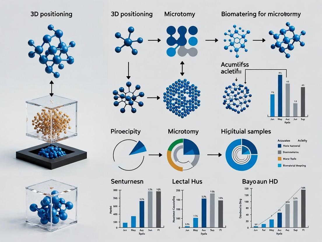
This article provides a comprehensive guide to 3D positioning and microtomy for biomaterial samples, essential for researchers in tissue engineering, drug development, and regenerative medicine.
Precision 3D Positioning and Microtomy: Advanced Techniques for Biomaterial Sectioning and Analysis
Abstract
This article provides a comprehensive guide to 3D positioning and microtomy for biomaterial samples, essential for researchers in tissue engineering, drug development, and regenerative medicine. We explore the fundamental principles of precise spatial orientation and thin-sectioning of complex biomaterials like scaffolds, hydrogels, and organoids. The content details cutting-edge methodological workflows, common troubleshooting strategies for challenging samples, and protocols for validating section quality and comparing techniques. This resource aims to equip scientists with the knowledge to obtain high-fidelity histological data from next-generation biomaterials, bridging the gap between material fabrication and meaningful biological insight.
The Essential Guide to 3D Biomaterial Architecture and Sectioning Fundamentals
Within the broader thesis on advancing 3D microtomy and spatial mapping, this application note details the critical role of precise 3D positioning in biomaterial analysis. The three-dimensional architecture of tissues, scaffolds, and implants dictates cellular behavior, drug diffusion, and integration outcomes. Analyzing biomaterials in 2D sections sacrifices this essential spatial context, leading to incomplete or misleading data. This document outlines protocols for 3D spatial analysis and provides the necessary toolkit for researchers in drug development and biomaterials science.
Quantitative Impact of Spatial Context: Key Metrics
Table 1: Comparative Analysis of 2D vs. 3D Biomaterial Assessment
| Analysis Parameter | 2D Section Analysis | 3D Spatial Positioning Analysis | Quantitative Improvement/Insight |
|---|---|---|---|
| Cell Infiltration Depth | Estimated from single plane | Measured volumetrically from registration of serial sections | Up to 300% more accurate mapping of cell distribution gradients. |
| Angiogenesis Metrics | Vessel count per area | Vessel length, branching points, and 3D network connectivity | 40-60% increase in detected vessel connections; enables tortuosity calculation. |
| Drug Release & Diffusion | Local concentration snapshots | Gradient mapping over time within the 3D construct | Enables kinetic modeling with R² >0.95 vs. ~0.7 in 2D approximations. |
| Biomaterial Degradation | Surface erosion measurement | Volumetric degradation rate and pattern anisotropy | Identifies heterogeneous degradation patterns missed in 90% of 2D samples. |
| Mechanical Property Mapping | Inferred from bulk testing | Correlated local stiffness (AFM) with 3D position | Reveals micromechanical gradients (±15 kPa variation) within a single scaffold. |
Experimental Protocols
Protocol 1: Serial Sectioning & 3D Reconstruction for Implant Analysis
Objective: To reconstruct the 3D spatial context of a polymer scaffold in vivo after a 4-week implantation. Materials: See "The Scientist's Toolkit" below. Method:
- Sample Preparation & Embedding: Perfuse-fix explanted scaffold-tissue construct. Dehydrate in graded ethanol series (50%, 70%, 95%, 100%). Infiltrate and embed in paraffin or optimal cutting temperature (OCT) compound for cryosectioning.
- Reference Marker Implantation: Before embedding, insert 3-5 sterile acupuncture needles coated with a fluorescent dye (e.g., DiI) at known, offset coordinates into the scaffold. These serve as fiducial markers for later alignment.
- Automated Serial Microtomy: Using a calibrated rotary microtome (for paraffin) or cryostat (for OCT), section the entire block at 5 µm thickness. Collect every section sequentially on charged glass slides or into well plates for high-content screening.
- Staining & Imaging: Perform automated immunofluorescence (e.g., for CD31/vessels, nuclei/DAPI, inflammatory markers) across all serial sections. Use a slide scanner with consistent focus and exposure settings.
- Image Registration & 3D Reconstruction:
- Import image stacks into software (e.g., Amira, Imaris, or Fiji/ImageJ with 3D plugins).
- Use the fiducial markers to perform rigid/affine registration, aligning all serial sections.
- Segment regions of interest (e.g., scaffold material, blood vessels, specific cell types) using thresholding and machine-learning classifiers.
- Render the segmented labels into a 3D volume for quantitative analysis (volume, distances, connectivity).
Protocol 2: Correlative 3D Positioning of Drug Particles via Mass Spectrometry Imaging (MSI)
Objective: To map the spatial distribution and metabolism of a drug compound within a 3D tissue-engineered model. Method:
- Sample Processing: Flash-freeze the 3D cell-laden biomaterial in liquid nitrogen-cooled isopentane. Embed in OCT.
- Cryosectioning for 3D Correlation: Section the block at 10 µm. Collect consecutive sections alternately on:
- Slide A: For MSI analysis (conductive indium tin oxide-coated slides).
- Slide B: For histological staining (H&E, immunofluorescence).
- Spatially-Registered Analysis:
- Perform Matrix-Assisted Laser Desorption/Ionization (MALDI)-MSI on Slide A to generate ion maps for the drug parent compound and its major metabolites.
- Image Slide B with standard microscopy techniques.
- Use landmark-based co-registration software to align the MSI data with the histological image from the adjacent section, creating a correlated map.
- Repeat alignment for the entire series to build a 3D model of drug distribution relative to tissue morphology.
Visualizations
Title: Workflow for Correlative 3D Drug Distribution Analysis
Title: Cycle of 3D Data Driving Biomaterial Development
The Scientist's Toolkit: Essential Reagents & Materials
Table 2: Key Research Reagent Solutions for 3D Spatial Analysis
| Item | Function & Importance |
|---|---|
| Fiducial Markers (e.g., Fluorescent Beads, DiI-coated needles) | Provide reference points across serial sections for accurate 3D image registration and alignment. Critical for maintaining spatial integrity. |
| Optimal Cutting Temperature (OCT) Compound | A water-soluble embedding medium for cryosectioning. Preserves native tissue state and is compatible with MSI, unlike paraffin. |
| Conductive ITO-Coated Microscope Slides | Essential substrate for MALDI-MSI. Allows for charge dissipation during ionization, enabling high-quality spatial metabolomics/drug imaging. |
| Automated Slide Staining System | Ensures consistent, reproducible immunofluorescence across hundreds of serial sections, removing a major source of variability in 3D studies. |
| Tissue Clearing Reagents (e.g., CUBIC, CLARITY) | Optional pre-processing step. Renders whole tissue/biomaterial constructs optically transparent for deeper imaging prior to sectioning, guiding region-of-interest selection. |
| 3D Image Analysis Software (e.g., Imaris, Amira, Arivis) | Specialized platforms for handling large serial-section datasets, performing segmentation, 3D rendering, and extracting quantitative spatial statistics. |
| High-Precision Microtome/Cryostat with Automatic Feeder | Enables reliable, consistent sectioning of an entire sample block into a complete series, which is the foundational step for any 3D reconstruction. |
Within the broader thesis on 3D positioning and analysis of biomaterial samples, microtomy serves as the foundational physical technique for accessing internal architectures. The precision of sectioning dictates the fidelity of subsequent 3D reconstructions, molecular mapping, and functional analysis. This document outlines core principles, application notes, and standardized protocols for paraffin and cryosectioning modalities critical for advanced biomaterials research.
Core Principles & Quantitative Comparisons
The choice between paraffin embedding and cryosectioning is dictated by sample nature, target analyte stability, and required resolution.
Table 1: Comparative Analysis of Microtomy Modalities for Biomaterials
| Parameter | Paraffin Microtomy | Cryomicrotomy (Complex Biomaterials) |
|---|---|---|
| Typical Section Thickness | 3–10 µm | 5–50 µm (Highly variable based on material) |
| Optimal Sample Temp. | Ambient (20–24°C) | -15°C to -30°C (Tissue); -20°C to -50°C (Stiff Composites) |
| Post-Sectioning Processing | Required (Deparaffinization, Rehydration) | Often direct to assay or fixation |
| Key Advantage | Superior morphological detail, thin sections | Preservation of labile molecules (lipids, antigens), no embedding for some materials |
| Primary Limitation | Heat & solvent exposure denatures many biomolecules | Sectioning artifacts (chatter, cracking) in heterogeneous materials |
| Best For | Fixed tissues, decalcified bone, soft polymers | Hydrogels, native bone, lipid-rich systems, metal-biomaterial composites |
Table 2: Impact of Knife Angle on Sectioning Artifacts (Empirical Data)
| Knife Clearance Angle | Result on Paraffin Sections | Result on Cryosections (at -20°C) |
|---|---|---|
| 3–5° | Compression, wrinkling | Severe chatter, shattering |
| 5–7° (Standard) | Acceptable, minor compression | Acceptable for homogeneous soft biomaterials |
| 8–10° | Good, less compression | Improved for dense, fibrous composites |
Protocols
Protocol 1: Paraffin Sectioning of Cell-Laden Hydrogel Biomaterials
Application Note: For 3D-cultured cell spheroids or soft polymers processed for histology.
- Fixation & Dehydration: Fix in 4% PFA for 24–48 hrs (dependent on scaffold thickness). Process through an ethanol series (70%, 95%, 100% x3), 1 hour each.
- Clearing & Infiltration: Clear in xylene or xylene-substitute (3 changes, 1 hr each). Infiltrate with molten paraffin wax (58–60°C) under vacuum (3 changes, 1–2 hrs each).
- Embedding & Blocking: Orient sample in mold. Solidify on cold plate.
- Trimming & Sectioning: Trim block face with razor to expose sample. Mount in microtome. Set thickness to 5–7 µm. Use a sharp, disposable metal blade.
- Section Transfer: Float ribbons on a 40°C water bath containing gelatin (0.1% w/v) to reduce folding. Mount on charged slides.
- Drying: Dry slides overnight at 37°C.
Protocol 2: Cryosectioning of Heterogeneous Bone-Biomaterial Implants
Application Note: For undecalcified bone-bioceramic composites requiring RNA/protein preservation.
- Sample Preparation: Freshly excise implant. Trim to <5 mm dimension. Do not fix if analyzing labile targets.
- Embedding & Orientation: Snap-freeze in liquid nitrogen-cooled isopentane for 2 min. Embed in Optimal Cutting Temperature (O.C.T.) compound on a pre-cooled (-20°C) specimen disc. Precisely orient for desired cross-section (critical for 3D positioning).
- Cryochamber Equilibration: Allow sample, chuck, and knife to equilibrate in cryostat to -25°C (adjust to -30°C for very dense composites).
- Trimming & Sectioning: Trim block face at 50 µm increments until full face is exposed. Set anti-roll plate with minimal gap. Section at 8–12 µm thickness with a slow, consistent cutting speed (1–2 mm/sec).
- Section Collection: For direct collection, use a room-temperature, charged slide to gently touch the section. For O.C.T.-embedded sections, use a fine brush to manipulate onto a cold slide.
- Immediate Processing: Fix slides in ice-cold acetone for 2 min or proceed directly to RNA extraction buffer.
The Scientist's Toolkit: Essential Research Reagent Solutions
Table 3: Key Materials for Advanced Biomaterial Microtomy
| Item | Function & Rationale |
|---|---|
| Charged/Adhesive Slides | Prevents section loss during rigorous staining protocols, especially for fragile biomaterials. |
| Optimal Cutting Temperature (O.C.T.) Compound | Water-soluble embedding medium that freezes to support tissue structure; formulation varies for hard/soft samples. |
| Cryostat Anti-Roll Plate | Critical for flattening sections during cryosectioning; alignment is paramount. |
| Disposable High-Profile Microtome Blades | Single-use ensures extreme sharpness for consistent paraffin sections, minimizing compression. |
| Poly-L-Lysine or Gelatin-Coated Slides | Enhances adhesion of paraffin sections, particularly for fatty or low-protein biomaterials. |
| Liquid Nitrogen-Cooled Isopentane | Enables rapid, uniform freezing of hydrated biomaterials, minimizing ice crystal damage. |
Visualized Workflows and Pathways
Paraffin Processing and Sectioning Workflow
Cryosectioning Method Decision Tree
Application Notes
The analysis of three-dimensional biomaterials like scaffolds, hydrogels, and native soft tissues is pivotal in tissue engineering, regenerative medicine, and drug development. However, their inherent physical properties—high porosity, variable stiffness, and substantial water content—pose significant challenges for histological processing and microtomy. These challenges must be addressed to ensure the structural and biomolecular integrity required for meaningful analysis within a 3D positioning and microtomy workflow.
1. Scaffolds (Synthetic & Natural):
- Primary Challenge: Porosity and structural rigidity/deformability. Rigid scaffolds (e.g., certain ceramics, hardened polymers) can shatter during sectioning. Soft, porous scaffolds often collapse during dehydration or tear during microtomy.
- Impact: Loss of 3D architecture, poor ribbon formation, and fragmented sections.
- Key Consideration: Optimal infiltration of paraffin or resin into pores is critical. Incomplete infiltration leads to sectioning artifacts.
2. Hydrogels:
- Primary Challenge: Extreme hydrophilicity and low mechanical strength. They undergo severe shrinkage (up to 70-80%) in standard ethanol dehydration series.
- Impact: Dramatic dimensional changes, altered microstructure, and diffusion of soluble factors, compromising spatial data accuracy.
- Key Consideration: Stabilization (e.g., with chemical crosslinkers) and the use of graduated, slow dehydration protocols or freeze-substitution are essential.
3. Soft Tissues (e.g., Engineered Tissue Constructs, Adipose, Brain):
- Primary Challenge: Lack of supportive endogenous matrix, leading to poor cohesion. Adipose tissues and lipid-rich constructs are particularly prone to leaching and fragmentation.
- Impact: Disintegration of the sample during processing, leading to loss of cellular localization within the 3D context.
- Key Consideration: Enhanced fixation and support during embedding (e.g., using agarose pre-embedding or celloidin bagging) is often required.
Comparative Data Summary
Table 1: Quantitative Challenges and Solutions for Biomaterial Microtomy
| Sample Type | Typical Elastic Modulus | Typical Water Content | Avg. Sectioning Thickness Range | Critical Processing Step | Recommended Embedding Medium |
|---|---|---|---|---|---|
| Porous Scaffold | 0.1 MPa - 2 GPa | 10% - 90% | 5 - 20 µm | Vacuum-assisted, extended infiltration | Paraffin (low-melt) or Glycol Methacrylate (GMA) resin |
| Hydrogel | 0.1 - 100 kPa | >90% | 10 - 50 µm (cryo) / 5 - 20 µm (resin) | Stabilization (e.g., 1-4% PFA/GA mix) | Optimal Cutting Temperature (O.C.T.) compound or GMA resin |
| Soft Tissue | 0.5 - 100 kPa | 60% - 80% | 5 - 30 µm | Agarose pre-embedding (2-4%) | Paraffin or O.C.T. compound |
Experimental Protocols
Protocol 1: Resin-Embedding for Fragile Porous Scaffolds Objective: To infiltrate and embed a porous, low-strength collagen scaffold for thin-section microtomy.
- Fixation: Immerse scaffold in 4% Paraformaldehyde (PFA) for 24h at 4°C.
- Washing: Rinse in 0.1M Phosphate Buffer (PB) 3 x 10 min.
- Dehydration: Gradual ethanol series: 50%, 70%, 80%, 90%, 95% (1h each), 100% (2 x 2h).
- Infiltration: Place in 1:1 mixture of 100% ethanol and Glycol Methacrylate (GMA) resin for 6h. Transfer to pure GMA resin under vacuum (25 inHg) for 48h with one resin change.
- Embedding: Orient scaffold in silicone mold filled with fresh GMA resin containing polymerization catalyst. Polymerize at 4°C under UV light for 48h.
- Sectioning: Trim block and cut 3-5 µm sections using a glass knife on a rotary microtome. Float sections on warm water (40°C) and collect on charged slides.
Protocol 2: Cryopreservation and Sectioning of Hydrogel Constructs Objective: To preserve hydrogel structure and cellular content for cryo-sectioning.
- Stabilization: Treat hydrogel with 2% PFA / 0.1% Glutaraldehyde (GA) in PBS for 2h at 4°C.
- Cryoprotection: Infiltrate with 15% Sucrose in PBS for 6h, then 30% Sucrose overnight at 4°C.
- Embedding: Position hydrogel in cryomold, surround with O.C.T. compound, and orient.
- Freezing: Slowly lower mold into isopentane chilled by liquid nitrogen to approximately -80°C until solid. Store at -80°C.
- Sectioning: Equilibrate block to -20°C in cryostat. Cut 10-20 µm sections using a sharp tungsten carbide blade. Pick up sections on superfrost slides at room temperature.
The Scientist's Toolkit
Table 2: Essential Research Reagent Solutions
| Reagent/Material | Primary Function | Application Context |
|---|---|---|
| Glycol Methacrylate (GMA) Resin | A hydrophilic, low-viscosity embedding medium that polymerizes at low temps with minimal shrinkage. | Ideal for embedding hydrogels and porous scaffolds where paraffin infiltration is insufficient. |
| Optimal Cutting Temperature (O.C.T.) Compound | A water-soluble glycol and resin polymer that provides structural support during freezing and cryo-sectioning. | Essential for cryopreservation and sectioning of hydrogels, soft tissues, and cellular constructs. |
| Agarose (Low Gelling Temperature) | Forms a supportive gel matrix around delicate samples to prevent disintegration during processing. | Pre-embedding for soft tissues and fragile engineered constructs prior to paraffin or resin processing. |
| Sucrose (15-30% in PBS) | Cryoprotectant that reduces ice crystal formation by displacing water during freezing. | Critical step for preserving ultrastructure in hydrogel and soft tissue samples for cryo-sectioning. |
| Paraformaldehyde (PFA) with Glutaraldehyde (GA) | Combined crosslinking fixative. PFA provides rapid penetration; GA enhances structural rigidity. | Stabilization of hydrogel matrices and extracellular proteins to resist processing-induced deformation. |
Visualizations
Resin Embedding Workflow for Porous Scaffolds
Biomaterial Challenges & Unified Processing Solution
The Role of Embedding Media and Support Systems in Preserving 3D Structure
Within the thesis on 3D positioning and microtomy for biomaterial research, maintaining the original three-dimensional architecture of samples during processing is paramount. Embedding media and support systems provide the structural reinforcement necessary to preserve spatial relationships during sectioning, enabling accurate downstream analysis crucial for drug development and fundamental research.
Application Notes
Quantitative Comparison of Common Embedding Media
The choice of embedding medium directly impacts morphological preservation, sectioning quality, and compatibility with stains or probes. Current research prioritizes media that balance structural support with molecular preservation.
Table 1: Properties of Common Embedding Media for 3D Biomaterial Sectioning
| Embedding Medium | Viscosity (cP, 25°C) | Polymerization Shrinkage (%) | Sectioning Thickness Range (µm) | Optimal Processing Temperature | Key Applications & Compatibility |
|---|---|---|---|---|---|
| Paraffin Wax | 10-15 (melted) | 8-15 | 2-10 | 55-60°C | Standard histology, H&E; poor for lipids/some antigens. |
| Glycol Methacrylate (GMA) | 15-25 | 5-8 | 0.5-3 | 4°C (UV) | High-resolution light microscopy, retains enzyme activity. |
| Low-Melting Agarose (4%) | ~1800 (40°C) | N/A (gels) | 20-100 (vibratome) | 37-45°C (liquefaction) | Pre-embedding for delicate tissues, maintains viability. |
| Polyacrylamide (PAA) 4-20% | Varies with % | 3-7 | 1-5 (cryostat) | 4°C (chemical) | CLARITY-based tissue clearing, hydrogel-tissue hybridization. |
| Optimal Cutting Temp (O.C.T.) | High (viscous) | Minimal (frozen) | 5-20 (cryostat) | -20 to -25°C (sectioning) | Cryosectioning, immunofluorescence, lipid-rich samples. |
| EPON/Araldite Resin | 500-1000 | 2-5 | 0.05-0.5 (ultrathin) | 60°C (thermal) | Electron microscopy, superior ultrastructural preservation. |
Support System Efficacy Data
Support systems work in concert with embedding media to prevent collapse, compression, or rolling during microtomy.
Table 2: Performance Metrics of Physical Support Systems
| Support System Material | Tensile Strength (MPa) | Flexibility (Elongation at Break %) | Adhesion to Common Media | Reusable? | Primary Function |
|---|---|---|---|---|---|
| Cellulose Acetate Filter Membranes | 40-60 | 15-30 | High (hydrophilic) | No | Prevents roll-up of thin sections, provides backing. |
| Polyester (PET) Mesh Grids | 150-200 | 70-100 | Moderate (requires coating) | Yes | 3D spatial indexing, supports serial sections. |
| Cyanoacrylate Glue Layer | 15-25 (bond) | 2-5 | Excellent (chemical bond) | No | Adheres sample to chuck, seals edges to prevent dehydration. |
| Gelatin-Coated Slides | N/A | N/A | High for cryosections | No | Adheres sections during staining, prevents wash-off. |
| Poly-L-lysine Coated Adhesive Tapes | 50-70 (tape backing) | 10-20 | Excellent (pressure-sensitive) | No | Section Transfer System: Enables intact collection of thin, fragile ribbons. |
Experimental Protocols
Protocol: Hydrogel-Embedding and Precision Microtomy for 3D Biomaterials
Objective: To preserve the 3D architecture of a soft, porous biomaterial scaffold (e.g., collagen hydrogel) for histological analysis.
I. Sample Preparation and Embedding
- Fixation: Immerse biomaterial sample in 4% paraformaldehyde in 0.1M phosphate buffer (pH 7.4) for 24 hours at 4°C.
- Washing: Rinse sample 3 x 1 hour in 0.1M phosphate buffer with 0.02% sodium azide.
- Dehydration: Pass sample through a graded ethanol series (30%, 50%, 70%, 85%, 95%, 100%, 100%) for 1 hour each at 4°C.
- Inflation & Pre-Infiltration: Place sample in 1:1 mixture of 100% ethanol and glycol methacrylate (GMA) monomer solution (containing 0.1% benzoyl peroxide) for 24 hours at 4°C under gentle agitation.
- Infiltration: Transfer to pure GMA monomer solution for 48 hours, with two changes, at 4°C.
- Embedding & Polymerization:
- Place sample in a silicone mold filled with fresh GMA monomer solution.
- Add polyethylene mold holder for stability.
- Catalyze polymerization with 0.5% N,N-dimethylaniline (in GMA) and 2.5% polyethylene glycol 400.
- Polymerize under UV light (365 nm) at 4°C for 48 hours.
II. Support-Mounted Microtomy
- Block Trimming: Using a glass knife on a precision microtome, trim the polymerized block to create a trapezoidal face around the sample.
- Support Application: Apply a thin layer of cyanoacrylate glue to the surface of a standard microtome specimen chuck. Firmly press the trimmed base of the GMA block onto the glued chuck. Allow 5 minutes for bond to set.
- Sectioning: Mount chuck onto a rotary microtome. Using a tungsten-carbide knife, cut sections at 2 µm thickness.
- Section Transfer & Support:
- For serial sectioning: Use a fine brush to float the ribbon of sections onto the surface of a water bath (40°C).
- For single sections: Use a polyester (PET) mesh grid (pre-coated with 0.1% gelatin) to carefully lift the section from the knife edge.
- Mounting: Place the mesh grid with adherent section onto a drop of distilled water on a glass slide. Gently heat slide (40°C) to evaporate water and adhere section.
Protocol: Cryo-embedding and Tape-Supported Sectioning for Lipid-Rich Samples
Objective: To preserve lipid content and antigenicity in a lipid-rich biomaterial (e.g., adipose-derived matrix) for immunofluorescence.
I. Cryo-embedding
- Cryoprotection: After fixation and washing, infiltrate sample with 15% sucrose (4 hours) followed by 30% sucrose (overnight) at 4°C.
- Embedding Mold Preparation: Fill a cryomold halfway with Optimal Cutting Temperature (O.C.T.) compound.
- Orientation & Freezing: Position sample in the mold. Completely fill mold with O.C.T., ensuring no bubbles. Slowly lower the mold onto the surface of a liquid nitrogen-chilled isopentane bath for rapid, uniform freezing. Store at -80°C.
II. Tape-Transfer System Microtomy
- Cryostat Preparation: Set cryostat chamber temperature to -20°C and object temperature to -18°C.
- Block Mounting: Adhere the frozen O.C.T. block to a pre-chilled cryostat chuck using a small amount of O.C.T. as glue. Allow to freeze solid.
- Tape Application: Trim block face. Prior to cutting the section, firmly apply a clear poly-L-lysine coated adhesive tape (CryoFilm) onto the trimmed block surface.
- Sectioning: Cut the section (5-10 µm thickness) with the tape adhered to its upper surface. The section is now supported entirely by the tape.
- Transfer: Remove the tape with the adherent section from the knife. Adhere the tape (section-side down) directly onto a charged glass slide.
- Fixation: Place slide on a warm plate (37°C) for 5 minutes to improve adhesion. Immediately fix in cold acetone for 2 minutes before staining.
Visualization: Workflows and Relationships
Decision Workflow for Embedding and Support in 3D Biomicrotomy
Embedding Solutions for Microtomy Challenges
The Scientist's Toolkit: Research Reagent Solutions
| Product/Category | Example Formulations/Brands | Primary Function in 3D Preservation |
|---|---|---|
| Hydrophilic Acrylic Resins | Glycol Methacrylate (GMA) Kits (e.g., Technovit 7100), Polyacrylamide (PAA) | Low shrinkage embedding; allows sectioning at 1-3 µm for high-resolution LM; preserves antigenicity. |
| Cryo-embedding Media | Optimal Cutting Temperature (O.C.T.) Compound, Tissue-Tek | Water-soluble glycols and resins that support tissue during snap-freezing and cryosectioning. |
| Adhesive Support Systems | CryoFilm Type 2C/3C, PEN Membrane Slides, Adhesive-Coated Microscope Slides | Provide a supporting substrate for fragile sections, preventing fragmentation and loss. |
| Spatial Indexing Grids | Polyester (PET) or Nickel Mesh Grids (e.g., ATUMtome substrates) | Physical grid applied to block face or section for tracking 3D location in serial section arrays. |
| Block Face Adhesives | Cyanoacrylate Glue (Super Glue), Two-part Epoxy | Securely binds embedded sample to microtome chuck, preventing dislodgement and vibration. |
| Section Floatation Bath Additives | Surfactants (e.g., Photo-Flo 200), Ethanol | Reduce water surface tension to minimize folding and distortion of floated paraffin ribbons. |
| Controlled Polymerization Catalysts | Benzoyl Peroxide (thermal), DMP-30 (epoxy), UV Initiators (e.g., DAPI analog) | Allow slow, controlled curing of resins to minimize exothermic heat and shrinkage artifacts. |
| Decalcification & Softening Agents | EDTA (pH 7.4), RDO Rapid Decalcifier | Treat mineralized biomaterials post-fixation to enable microtomy without compromising soft matrix. |
Step-by-Step Protocols: Optimized Workflows for Biomaterial Embedding and Sectioning
This protocol details the first critical workflow in a comprehensive thesis on 3D spatial targeting for biomaterial analysis. Precise orientation and embedding of 3D scaffolds (e.g., hydrogels, decellularized matrices, synthetic polymer networks) is the foundational step that enables subsequent accurate microtomy and volumetric analysis of cellular events, drug distribution, and material integration. Failure at this stage compromises all downstream spatial data, making reproducibility and geometric fidelity paramount.
Application Notes: Rationale and Key Principles
Objective: To immobilize a 3D scaffold sample within a supporting medium (e.g., resin, OCT, paraffin) while maintaining a known, precise geometric orientation relative to its original 3D structure, and to create embedding landmarks for spatial targeting.
Critical Challenges:
- Deformation: Minimizing scaffold shrinkage/swelling during fixation and processing.
- Landmark Loss: Preserving intrinsic sample landmarks (e.g., a specific corner, injection point) or creating artificial ones.
- Medium Infiltration: Ensuring complete infiltration of the embedding medium without creating bubbles or voids that hinder sectioning.
Spatial Targeting Relevance: Correct orientation allows for correlated imaging, where a region of interest (ROI) identified via non-destructive imaging (e.g., micro-CT, confocal) can be systematically located and sectioned for high-resolution histology or molecular analysis.
Detailed Protocol: Oriented Embedding of 3D Scaffolds
Materials and Equipment
Table 1: Research Reagent Solutions & Essential Materials
| Item | Function/Brief Explanation |
|---|---|
| Slow-Curing, Low-Shrinkage Epoxy Resin (e.g., EPON 812 substitute) | Infiltrates porous scaffolds, providing rigid support for thin microtomy; low exothermic heat minimizes damage. |
| Cryostat Embedding Medium (O.C.T.) | For frozen sectioning; preserves labile antigens and fluorescent proteins. |
| Biocompatible Fluorescent Microbeads (1-10µm) | Injected or placed as fiducial markers for precise 3D registration and landmarking. |
| 3D-Printed Orientation Mold (Polystyrene) | Custom mold with registration notches/keyways to hold sample in defined XYZ orientation during embedding. |
| Tri-Angulation Reference Object (TARO) | A small, inert physical object (e.g., metal pin, colored polymer cube) co-embedded adjacent to the sample as a macroscopic directional guide during trimming. |
| Vacuum Infiltration Chamber | Removes air from scaffold pores to ensure complete resin/OCT infiltration. |
| Tissue-Tek Uni-Cassette with Coordinate Grid | Cassette with an imprinted grid allows for manual logging of sample orientation sketches. |
Step-by-Step Methodology
Day 1: Fixation and Landmarking
- Primary Fixation: Immerse scaffold in 4% paraformaldehyde (PFA) in 0.1M phosphate buffer (pH 7.4) for 18-24 hours at 4°C.
- Fiducial Marker Application: Using a microinjector, deposit 3-5 distinct lines or points of fluorescent microbeads at known, asymmetric locations on the scaffold surface. Record their 3D coordinates via confocal or micro-CT imaging.
- Dehydration: For resin embedding, perform graded ethanol dehydration (50%, 70%, 90%, 100% x3), 1 hour per step.
Day 2: Infiltration and Casting
- Resin Infiltration: Place dehydrated sample in a 1:1 mixture of ethanol:resin for 4 hours, then pure resin for 8 hours under vacuum (25 inHg) at room temperature.
- Orientation in Mold:
- Fill the custom 3D-printed orientation mold halfway with fresh resin.
- Under a stereomicroscope, use fine forceps to place the sample. Align the pre-marked fiducial points with the mold's registration notches.
- Place the Tri-Angulation Reference Object (TARO) in a corner of the mold, noting its position relative to the sample.
- Completely fill the mold with resin.
- Polymerization: Cure at 60°C for 48 hours.
Day 3: Block Trimming and Documentation
- Remove the polymerized block from the mold.
- Trimming: On a precision trimmer, roughly trim the block, leaving the TARO object intact. Then, carefully trim the block face to expose the scaffold surface, using the TARO and mold notches as guides to maintain the correct orientation.
- Documentation: Photograph the final trimmed block face alongside a schematic drawing in a lab notebook. Note the relationship between the exposed scaffold face and the cassette's coordinate grid.
Data Presentation
Table 2: Quantitative Comparison of Embedding Media for Spatial Targeting
| Parameter | Slow-Curing Epoxy Resin | Cryo-Embedding Medium (OCT) | Paraffin |
|---|---|---|---|
| Dimensional Change | ~2% shrinkage | ~5-15% expansion/ice artifact | ~30% shrinkage |
| Section Thickness Range | 0.5 - 5 µm | 5 - 30 µm | 3 - 10 µm |
| Spatial Fidelity (Landmark Preservation) | Excellent (95-98%) | Good, if snap-frozen (85-90%) | Poor (70-80%) |
| Best For | Ultrastructure, precise nano/microtomy | Labile biomarkers, fluorescence | High-throughput histology |
| Compatible with 3D Landmarking (Beads) | Yes | Yes, if beads are freeze-tolerant | No (lost during processing) |
Visualized Workflows and Pathways
Title: Precise Scaffold Embedding and Orientation Workflow
Title: Thesis Workflow Dependency for Spatial Mapping
Application Notes
Cryo-preparation and sectioning are critical techniques for the histological and biomolecular analysis of hydrated, delicate biomaterial samples, such as synthetic hydrogels and cell-laden constructs. Within the broader thesis on 3D positioning and microtomy for biomaterial research, this workflow enables the precise spatial preservation of cellular architecture and extracellular matrix (ECM) components within a 3D context. The primary challenge is to prevent ice crystal formation that can distort ultrastructure, while achieving sections with sufficient integrity for downstream applications like immunostaining, in situ hybridization, or correlative microscopy.
Key Advantages:
- Preservation of Native State: Rapid freezing vitrifies water, locking biomolecules and cellular organelles in their near-native 3D positions.
- Compatibility with Biomaterials: Maintains the structural integrity of soft, water-rich hydrogels that would collapse under conventional paraffin processing.
- Retention of Antigenicity: Superior for immunohistochemistry compared to chemical fixation, which can mask epitopes.
Typical Quantitative Outcomes: The success of cryo-sectioning is quantitatively assessed by section integrity, thickness consistency, and antigen preservation.
Table 1: Quantitative Metrics for Cryo-Sectioning Quality Assessment
| Metric | Target Range/Outcome | Measurement Method | Impact of Deviation |
|---|---|---|---|
| Section Thickness | 5 - 20 µm (for light microscopy) | Microtome setting verification; interference colors | Too thin: fragmentation. Too thick: poor imaging clarity. |
| Section Integrity | >90% contiguous area | Microscopic visual analysis | Cracks/folds compromise analysis of 3D positioning. |
| Cellular Morphology Preservation | Minimal vacuolization, distinct membranes | Histological scoring (e.g., H&E) | Indicates poor freezing or fixation, invalidating spatial data. |
| Antigen Preservation Score | High fluorescence intensity, low background | Quantitative fluorescence microscopy | Poor scores hinder biomarker colocalization studies. |
Detailed Protocol: Cryo-Preparation and Sectioning
This protocol details the steps from sample harvest to mounted cryo-sections ready for staining.
Materials & Reagents
Table 2: Research Reagent Solutions & Essential Materials
| Item | Function/Explanation |
|---|---|
| Optimal Cutting Temperature (OCT) Compound | A water-soluble glycol and resin mixture that infiltrates and supports the sample during freezing and sectioning. It freezes to a consistent matrix. |
| Cryo-Molds (e.g., Peel-A-Way) | Disposable molds for embedding samples in OCT. |
| Isopentane (2-Methylbutane) | A secondary coolant chilled by liquid nitrogen. Its higher boiling point than LN2 allows for rapid, non-insulating freezing, minimizing ice crystal damage. |
| Liquid Nitrogen | Primary coolant for super-cooling isopentane. |
| Cryostat | A refrigerated microtome housed in a thermally controlled chamber (typically -20°C). |
| Poly-L-Lysine or Charged Microscope Slides | Coating ensures adherent sections remain fixed during staining procedures. |
| Cryoprotectant Solution (e.g., 15-30% Sucrose in PBS) | Infiltrates tissue prior to freezing, displacing water and further suppressing ice crystal formation. |
| Fine Forceps & Brushes | For manipulating frozen blocks and delicate sections. |
Procedure
Part A: Sample Preparation & Cryo-Embedding
- Harvest & Fixation: Excise hydrogel/cell construct. For optimal morphology, immerse in 4% paraformaldehyde (PFA) in PBS for 4-24 hours at 4°C.
- Cryoprotection (Optional but Recommended): Transfer fixed sample to 15-30% sucrose in PBS. Incubate at 4°C until the sample sinks (4-24 hours), indicating sucrose infiltration.
- Embedding: a. Fill a cryo-mold partially with OCT. b. Orient the sample within the mold using fine tools, ensuring the desired cutting plane aligns with the mold base. c. Completely cover the sample with fresh OCT.
- Snap-Freezing: a. Prepare a freezing bath by partially filling a metal beaker with isopentane. Slowly add liquid nitrogen until the isopentane begins to frost. b. Using forceps, submerge the bottom of the cryo-mold into the super-cooled isopentane for 30-60 seconds until the OCT is completely opaque and frozen. c. Immediately transfer the frozen block to dry ice or a -80°C freezer for storage.
Part B: Cryo-Sectioning
- Cryostat Preparation: Set the cryostat chamber temperature to -20°C to -22°C and the object disk temperature to -18°C to -20°C. Allow to equilibrate.
- Block Mounting: Apply a small amount of OCT to a cryostat specimen disk. Adhere the frozen block, ensuring the cutting face is parallel to the microtome blade. Allow to freeze firmly.
- Trimming & Sectioning: a. Trim the block face with coarse sections (e.g., 30-50 µm) until the full sample surface is exposed. b. Set the section thickness (e.g., 10 µm). c. Begin sectioning using a smooth, steady pace. Use an anti-roll plate or a fine brush to gently guide the ribbon of sections as they form.
- Section Mounting: Bring a room-temperature charged slide close to, but not touching, the section. The thermal gradient will cause the section to adhere to the slide upon gentle contact. Air-dry slides for 30-60 minutes before staining or storage at -80°C.
Visualized Workflows
Cryo-Preparation and Sectioning Workflow
Impact of Cryo-Prep on 3D Spatial Data Validity
Within the broader thesis on 3D Positioning and Microtomy for Biomaterial Samples Research, this application note addresses a critical technical challenge: the longitudinal, spatial analysis of complex 3D cell cultures. Organoids and spheroids recapitulate tissue microenvironments, but their 3D architecture obscures internal cellular and molecular heterogeneity. This protocol details a method for precise spatial positioning, serial sectioning, and multi-modal analysis of the same sample over a simulated time-course, enabling the study of dynamic processes like drug response or differentiation within a 3D context.
Research Reagent Solutions & Essential Materials
| Item Name | Function/Brief Explanation |
|---|---|
| Low-Melt Agarose (2-3%) | Embedding matrix for gentle immobilization of live or fixed spheroids/organoids without morphological distortion. |
| Optimal Cutting Temperature (OCT) Compound | Water-soluble embedding medium for cryosectioning; provides structural support during sectioning at -20°C. |
| Cryomolds & Specimen Discs | For orienting and positioning the agarose/OCT-embedded sample in a defined 3D coordinate system on the microtome. |
| Fluorescent Cell Tracking Dyes (e.g., CFSE) | For pre-labeling cell populations to track their spatial fate and proliferation longitudinally in sectioned samples. |
| Phosphate-Buffered Saline (PBS) | Standard solution for washing samples and preparing reagents. |
| Paraformaldehyde (4%, PFA) | Common fixative for cross-linking proteins and preserving tissue architecture for immunohistochemistry. |
| Sucrose (30% in PBS) | Cryoprotectant; prevents ice crystal formation during the freezing process for cryosectioning. |
| Permeabilization Buffer (e.g., Triton X-100) | Allows antibodies and dyes to penetrate the dense extracellular matrix of the 3D sample. |
| Blocking Serum | Reduces non-specific antibody binding, critical for low-background imaging in immunohistochemistry. |
| Mounting Medium with DAPI | Preserves sections under a coverslip and provides a nuclear counterstain for orientation. |
Table 1: Comparison of Key Parameters for Organoid/Spheroid Sectioning Methodologies
| Parameter | Vibratome Sectioning | Cryostat Sectioning | Paraffin Microtomy |
|---|---|---|---|
| Typical Section Thickness | 50 - 500 µm | 5 - 30 µm | 3 - 10 µm |
| Sample State | Fixed, optionally live | Fixed, frozen (unfixed possible) | Fixed, dehydrated, paraffin-embedded |
| Antigen Preservation | Excellent | Good to Excellent | Variable (requires antigen retrieval) |
| RNA Preservation | Good (with RNase inhibitors) | Excellent (if snap-frozen) | Poor |
| Processing Time | Medium (hours) | Fast (hours) | Slow (days) |
| Suitability for Longitudinal In-Sample Analysis | High (thick sections for 3D imaging) | Medium (sequential thin sections) | Low (destructive) |
| Key Advantage | Thick sections for deep 3D imaging | Speed & compatibility with lipids/RNA | Superior morphology & archival stability |
Experimental Protocol: Longitudinal Analysis via Serial Cryosectioning
Protocol: Sequential Cryosectioning and Staining of Spheroids for Pseudo-Longitudinal Analysis
Objective: To analyze spatial heterogeneity and biomarker distribution within the same organoid at different "time points" (simulated via sequential sectioning).
I. Sample Preparation & 3D Positioning
- Fixation: Transfer mature spheroids/organoids to 4% PFA for 45-60 minutes at 4°C.
- Cryoprotection: Wash 3x with PBS. Infiltrate with 30% sucrose in PBS overnight at 4°C until samples sink.
- Embedding & Orientation:
- Place a drop of OCT in a cryomold.
- Using a wide-bore pipette, transfer the spheroid in minimal sucrose solution to the mold.
- Under a stereomicroscope, use fine tools to position the spheroid in the desired orientation (e.g., apical surface up).
- Surround with OCT, ensuring no bubbles. Place a specimen disc on top.
- Slowly lower onto a slurry of dry ice/isopentane or place in pre-cooled cryostat chamber for frozen alignment.
II. Serial Sectioning and Section Management
- Microtomy: Mount the frozen block on the cryostat. Trim until the spheroid center is reached.
- Section Collection: Cut serial sections at 10-16 µm thickness.
- Section Mapping: Collect sequential sections on alternating slides (e.g., Slide A: sections 1, 4, 7...; Slide B: sections 2, 5, 8...; Slide C: sections 3, 6, 9...). This creates a series of slides representing different "depths" or time-points within the same spheroid.
- Storage: Store slides at -80°C.
III. Multi-modal Staining & Imaging
- Staining Scheme: Apply different stains to the sequential slide series:
- Slide Series A (Depth 1): Hematoxylin & Eosin (H&E) for core morphology.
- Slide Series B (Depth 2): Immunofluorescence for proliferative marker (e.g., Ki67).
- Slide Series C (Depth 3): Immunofluorescence for differentiation marker (e.g., β-III tubulin for neural organoids).
- Co-registration: Use fiduciary markers (e.g., labeled agarose) and software to digitally align images from all series, reconstructing a composite 3D map of the original spheroid.
Visualization Diagrams
Workflow for Pseudo-Longitudinal Sectioning Analysis
Logical Framework: From Thesis to Impact
This document details advanced protocols for the sectioning and handling of fragile biomaterial samples—such as hydrogels, decellularized matrices, and low-temperature polymerized composites—within a broader thesis on 3D positioning and serial microtomy for volumetric analysis. The integrity of these sections is paramount for downstream applications including multi-modal imaging (e.g., SEM, confocal), spatial transcriptomics, and high-resolution histology. Tape-transfer systems and conductive adhesive methods address the primary challenges of section wrinkling, fragmentation, and charging artifacts.
Quantitative Comparison of Sectioning Support Systems
Table 1: Performance Metrics of Fragile Section Support Methods
| Method / Parameter | Section Integrity Score (1-5) | Max Section Thickness (µm) | Compatibility with IHC/FISH | Conductivity (S/cm, approx.) | Primary Artifact Risk |
|---|---|---|---|---|---|
| Cryofilm (Type 2C) | 4.5 | 60 | High | Very Low (< 10^-15) | Folding at ribbon edges |
| Conductive Carbon Tape | 3.5 | 30 | Moderate | High (~ 10^2) | Adhesive bleed-through |
| ITO-Coated Slides | 4.0 | 20 | Very High | Intermediate (~ 10^-3) | Partial adhesion failure |
| Poly-L-Lysine Adhesive | 3.0 | 10 | Very High | Very Low (< 10^-15) | Section stretching |
| Cryo-Tape-Transfer System | 5.0 | 100 | Low (pre-embedding) | Adjustable (with coating) | Tape adhesive residue |
Experimental Protocols
Protocol 3.1: Tape-Transfer System for Serial Sectioning of Hydrogel-Based Biomaterials
Objective: To obtain contiguous, wrinkle-free serial sections of a hydrated, low-density polymer scaffold (e.g., 1% alginate + cell matrix) for 3D reconstruction.
Materials: Cryostat (-20°C to -30°C), Cryofilm Type 2C (Section-Lab), specimen mount, conductive slide (e.g., ITO-coated), freeze spray, fine brush, DI water.
Method:
- Embedding & Mounting: Infiltrate the fixed hydrogel sample with OCT compound minimally. Mount onto the cryostat chuck and freeze completely at -25°C.
- Film Application: Adhere a strip of Cryofilm to the block surface ahead of the knife. Ensure no air bubbles are trapped.
- Sectioning: Cut sections at 10-40 µm thickness with a slow, consistent cutting speed (1-2 mm/sec). The section will adhere directly to the Cryofilm.
- Transfer: Float the film+section onto a droplet of DI water on a conductive slide. Gently tease the film away from the section, leaving the section flat on the slide.
- Drying: Air-dry for 30 minutes, then vacuum-dry for 1 hour prior to staining or coating.
Protocol 3.2: Conductive Adhesive Mounting for Scanning Electron Microscopy (SEM) of Mineralized Fragile Sections
Objective: To mount a fragile, non-conductive mineral-coated collagen section for high-vacuum SEM without charging or distortion.
Materials: Conductive carbon adhesive tape (e.g., Ted Pella), silver epoxy, SEM stub, vacuum sputter coater, fine-tip applicator, low-lint wipes.
Method:
- Stub Preparation: Apply a strip of conductive carbon tape to the surface of an aluminum SEM stub. Smooth out with a rod to ensure uniform contact.
- Section Retrieval: Using a fine brush or eyelash tool, lift the floating cryo- or microtome section from the water bath.
- Mounting: Gently place the section onto the center of the carbon tape. Carefully blot excess peripheral water with a filter paper wedge.
- Curing: Allow the assembly to dry in a desiccator for 2 hours.
- Secondary Conduction: Apply a thin bead of silver epoxy from the edge of the section to the bare metal stub to create a conductive bridge.
- Coating: After the epoxy cures (per manufacturer instructions), sputter-coat the entire assembly with a 10-15 nm layer of gold/palladium.
Visualization: Workflow & Decision Pathway
Diagram 1: Workflow for Handling Fragile Biomaterial Sections
Diagram 2: Conductive Adhesive Method Signaling Pathway
The Scientist's Toolkit: Research Reagent Solutions
Table 2: Essential Materials for Advanced Section Handling
| Item | Manufacturer/Example | Function in Protocol |
|---|---|---|
| Cryofilm Type 2C | Section-Lab, Leica | Specialized polyethylene film that supports fragile sections during cryosectioning, preventing compression and folds. |
| Conductive Carbon Tape | Ted Pella, SPI Supplies | Provides immediate adhesive contact and electrical conductivity for SEM sample mounting. |
| Silver Epoxy | Ted Pella, MG Chemicals | Creates a high-fidelity, low-resistance conductive path from sample to stub to prevent charging. |
| ITO-Coated Slides | Delta Technologies, Matsunami | Glass slides with an indium tin oxide coating; conductive for SEM prep and compatible with optical microscopy. |
| Cryostat w/ Tape-Transfer Arm | Leica, Thermo Scientific | Microtome system with dedicated hardware for automated tape application and section transfer. |
| Low-Temperature Embedding Matrix (OCT) | Sakura, Tissue-Tek | Optimal cutting temperature compound for supporting hydrogel samples during freezing. |
| Anti-Static Gun | Simco-Ion, Exair | Neutralizes static charge on slides, tapes, and tools to prevent section attraction and dust contamination. |
Within the broader thesis on 3D positioning and microtomy for biomaterial samples, the precise anatomical orientation of tissue sections is paramount. Biomaterials, such as bone scaffolds, neural implants, or drug-eluting matrices, interact with host tissues in complex three-dimensional ways. Traditional planar histology risks missing critical regions of interest (ROIs), such as the exact interface between implant and tissue, specific vascularization patterns, or localized inflammatory responses. Integrating pre-sectioning volumetric imaging via micro-Computed Tomography (µCT) or Magnetic Resonance Imaging (MRI) provides a 3D map to strategically guide the microtomy process. This protocol details the application of registered 3D image data to inform and optimize physical sectioning planes, ensuring histological analysis targets the most scientifically relevant spatial coordinates.
Application Notes: µCT vs. MRI for Biomaterial Guidance
The choice between µCT and MRI as a guidance modality depends on sample properties and the research question.
Table 1: Comparative Guide to Imaging Modalities for Sectioning Planning
| Feature | µCT (Micro-Computed Tomography) | MRI (Magnetic Resonance Imaging) |
|---|---|---|
| Primary Contrast | X-ray attenuation (density). Excellent for mineralized tissue, dense biomaterials, scaffold architecture. | Proton density, T1/T2 relaxation. Superior for soft tissues, hydrogel interfaces, edema, fluid-filled spaces. |
| Spatial Resolution | Very high (typically 1-10 µm isotropically). | High to moderate (typically 10-50 µm isotropically for preclinical systems). |
| Key Application in Biomaterials | Visualizing bone-ingrowth into porous scaffolds, locating metallic implant fragments, assessing biomaterial degradation (if density changes). | Visualizing soft tissue integration, fibrous capsule formation around implants, assessing hydrogel hydration/degradation, locating non-calcified ROI. |
| Sample Preparation | Often requires fixation. Can be stained with contrast agents (e.g., phosphotungstic acid) for soft tissue enhancement. | Requires fixation and immersion in proton-free fluid (e.g., perfluoropolyether) or use of specialized coils for optimal signal. |
| Integration with Histology | Excellent for co-registration with hard tissue histology (e.g., von Kossa, TRAP). Landmarks (e.g., bone spicules, implant edges) are easily matched. | Excellent for co-registration with soft tissue stains (e.g., H&E, Masson's Trichrome, immunohistochemistry). |
| Quantitative Data for Planning | Precise 3D coordinates of biomaterial boundaries, pore positions, mineralized tissue volumes. | 3D maps of tissue morphology, fluid boundaries, and contrast-enhanced regions. |
Detailed Experimental Protocol
Protocol Title: Registration of Volumetric Image Data to Physical Sample for Guided Microtomy
Objective: To use pre-acquired 3D µCT or MRI data to define the optimal cutting plane for histological sectioning of a biomaterial-tissue sample.
Materials & Reagents:
- Fixed biomaterial-tissue sample (e.g., scaffold explant).
- Pre-acquired µCT or MRI dataset of the fixed sample.
- Embedding medium (e.g., paraffin, PMMA, or optimal cutting temperature (OCT) compound).
- Orientation aids: Nylon sutures (9-0), India ink dots, laser-engraved fiduciary marks on embedding cassette.
- Laboratory µCT system (e.g., Bruker SkyScan, Scanco µCT) or preclinical MRI system (e.g., Bruker BioSpec, Agilent).
- Image processing software (e.g., Amira, 3D Slicer, Fiji/ImageJ).
- Precision microtome or cryostat.
- Sample chuck compatible with microtomy and optionally with the imaging stage.
Step-by-Step Workflow:
Primary 3D Imaging:
- Fix the explanted biomaterial-tissue construct thoroughly.
- For µCT: Place sample in a radiolucent tube. If soft tissue contrast is needed, stain the sample with a radio-opaque agent (e.g., 1% phosphotungstic acid in water) for 24-72 hours. Scan at a resolution sufficient to resolve the target features (e.g., 5 µm isotropic voxel size). Reconstruct the projection data to obtain a 3D volume.
- For MRI: Place the fixed sample in a tube filled with a proton-free fluid (e.g., perfluoropolyether) to eliminate background signal. Use a T2-weighted or proton density-weighted 3D sequence to acquire the volumetric data.
Digital Plane Planning:
- Import the 3D volume into image analysis software (e.g., 3D Slicer).
- Use multiplanar reconstruction (MPR) viewers to navigate through the volume.
- Identify the Region of Interest (ROI): e.g., "the central plane of the maximum defect infiltration," or "the plane tangential to the implant-tissue interface showing the thickest fibrous capsule."
- Define the desired sectioning plane digitally. The software will provide the plane's equation or orientation angles (azimuth, elevation) relative to the sample's coordinate system.
Physical Registration and Embedding:
- Critical Step: Establish physical fiducials on the sample that are visible in both the 3D image and reality. Before embedding, carefully place 2-3 small India ink dots or suture knots at distinct, non-planar locations on the sample surface.
- Re-image the sample with fiducials in place using a quick, low-resolution scan. This creates a "fiducial map" dataset.
- In software, co-register the high-resolution scan (Step 1) with the fiducial map scan. This links the digital coordinate system to the physical fiducials.
- Embed the sample in the chosen medium. Use an embedding mold/cassette with laser-etched registration marks (e.g., a corner notch or engraved crosshairs). Precisely align the physical sample fiducials relative to these cassette marks under a dissecting microscope.
Coordinate Transfer & Block Trimming:
- The digital cutting plane is now defined relative to the fiducials, which are in a known relationship to the embedding cassette.
- Calculate the necessary angles and depths for trimming the embedding block to bring the pre-defined plane parallel to the future microtome knife face.
- Using a precision trimmer or microtome, gradually trim the block face to the calculated orientation. Periodically, image the block face with a macro-photography setup or a low-resolution µCT scan to compare its position with the digital target plane.
Guided Sectioning:
- Mount the precisely trimmed block on the microtome.
- Perform sectioning. The initial sections will correspond directly to the digitally planned plane.
- Validation: For the first few sections (e.g., ribbons 1-3), acquire brightfield scans. These can be digitally registered back to the original 3D volume to confirm plane accuracy, completing the feedback loop.
Diagram 1: Workflow for Imaging-Guided Microtomy
The Scientist's Toolkit: Research Reagent Solutions
Table 2: Essential Materials for Imaging-Guided Sectioning
| Item | Function in Protocol |
|---|---|
| Phosphotungstic Acid (PTA) 1% Solution | Radio-opaque contrast agent for µCT. Stains soft tissue (e.g., collagen, cells) within biomaterial explants, enabling visualization for plane planning. |
| Perfluoropolyether (e.g., Fomblin) | Proton-free immersion fluid for MRI. Eliminates background signal from surroundings, drastically improving signal-to-noise ratio for ex vivo samples. |
| India Ink / Surgical Marking Dye | Provides high-contrast, indelible physical fiducial marks on the sample surface, visible in both 3D images and during embedding. |
| Laser-Engraved Embedding Cassettes | Cassettes with permanent, precise registration marks (notches, crosses). Provide a stable coordinate reference system for aligning the sample. |
| Poly(methyl methacrylate) (PMMA) Embedding Kit | For hard biomaterial/bone samples requiring non-decalcified sectioning. Allows precise trimming and is compatible with µCT imaging. |
| 3D Image Co-registration Software (e.g., 3D Slicer) | Open-source platform for loading multi-modal volumes, defining planes, and performing landmark-based co-registration of image data. |
| High-Precision Sample Trimmer | Enables the removal of embedding material at controlled, sub-millimeter increments to accurately approach the digitally-planned cutting face. |
Solving Common Challenges: Expert Tips for Perfect Biomaterial Sections
Preventing Sample Deformation, Cracking, and Detachment from the Block
Within the broader thesis on precision 3D positioning and microtomy for biomaterial research, sample integrity is paramount. Artifacts such as deformation, cracking, and detachment from the specimen block fundamentally compromise downstream histological, immunohistochemical, and ultrastructural analyses. These artifacts introduce non-biological variances, obscuring true biomaterial performance in drug development and basic research. This document outlines current protocols and material solutions to ensure sample integrity from embedding through to sectioning.
Table 1: Incidence of Sample Integrity Artifacts Under Different Protocols
| Protocol / Condition | Deformation Rate (%) | Micro-Crack Incidence (%) | Complete Detachment Rate (%) | Key Mitigating Factor |
|---|---|---|---|---|
| Standard Paraffin Embedding (70°C) | 15-25 | 5-10 | 1-5 | High infiltration temperature |
| Low-Temperature Paraffin (56°C) | 5-12 | 2-5 | 0.5-2 | Reduced thermal stress |
| Glycol Methacrylate (GMA) Resin | 2-8 | 1-3 | <1 | Low polymerization heat |
| Cryosectioning (-20°C) | 10-30* | 15-40* | 5-15* | Optimal cutting temperature |
| Cryosectioning with Optimal CTT | 3-10 | 2-8 | <2 | Precise thermal management |
| Acrylamide-Based Hydrogel Embedding | 1-5 | <1 | <0.5 | Tissue-hydrogel homology |
Highly sample-dependent; *Cryosectioning Temperature Transition (CTT) protocol.
Table 2: Efficacy of Adhesion Promoters on Glass Slides (Peel-Off Force Measurement)
| Adhesive Coating | Mean Peel-Off Force (mN) | Standard Deviation | Best For Sample Type |
|---|---|---|---|
| Poly-L-Lysine | 45 | ±8 | General histology |
| APES (3-Aminopropyltriethoxysilane) | 72 | ±12 | Difficult tissues (fatty, decalcified) |
| Chrome-Alum Gelatin | 38 | ±10 | Routine cryosections |
| Positively Charged (Proprietary) | 85 | ±9 | FFPE sections for IHC/ISH |
| Electrostatic Adhesive | 110 | ±15 | Hard-to-adhere biomaterials |
Detailed Experimental Protocols
Protocol 3.1: Optimized Low-Temperature Paraffin Embedding for Delicate Biomaterials
Objective: To minimize thermal stress-induced deformation and detachment.
- Dehydration & Clearing: Process tissue through graded ethanol series (70%, 80%, 95%, 100%, 100%) for 1 hour each. Use low-toxicity clearing agent (e.g., Neo-Clear) at room temperature, 2 changes, 1 hour each.
- Low-Temperature Infiltration: Use a low-melting point paraffin (52-54°C). Place vials in oven at 55°C. Perform 3 infiltrations: Paraffin I (1 hour), Paraffin II (1 hour), Paraffin III (2 hours). Do not exceed 56°C.
- Orientation & 3D Positioning: Use a pre-warmed (52°C) precision alignment mold. Under a stereomicroscope, orient the sample using micro-manipulators. Record 3D coordinates relative to block face for future reference.
- Cooling Protocol: Solidify block on a pre-cooled (4°C) thermoelectric plate for 30 minutes, then transfer to -20°C for 1 hour. This ensures gradual crystallization.
Protocol 3.2: Cryo-Embedding and Sectioning with Controlled Temperature Transition (CTT)
Objective: To prevent freeze-cracking and improve adhesion during cryosectioning.
- Cryoprotection & Embedding Medium: Infuse sample with 15% sucrose (4°C, 12 hours), then 30% sucrose (4°C, 12 hours). Embed in Optimal Cutting Temperature (O.C.T.) compound mixed 9:1 with Polyvinyl Alcohol (PVA) for improved cohesion.
- Controlled Freezing: Place mold on a liquid nitrogen-cooled isopentane slurry (-80°C) for 1 minute, ensuring even contact. Do NOT submerge directly in LN2.
- Storage & Equilibration: Store at -80°C. Prior to sectioning, equilibrate block in cryostat chamber (-20°C) for exactly 30 minutes to reach optimal, uniform cutting temperature.
- Precision Sectioning: Use a cryostat with anti-roll plate and adhesive tape transfer system (e.g., CryoJane). Set section thickness to 5-10 µm. For fragile samples, use a slow cutting speed. Apply adhesive tape to block face before cutting; the section adheres directly to tape upon separation.
Protocol 3.3: Acrylamide-Based Hydrogel Embedding for High-Water-Content Biomaterials
Objective: To match sample and matrix mechanical properties, eliminating differential shrinkage.
- Hydrogel Solution Preparation: Prepare 4% acrylamide, 0.05% bis-acrylamide, 0.25% VA-044 initiator in 1x PBS. Degas for 15 minutes.
- Sample Infiltration: Immerse fixed sample in hydrogel solution at 4°C for 24-48 hours, depending on sample size.
- Gelation: Transfer to a gas-tight chamber, flush with nitrogen gas for 2 minutes, and polymerize at 37°C for 2-3 hours.
- Sectioning: The polymerized hydrogel block can be sectioned on a vibratome (50-200 µm) or, after dehydration, on a standard microtome. Intrinsic bonding prevents detachment.
Visualization Diagrams
The Scientist's Toolkit: Essential Reagent Solutions
Table 3: Key Research Reagents for Sample Integrity
| Reagent / Material | Primary Function | Key Consideration for Integrity |
|---|---|---|
| Low-Melt Paraffin (52-54°C) | Embedding medium for delicate tissues. | Reduces thermal stress and shrinkage artifacts during infiltration. |
| O.C.T. Compound with PVA Additive | Cryo-embedding matrix. | PVA increases cohesion, reducing fragmentation during cryosectioning. |
| 4% Acrylamide Hydrogel Kit | Tissue embedding for matrix matching. | Minimizes mechanical mismatch; ideal for soft, hydrated biomaterials. |
| 3-Aminopropyltriethoxysilane (APES) | Slide adhesive for section mounting. | Forms covalent bonds with tissue silanol groups, preventing detachment. |
| Poly-L-Lysine Solution (0.1% w/v) | Electrostatic slide coating. | Provides uniform positive charge for general section adhesion. |
| Diagnostic Grade Isopentane | Cryogen for controlled freezing. | Chills to -80°C without boiling layer, preventing rapid cracking. |
| Precision Microtome Blades (Disposable) | Sectioning. | Guaranteed sharpness for clean cuts with minimal drag and compression. |
| Tissue-Tek Cryomold (Disposable) | Specimen block molds. | Consistent size and non-stick surface for easy, damage-free block release. |
Optimizing Knife Angle, Speed, and Temperature for Heterogeneous Material Properties
Within the broader thesis on "High-Precision 3D Positioning and Automated Microtomy for Spatial Omics of Biomaterial Implants," the sectioning of heterogeneous biomaterials (e.g., polymer scaffolds with embedded cells, mineralized composites, hydrogel-tissue interfaces) presents unique challenges. This document provides application notes and protocols for optimizing microtomy parameters to achieve pristine sections for downstream histological, immunohistochemical, and spatial transcriptomic analysis.
Quantitative Parameter Optimization Data
The following table synthesizes current research on the effects of key microtomy parameters on section quality for common heterogeneous biomaterials.
Table 1: Optimized Microtomy Parameters for Heterogeneous Biomaterials
| Material Type (Example) | Recommended Knife Angle | Sectioning Speed (mm/s) | Sample Temperature (°C) | Knife Type | Key Outcome Metric |
|---|---|---|---|---|---|
| Decellularized ECM Scaffold + Seeded Cells | 5° - 7° | 0.4 - 0.6 | -20 to -25 (Cryo) | Tungsten Carbide | Ribbon continuity, cell retention >95% |
| Polymer-Hydrogel Composite | 3° - 5° | 0.2 - 0.4 | -15 to -20 (Cryo) | Low-Profile Steel | Minimized polymer curl, uniform thickness |
| Calcified Tissue-Implant Interface | 4° - 6° | 0.1 - 0.3 | -10 to -15 (Cryo) | Diamond | Reduced chatter, intact mineral layer |
| Soft Hydrogel (High Water Content) | 8° - 10° | 0.8 - 1.2 | -25 to -30 (Cryo) | Disposable Steel | Prevention of compression, >90% structural fidelity |
| Dense PCL/PLA Scaffold | 2° - 4° | 0.3 - 0.5 | Room Temp (Resin-Embedded) | Glass | Smooth surface, thickness CV <5% |
Experimental Protocols
Protocol 2.1: Systematic Calibration of Knife Angle for a Novel Biomaterial
Objective: To determine the optimal clearance angle for a new heterogeneous sample to minimize compression and chatter. Materials: Cryostat or microtome, sample blocks, tungsten carbide knife, adhesive tape or slides, forceps. Procedure:
- Mounting: Secure the sample block and knife in the microtome. Set the specimen temperature to the expected operating point (e.g., -20°C).
- Initial Setting: Set the knife angle to a standard 5°.
- Test Sectioning: Cut five consecutive sections at a slow speed (0.3 mm/s). Collect sections on slides or tape.
- Iteration: Incrementally adjust the knife angle by 1° (from 3° to 10°). At each angle, repeat step 3.
- Analysis: Image all sections under phase-contrast microscopy. Assess for:
- Compression: Ratio of section length post-cut to immediately before detachment.
- Chatter: Presence of periodic thickness variations via optical profilometry.
- Integrity: Visual inspection for tears or folds.
- Optimization: Select the angle yielding the highest section integrity score (a composite of the above metrics).
Protocol 2.2: Integrated Speed-Temperature Profiling
Objective: To define the coupled relationship between sectioning speed and sample temperature for a temperature-sensitive hydrogel composite. Materials: Cryostat with precise temperature control, thermocouple, high-speed camera (optional), disposable steel knives. Procedure:
- Temperature Equilibration: Equilibrate the sample block at -15°C in the cryostat chamber for 2 hours.
- Speed Matrix: At the base temperature (-15°C), cut sections at speeds of 0.2, 0.5, 0.8, and 1.0 mm/s.
- Temperature Matrix: Repeat the speed matrix at sample temperatures of -10°C, -20°C, and -25°C.
- Data Collection: For each condition, record:
- Section morphology (images).
- Required cutting force (if instrumented microtome is available).
- Block face temperature change during cutting (using embedded micro-thermocouple).
- Mapping: Create a 2D heat map identifying the "sweet spot" (speed/temperature combinations) that produce continuous, non-compressed sections.
Visualization of Experimental Workflow and System Interplay
Diagram Title: Microtomy Optimization Feedback Loop for Biomaterials
Diagram Title: Protocol Workflow for Optimized Serial Sectioning
The Scientist's Toolkit: Research Reagent Solutions
Table 2: Essential Materials for Biomaterial Microtomy
| Item | Function & Rationale |
|---|---|
| Optimal Cutting Temperature (OCT) Compound | Water-soluble embedding medium for cryosectioning. Provides structural support to heterogeneous samples during freezing and sectioning. |
| Polyethylene Glycol (PEG) Embedding Matrix | Alternative to OCT for some polymer scaffolds; improves cutting consistency at room temperature or mild cooling. |
| Cryostat with Peltier Cooling & Vibration Isolation | Provides precise, stable sample temperature control. Vibration isolation is critical for minimizing chatter at low sectioning speeds. |
| Low-Profile Disposable Steel Knives | Cost-effective for soft, abrasive materials like hydrogels; fresh edge for each experiment reduces variable tearing. |
| Tungsten Carbide-Coated Knives | Essential for cutting harder composites or mineralized interfaces. Maintains a sharp edge longer than steel. |
| HistoBond+ or Poly-L-Lysine Coated Slides | Ensures robust adhesion of challenging biomaterial sections during staining and washing protocols. |
| Conductive Adhesive Tape (e.g., PEN foil slides) | For tape-based collection systems, crucial for maintaining ribbon continuity of fragile sections for spatial transcriptomics. |
| Precision Angle Gauge | Allows for manual verification and calibration of the knife clearance angle set on the microtome. |
| Digital Thickness Probe | Non-contact measurement of section thickness for validation and quality control (QC). |
| RNAse/DNAse Inactivation Solutions | If sections are for genomic analysis, these are mandatory to preserve nucleic acid integrity during the sectioning process. |
Managing Section Wrinkling, Chattering, and Compression Artifacts
Precise 3D positioning of biomaterial samples—from engineered tissues to disease models—is foundational for volumetric analysis. The fidelity of this 3D reconstruction is wholly dependent on the quality of serial sections produced by microtomy. Artifacts such as wrinkling (non-planar folds), chattering (parallel ridges), and compression (axial shortening) distort the sample's geometry, invalidating spatial measurements and compromising downstream analyses like immunohistochemistry or in situ hybridization. This document provides application notes and standardized protocols to identify, mitigate, and correct these critical sectioning artifacts, ensuring data integrity for research and drug development.
Artifact Characterization and Quantitative Impact
The following table summarizes the primary causes and quantitative impacts of key microtomy artifacts, as established in recent literature.
Table 1: Characterization and Impact of Sectioning Artifacts
| Artifact | Primary Causes | Typical Severity Metric | Impact on 3D Analysis |
|---|---|---|---|
| Wrinkling | Improper knife angle, static charge, slow sectioning speed, uneven embedding medium. | % Area of section affected by folds (Image analysis). | Disrupts tissue continuity, introduces false voids, prevents accurate registration. |
| Chattering | Dull knife, excessive clearance angle, loose microtome parts, overly hard sample. | Amplitude/Frequency of ridges (µm/µm, via profilometry). | Creates periodic density artifacts, obscures fine cellular structures, skews morphometry. |
| Compression | Knife dullness, too-small clearance angle, thick sectioning of soft samples. | Axial Compression Ratio (Sectioned thickness / Floating thickness). | Distorts anisotropic structures, reduces measured lengths along cutting axis, compromises volumetric calculations. |
Experimental Protocols for Artifact Mitigation
Protocol 3.1: Optimized Microtomy for Paraffin-Embedded Biomaterials
This protocol is designed to minimize all three artifacts in paraffin-embedded soft tissues and biomaterial scaffolds.
Materials:
- Precision rotary microtome
- Low-profile, high-quality disposable steel blades or tungsten carbide knives
- Static dissipative brush or ionizer
- Temperature-controlled water bath (42-45°C)
- Poly-L-lysine or charged adhesive slides
- Forceps, fine brush
Method:
- Knife Installation: Set the knife clearance angle to 5-7°. Ensure all microtome clamps and locks are securely fastened.
- Sample Block Trimming: Roughly trim the block face with a separate, coarse knife. For the final facing, take 10-20 µm sections until the entire sample face is fully exposed and smooth.
- Sectioning: a. Set section thickness (e.g., 5 µm). b. Maintain a steady, moderate cutting speed (no jerking). A consistent rhythm is key. c. Use the brush to gently guide the ribbon from the knife edge, minimizing handling. d. Pass the static dissipative brush over the ribbon if static-induced wrinkling is observed.
- Floating Out: a. Using forceps, place the ribbon shiny side down onto the water bath surface (45°C) for 30-60 seconds. b. Observe for expansion. A properly cut section will expand slightly, reducing compression.
- Mounting: a. Submerge a charged slide under the selected section. b. Gently lift the slide, allowing the section to drape onto it. c. Drain excess water and dry upright at 37°C overnight.
Protocol 3.2: Diagnostic Test for Chattering Source Identification
This protocol systematically identifies the mechanical source of chattering.
Materials:
- Microtome in normal operating condition
- New, certified knife blade
- Standardized test block (e.g., uniform wax or polymer block)
Method:
- Install a brand-new knife blade, setting the clearance angle to 5°.
- Cut 5 µm sections from the standardized test block. Observe. If chattering is absent, the cause was likely a dull knife. If chattering persists, proceed.
- Test for Loose Components: While cutting, apply gentle, varying hand pressure to the knife holder, then the block holder. Note if chattering amplitude changes. A change indicates a loose component or insufficient rigidity in the pressured part.
- Test for Speed Dependence: Cut sections at deliberately slow, medium, and fast speeds. Consistent chattering suggests a resonance issue; speed-dependent chattering suggests a feed mechanism problem.
- Document the frequency (ridges per unit length) and amplitude of the chatter under each condition using microscopy.
Protocol 3.3: Digital Correction of Compression Artifacts
A computational protocol to restore original geometry prior to 3D reconstruction.
Materials:
- ImageJ/Fiji or similar software with registration plugins (e.g., StackReg, bUnwarpJ).
- Serial section image stack.
Method:
- Calibration: Image a stage micrometer at the same magnification as your sections.
- Measure Compression: a. For a sample with known isotropic structures (e.g., spherical beads), measure their diameter along the cutting axis (Y) and perpendicular to it (X). b. Calculate the Compression Factor (CF) = X/Y. A CF > 1 indicates compression.
- Apply Affine Transformation:
a. In Fiji, run
Process › Transform › Scale...on the image stack. b. SetY Scaling Factorto your measured CF. SetX Scaling Factorto 1. c. SelectInterpolation method: Bicubic. - Re-align Stack: Use "StackReg" or "Linear Stack Alignment with SIFT" to re-register the geometrically corrected sections.
Visualizing the Diagnostic and Mitigation Workflow
Title: Diagnostic and Mitigation Workflow for Sectioning Artifacts
The Scientist's Toolkit: Key Reagents & Materials
Table 2: Essential Research Reagent Solutions for Artifact-Free Microtomy
| Item | Function & Rationale |
|---|---|
| High-Profile Disposable Steel Blades | Provides a long, sharp cutting edge for consistent sectioning. Disposable nature ensures reproducible sharpness, critical for preventing chatter and compression. |
| Tungsten Carbide or Diamond Knives | For extremely hard or brittle samples (e.g., bone-biomaterial composites). Superior wear resistance maintains a sharp edge far longer than steel. |
| Static Dissipative Brushes/Ionizers | Neutralizes static charge on ribbons, preventing them from clinging and wrinkling. Essential in low-humidity environments. |
| Charged or Adhesive-Coated Slides | (e.g., Poly-L-lysine, silane). Provides strong electrostatic or covalent bonding of the section, preventing detachment during harsh staining protocols and minimizing fold artifacts during mounting. |
| Temperature-Controlled Water Bath | Maintains optimal temperature (typically 5-10°C below embedding medium melting point) for gentle ribbon expansion to relieve compression without causing melting or distortion. |
| Standardized Polymer Test Blocks | Used for diagnostic microtome performance checks and knife evaluation. Provides a uniform material to isolate instrument variables from sample heterogeneity. |
| Histology Grade Solvents & Paraffin | Ensures pure, consistent embedding medium properties. Impurities can alter block hardness, leading to uneven cutting and chatter. |
Strategies for Sectioning Biomaterials with Extreme Hardness or Porosity
Within a thesis on 3D positioning and microtomy for biomaterial samples research, a critical challenge arises when preparing histological sections from biomaterials with extreme mechanical properties. Hard ceramics, metal implants, highly cross-linked polymers, and highly porous scaffolds (e.g., >80% porosity) defy conventional microtomy. This document outlines integrated strategies for the precise sectioning of such materials, enabling downstream analysis of cell-biomaterial interactions, integration, and drug release profiles.
Extreme Hardness (>5 GPa Vickers hardness): Causes knife chattering, excessive wear, and sample shattering. Strategy: Utilize decalcification-free, abrasive sectioning methods or ultra-hard cutting tools.
Extreme Porosity (>80% void volume): Leads to collapse, shredding, and loss of architectural integrity during cutting. Strategy: Employ robust embedding and infiltration protocols to provide mechanical support.
An integrated approach combining sample preparation, tool selection, and 3D positioning is paramount.
Key Research Reagent Solutions
| Reagent / Material | Primary Function | Application Notes |
|---|---|---|
| Glycol Methacrylate (GMA) | Low-viscosity, polymerizing embedding resin | Infiltrates porous networks; cures at low temps to minimize shrinkage; suitable for hard composites. |
| Polyethylene Glycol (PEG) 2000 | Water-soluble supporting matrix | Used for cryo-sectioning of porous, hydrated scaffolds; washed out post-sectioning. |
| Diamond-Coated Microtome Knives | Ultra-hard cutting edge | For direct sectioning of calcified tissues, bioceramics, and metal-polymer composites. |
| Abrasive Grinding/Polishing System | Precision surface removal | For planar grinding of ultra-hard materials to create thin slabs (20-100 µm) for analysis. |
| Slow-Cure Epoxy (e.g., EpoTek 301) | High-strength, low-shrinkage embedding | Provides rigid support for porous, fragile scaffolds prior to sawing/grinding. |
| Fluorescent Impregnation Dyes (e.g., Alizarin Red) | Contrast agent for porous matrices | Pre-embedding infiltration to visualize pore architecture in situ during/after sectioning. |
| High-Performance Adhesive Tape System | Sample support during cutting | "Tape-assisted" sectioning to hold fragmented pieces of porous materials together on a slide. |
Experimental Protocols
Protocol 1: Resin-Embedding and Diamond-Knife Sectioning for Hard Composites
Objective: Obtain 5-10 µm sections from hydroxyapatite-reinforced polymer or bioceramic samples.
- Dehydration & Infiltration: Dehydrate fixed sample in graded ethanol (70%, 95%, 100%). Infiltrate with increasing concentrations of GMA resin (e.g., 75%, 100%) under vacuum (25 inHg) for 24 hours each.
- Embedding & Polymerization: Orient sample in mold using a precision 3D positioning stage. Fill with GMA+ catalyst. Polymerize at 4°C under UV light (360 nm) for 48 hours.
- Block Trimming & Facing: Roughly trim the polymerized block with a precision saw. Use a glass or diamond knife on a rotary microtome to "face" the block until the sample surface is fully exposed.
- Diamond-Knife Sectioning: Mount a histo-diamond knife (45° angle) on a motorized, precision microtome. Set sectioning window to 5-10 µm. Use a very slow cutting speed (0.5-1 mm/sec). Critical: Use a dry sectioning method or a low-alcohol solution as a lubricant/floatation bath.
- Section Collection: Carefully lift sections using anti-static, adhesive-coated slides or a fine brush. Air-dry and adhere to slide using gentle heat (60°C for 1 hour).
Protocol 2: Cryo-Tape-Supported Sectioning of Ultra-Porous Scaffolds
Objective: Obtain intact 20-30 µm sections from a collagen scaffold with >90% porosity.
- Impregnation & Cryoprotection: Immerse fixed scaffold in 15% sucrose (4 hrs), then 30% sucrose (overnight). Infiltrate with Optimal Cutting Temperature (OCT) compound + 30% sucrose (1:1) for 6 hours.
- Embedding & Freezing: Position scaffold in mold on a 3D cryo-stage. Surround with OCT. Rapidly freeze in isopentane cooled by liquid nitrogen (-160°C). Store at -80°C.
- Cryo-Microtomy Setup: Equip cryostat with a fresh, low-profile stainless steel blade or tungsten carbide blade. Pre-cool chamber to -25°C. Install a proprietary adhesive tape (e.g., Cryofilm) over the block surface and cutting window.
- Tape-Assisted Sectioning: Advance the block to contact the tape. Cut section (20-30 µm). The tape fully supports the section, preventing fragmentation.
- Section Transfer & Staining: Place a charged glass slide onto the section+tape. Apply gentle pressure and illuminate with UV to cross-link adhesive. Carefully peel the tape away, leaving the section on the slide. Fix and stain immediately.
Table 1: Performance Comparison of Sectioning Methods for Hard Biomaterials
| Method | Typical Thickness Range | Suitable Hardness | Preservation of Material Interface | Relative Cost |
|---|---|---|---|---|
| Diamond Knife Microtomy | 3-10 µm | Up to 8 GPa | Excellent | High |
| Precision Sawing | 50-1000 µm | Any | Good | Medium |
| Abrasive Grinding/Polishing | 20-100 µm | Any | Very Good | Medium-High |
| Focused Ion Beam (FIB) Milling | 0.1-5 µm | Any | Excellent (but tiny area) | Very High |
Table 2: Efficacy of Support Methods for Porous Biomaterials (>80% Porosity)
| Support Method | Recommended Porosity | Key Advantage | Key Limitation |
|---|---|---|---|
| PEG Cryo-Embedding | 80-95% | Water-soluble, no resin | Requires stable cryo-system |
| GMA Resin Infiltration | 70-90% | Excellent for histology | Long infiltration times needed |
| Tape-Supported Sectioning | Any, esp. >90% | Prevents fragment collapse | Special tape and UV system required |
| Slow-Cure Epoxy Embedding | 50-85% | Extreme mechanical support | Not suitable for many stains |
Visualized Workflows
Title: Workflow for Sectioning Hard Biomaterials
Title: Workflow for Sectioning Porous Biomaterials
Preserving Antigenicity and RNA Integrity for Downstream IHC/ISH Applications
Within the context of 3D biomaterial sample research, optimal tissue processing is paramount for successful downstream immunohistochemistry (IHC) and in situ hybridization (ISH) analyses. The primary challenge lies in preserving both protein epitopes (antigenicity) and nucleic acid integrity simultaneously, as many fixatives and processing conditions that protect one can degrade the other. This application note details validated protocols for preparing tissue sections from complex 3D biomaterial constructs for high-fidelity molecular imaging.
The table below summarizes critical parameters affecting antigen and RNA preservation during tissue processing, based on current literature.
Table 1: Impact of Fixation and Processing Parameters on Biomarker Preservation
| Parameter | Optimal Condition for Antigenicity | Optimal Condition for RNA Integrity | Compromise Protocol Recommendation |
|---|---|---|---|
| Primary Fixative | Neutral Buffered Formalin (NBF), 24-48 hrs | Fresh Frozen (unfixed) or PAXgene | Zinc-based fixatives or short NBF (6-24 hrs) |
| Fixation Duration | 18-72 hours (NBF) | <24 hours (NBF) or immediate freezing | 6-24 hours (NBF) at 4°C |
| Fixation Temperature | Room Temperature | 4°C | 4°C |
| Tissue Processor | Standard Paraffin Embedding | Rapid, low-temperature processing | Controlled, sub-40°C paraffin processing |
| Embedding Medium | Paraffin Wax | Optimal Cutting Temperature (O.C.T.) Compound | Low-melting-point paraffin or specialized resins |
| Sectioning | 3-5 µm microtomy | 5-10 µm cryotomy | 4-5 µm microtomy with charged slides |
| Slide Storage | Room temperature, desiccated | -80°C, desiccated | 4°C short-term; -20°C or -80°C long-term |
Protocols
Protocol 1: Concurrent Antigen and RNA Preservation for 3D Biomaterial Samples
This protocol is optimized for porous scaffolds or hydrogel-embedded cell cultures.
Materials:
- Pre-cooled 1X Phosphate-Buffered Saline (PBS), pH 7.4
- Pre-cooled 10% Neutral Buffered Formalin (NBF)
- Ethanol series (70%, 95%, 100%)
- Xylene or xylene substitute
- Low-melting-point (52-54°C) paraffin wax
- Positively charged or adhesive glass slides
- RNase-free tubes, reagents, and barriers
Procedure:
- Harvest & Immediate Fixation: Excise biomaterial sample and immediately submerge in a 20:1 volume of pre-cooled (4°C) 10% NBF. Agitate gently on a rotator at 4°C for 6-24 hours. Critical Step: Do not exceed 24 hours for optimal RNA.
- Dehydration: Transfer tissue to a cassette and process through a graded series of pre-cooled ethanol: 70% (1 hr, 4°C), 95% (1 hr, 4°C), 100% (2 x 1 hr, RT).
- Clearing: Process through two changes of xylene or xylene substitute (1 hr each, RT).
- Infiltration & Embedding: Infiltrate with low-melting-point paraffin wax at 52-54°C under vacuum (2 x 2 hours). Embed in fresh wax, orienting the biomaterial for desired cross-section.
- Sectioning: Section on a standard microtome at 4-5 µm thickness. Use a fresh, clean blade for each sample. Float sections on a 42°C RNase-free water bath.
- Mounting: Mount sections on positively charged slides. Air-dry sections upright for 30 minutes, then place on a 42°C hot plate for 1 hour to enhance adhesion.
- Storage: For immediate ISH, store slides at 4°C in a desiccated slide box. For long-term storage (>1 week) or for optimal RNA preservation, store slides at -80°C in sealed boxes with desiccant.
Protocol 2: Antigen Retrieval for IHC Following Combined Preservation
This protocol is for formalin-fixed, paraffin-embedded (FFPE) sections prepared via Protocol 1.
Materials:
- Target Retrieval Solution (Citrate pH 6.0 or EDTA/TRIS pH 9.0)
- Pressure cooker, steamer, or water bath
- Humidity chamber
Procedure:
- Dewax & Rehydrate: Bake slides at 60°C for 20 min. Deparaffinize in xylene (2 x 10 min), rehydrate through graded ethanol (100%, 95%, 70%) to RNase-free water.
- Retrieval: Place slides in preheated target retrieval solution (95-100°C) in a pressure cooker (15 min at full pressure), steamer (30 min), or water bath (20-40 min). Note: Heat-induced epitope retrieval (HIER) is generally compatible with RNA ISH if slides are not allowed to dry afterward.
- Cooling: Allow the container to cool at room temperature for 30 minutes.
- Rinse: Rinse slides gently in 1X PBS, pH 7.4. Proceed immediately to IHC staining protocol, ensuring slides do not dry.
Visualized Workflows
Workflow for Preserving Samples for IHC and ISH
Mechanism of Heat-Induced Antigen Retrieval
The Scientist's Toolkit: Research Reagent Solutions
Table 2: Essential Reagents for Concurrent Antigen/RNA Preservation
| Reagent/Material | Primary Function in Protocol | Key Consideration for 3D Samples |
|---|---|---|
| Neutral Buffered Formalin (NBF), 4°C | Crosslinks proteins to preserve morphology and immobilize biomolecules. | Cold temperature slows fixation, allowing better penetration into dense biomaterials while reducing RNA degradation. |
| RNase Inhibitors (e.g., RNaseZap) | Decontaminates surfaces and tools to prevent RNA degradation. | Critical when sectioning porous materials that can harbor RNases. Treat microtome, water bath, and workspace. |
| Low-Melting-Point Paraffin (52-54°C) | Tissue infiltration and embedding medium. | Lower heat exposure during embedding compared to standard paraffin (56-58°C) helps preserve RNA and heat-labile epitopes. |
| Positively Charged Slides | Electrostatic attachment of tissue sections. | Prevents section loss during stringent ISH washing steps. Essential for thin sections from friable biomaterials. |
| HIER Buffer (Citrate pH 6.0/EDTA pH 9.0) | Breaks protein crosslinks formed by formalin to unmask epitopes. | Choice of pH is target-dependent. EDTA (pH 9.0) is stronger and often needed for transcription factors or nuclear antigens. |
| RNAscope or BaseScope Assay Kits | Signal-amplifying ISH technology for FFPE RNA detection. | Highly sensitive and specific; effective for detecting mRNA in partially degraded RNA from fixed 3D samples. |
| Hydrophobic Barrier Pen | Creates a barrier around tissue to minimize reagent volume and isolate samples. | Conserves expensive probes/antibodies on small biomaterial sections and prevents cross-contamination. |
Ensuring Data Fidelity: How to Validate and Compare Sectioning Techniques
Quantitative Metrics for Assessing Section Quality, Thickness Uniformity, and Structural Integrity
Within the broader thesis on 3D positioning and microtomy for biomaterial samples research, the precision of sectioning is paramount. This document establishes standardized quantitative metrics and protocols for the objective assessment of histological and biomaterial section quality. Consistent, high-quality sections are foundational for accurate downstream analysis in drug development and basic research, enabling reliable correlation of structure with function.
Core Quantitative Metrics: Definitions and Standards
The following metrics are essential for objective assessment. Data from recent studies and methodological papers are summarized in Table 1.
Table 1: Core Quantitative Metrics for Section Assessment
| Metric | Definition | Ideal Value (Typical) | Measurement Tool | Critical for |
|---|---|---|---|---|
| Thickness Uniformity (Coefficient of Variation, CV) | (Standard Deviation of Thickness / Mean Thickness) x 100%. Measured across multiple points on a section. | < 5% | Laser Scanning Micrometer, Spectroscopic Reflectometry, Toluidine Blue Staining with Densitometry | Structural integrity, quantitative staining |
| Mean Absolute Thickness Deviation | Absolute difference between the target section thickness and the empirically measured mean thickness. | < 10% of target thickness | As above | 3D reconstruction fidelity |
| Section Integrity Score | Semi-quantitative score (e.g., 0-5) based on the presence of folds, tears, chatter (knife marks), or compression artifacts. | 4-5 (Minimal artifacts) | Digital Microscopy, Image Analysis (Fourier Transform for chatter) | Morphological analysis, immunohistochemistry |
| Tissue/Scaffold Retention | Percentage of the original sample area retained in the section without loss. | > 95% | Brightfield Microscopy with threshold-based area analysis | Biomaterial integration studies |
| Roughness (Ra) | Arithmetical mean deviation of the section surface profile from a mean line. | < 0.5 µm (for smooth polymers) | Atomic Force Microscopy (AFM), White Light Interferometry | Interface studies, coating uniformity |
Detailed Experimental Protocols
Protocol 3.1: Spectroscopic Reflectometry for Thickness Mapping
Objective: To non-destructively measure the thickness and uniformity of transparent or semi-transparent sections (e.g., resin-embedded biomaterials, hydrogels).
Materials:
- Microtomed section on glass slide
- Spectroscopic reflectometer with spot size < 50 µm
- XYZ motorized stage
- Calibration standards (known thickness)
Procedure:
- Calibration: Measure a blank glass slide and a known standard to establish the instrument's response curve.
- Stage Programming: Define a grid pattern over the section (e.g., 5x5 points, avoiding obvious artifacts).
- Data Acquisition: At each point, acquire the reflectance spectrum. The built-in software fits the interference pattern to a model, calculating thickness based on the optical path difference.
- Analysis: Export thickness values for all points. Calculate the mean thickness, standard deviation, and Coefficient of Variation (CV). Generate a 2D thickness map.
Protocol 3.2: Image Analysis-Based Integrity Scoring
Objective: To assign a quantitative integrity score to a stained section using digital image analysis.
Materials:
- Digitally scanned whole-slide image (WSI) of stained section (H&E, Toluidine Blue)
- Image analysis software (e.g., Fiji/ImageJ, QuPath)
Procedure:
- Pre-processing: Apply a mild Gaussian blur to reduce high-frequency noise. Convert to grayscale if using color.
- Artifact Detection:
- Folds/Tears: Use a morphological top-hat filter to highlight thin, bright linear structures (folds). Threshold and quantify the total area.
- Chatter: Apply a 2D Fast Fourier Transform (FFT). Regular, periodic chatter appears as distinct peaks in the FFT power spectrum away from the central DC peak. The intensity and spread of these peaks are quantified.
- Compression: Analyze the aspect ratio of naturally isotropic structures (e.g., cell nuclei, sponge pores in scaffolds). Deviations indicate anisotropic compression.
- Scoring Algorithm: Define a weighted composite score (0-5).
- 5: No artifacts detected by automated analysis.
- 4: Minor artifacts (<2% area affected).
- 3: Moderate artifacts (2-5% area affected).
- 2: Significant artifacts (5-10% area affected).
- 1: Extensive artifacts (>10% area).
- 0: Section unusable.
Visualizing the Assessment Workflow
Diagram Title: Integrated Quality Assessment Workflow for Biomaterial Sections
The Scientist's Toolkit: Essential Reagents & Materials
Table 2: Research Reagent Solutions for High-Quality Sectioning
| Item | Function & Rationale |
|---|---|
| Low-Melting Point Agarose (2-4%) | Pre-embedding matrix for fragile hydrogel samples, providing mechanical support during primary fixation and processing without damaging the 3D structure. |
| Gradient-Responsive Resin Kits (e.g., LR White, Technovit 7100) | Infiltration and embedding media designed for hydrophilic biomaterials and polymers. Maintains antigenicity for immunolabeling and minimizes shrinkage artifacts. |
| Diamond-Coated Microtome Knives | Essential for cutting hard composite biomaterials or highly cross-linked polymers. Provides superior edge longevity and minimizes chatter compared to glass or steel. |
| Adhesive-Coated Slides (e.g., Poly-L-Lysine, APES) | Prevents section detachment during harsh staining protocols, crucial for maintaining tissue/scaffold retention metrics. |
| Conductive Adhesive Tabs (for SEM) | Used to mount cryo- or microtomed sections for SEM/EDX analysis, preventing charging and ensuring accurate spatial positioning of elemental maps. |
| Optical Clearing Agents (e.g., SeeDB, FRUIT) | Used post-sectioning on thick slices (>50 µm) to reduce light scattering, enabling deeper, more accurate confocal imaging for 3D reconstruction. |
| Fluorescent Nanobeads (Size Standard) | Mixed into embedding media or applied to the block face as fiducial markers. Allows for precise monitoring of section thickness and deformation via fluorescence microscopy. |
This analysis provides a structured comparison of three primary embedding methodologies—paraffin, cryo (frozen), and plastic (resin)—within the context of 3D positioning and microtomy for advanced biomaterial research. The optimal embedding medium is dictated by the biomaterial class and the analytical endpoints, balancing structural preservation, antigenicity, and sectioning requirements for high-resolution 2D and 3D reconstruction.
Core Principles and Comparison
Embedding stabilizes biomaterials for sectioning, providing crucial 3D positional context for heterogeneous samples like tissue-engineered constructs, organoids, and implanted devices.
Table 1: Quantitative Comparison of Embedding Modalities
| Parameter | Paraffin Embedding | Cryoembedding (Frozen) | Plastic/Resin Embedding |
|---|---|---|---|
| Processing Temperature | 55-65°C (infiltration) | -20°C to -150°C | 4°C to 60°C (depends on resin) |
| Typical Section Thickness | 2-10 μm | 5-20 μm (cryostat); 50-1000 nm (cryo-ultramicrotomy) | 0.5-2 μm (semi-thin); 50-100 nm (ultra-thin) |
| Processing Time | 12-24 hours (standard) | < 1 hour (snap-freeze to sectioning) | 2-7 days (epoxy); 1-2 days (acrylic) |
| Structural Preservation | Moderate; shrinkage artifacts (~30%) | Excellent; minimal chemical alteration | Excellent; superior ultrastructure |
| Antigenicity Preservation | Poor (requires antigen retrieval) | Excellent (no fixation or mild fix) | Poor (epoxy); Moderate (acrylic, LR White) |
| Lipid Retention | Poor (solvent-cleared) | Excellent (no solvents) | Moderate to Poor |
| Resin Hardness (Shore D) | N/A (wax) | N/A (frozen tissue) | 75-85 (Epoxy); 60-70 (MMA) |
| Primary Application | High-throughput histology, IHC (with AR) | Enzyme histochemistry, lipid studies, labile antigens, IHC without AR | Ultrastructure (TEM), high-resolution LM, undecalcified bone/metal |
| Compatibility with Hard Materials | Poor (soft tissues only) | Moderate (can embed bone with cryoprotectant) | Excellent (bones, teeth, polymers, implants) |
Application Notes by Biomaterial Class
Soft Tissues & Organoids
- Paraffin: Standard for histopathology. Enables serial sectioning for 3D reconstruction of organoid architecture. Requires optimization of fixation and dehydration to minimize organoid collapse.
- Cryo: Method of choice for fresh organoids, especially for single-cell RNA-seq validation via in situ hybridization or detection of phosphorylated signaling proteins.
- Plastic (Acrylic, e.g., LR White): Suitable for correlative light and electron microscopy (CLEM) of organoids, bridging macro- and nano-scale 3D positioning.
Mineralized Tissues (Bone, Calcified Cartilage)
- Paraffin: Only possible after complete decalcification (weeks), which destroys mineral phase and some matrix epitopes.
- Cryo: Possible with sucrose/PVP cryoprotection and specialized cryostats (e.g., Kawamoto film method). Allows enzyme-based osteoclast activity assays (TRAP).
- Plastic (Methyl Methacrylate - MMA): Gold standard. Allows sectioning of undecalcified bone with implanted biomaterials. Preserves bone-implant interface for 3D micro-CT correlation.
Biomaterial Scaffolds & Hybrid Constructs
- Paraffin: Poor. Polymers (PGA, PLGA) often dissolve in xylene/ethanol.
- Cryo: Good for hydrogel-based scaffolds. Maintains hydration state and biomolecule distribution. Ice crystal artifact must be controlled.
- Plastic (Glycol Methacrylate - GMA or Epoxy): Essential for porous polymer or ceramic scaffolds. Infiltrates pores, allowing sectioning through the scaffold-tissue interface for cell infiltration analysis.
Detailed Protocols for Key Experiments
Protocol 1: Cryoembedding for Labile Antigen Detection in Hydrogel-Cell Constructs
Objective: Preserve phosphorylation state of signaling proteins (e.g., p-ERK) in a 3D bioprinted construct for immunofluorescence. Workflow Diagram:
Diagram Title: Cryo-Workflow for Labile Antigens
Key Reagents:
- Optimal Cutting Temperature (OCT) Compound: Water-soluble polymer for sample mounting and cryoprotection.
- LN2-cooled Isopentane: Enables ultra-rapid cooling, minimizing destructive ice crystal formation.
- Acetone (pre-chilled): Post-sectioning fixation that preserves most epitopes without heat-induced retrieval.
Protocol 2: MMA Embedding for Undecalcified Bone-Implant Interface Analysis
Objective: Generate sections containing metal/polymer implants and adjacent bone for histomorphometry. Workflow Diagram:
Diagram Title: MMA Embedding for Hard Biomaterials
Key Reagents:
- Unstabilized Methyl Methacrylate (MMA): Must be free of hydroquinone inhibitor to allow polymerization.
- Benzoyl Peroxide (Initator): Catalyst for polymerization reaction.
- Polyethylene Glycol 400 (PEG 400): Optional plasticizer added to infiltration mix to reduce brittleness.
Protocol 3: Paraffin Embedding for 3D Reconstruction of Serial Sections
Objective: Generate a complete series of sections from a tissue-engineered construct for digital 3D model assembly. Critical Step Rationale Diagram:
Diagram Title: Paraffin Serial Sectioning Success Factors
Key Reagents:
- Histological Grade Paraffin Wax: Low-melt (~56°C) waxes are preferable for delicate constructs.
- Xylene or Xylene Substitutes (e.g., Limonene): Clearing agents that are miscible with both ethanol and paraffin.
- Poly-L-Lysine or Charged Slides: Ensure section adhesion throughout staining and scanning.
The Scientist's Toolkit: Essential Research Reagent Solutions
Table 2: Key Reagents for Biomaterial Embedding and Analysis
| Reagent / Solution | Primary Function | Critical Application Note |
|---|---|---|
| 10% Neutral Buffered Formalin (NBF) | Cross-linking fixative for paraffin and plastic. | Standard fixation time is 24-72h; over-fixation (weeks) severely hampers IHC. |
| Periodate-Lysine-Paraformaldehyde (PLP) | Superior fixative for carbohydrate and glycoprotein preservation. | Ideal for hydrogel-based biomaterials and extracellular matrix studies. |
| Sucrose (10-30%) in PBS | Cryoprotectant for frozen sectioning. | Infiltrates tissue prior to OCT embedding to reduce ice crystal artifacts. |
| Optimal Cutting Temperature (OCT) Compound | Water-soluble embedding matrix for cryosectioning. | Must be kept frozen; excess can interfere with downstream assays (e.g., RNA-seq). |
| Unstabilized Methyl Methacrylate (MMA) | Hard plastic resin for mineralized tissues. | Highly flammable and volatile. Must be handled in fume hood, polymerized under inert gas. |
| Glycol Methacrylate (GMA) | Water-miscible plastic resin for sensitive IHC. | Allows embedding at low temps (-20°C) and preserves many antigens without AR. |
| Citrate Buffer (pH 6.0) or EDTA (pH 8.0/9.0) | Antigen Retrieval solutions for paraffin sections. | Heat-induced epitope retrieval (HIER) is mandatory for most IHC on paraffin. |
| Toluidine Blue / Basic Fuchsin | Polychromatic stains for plastic (MMA/GMA) sections. | Provide excellent contrast for cellular details on semi-thin (1µm) plastic sections. |
Within the broader thesis on 3D positioning and microtomy for biomaterial samples research, validating the spatial fidelity of 2D sections against their original 3D volume is paramount. This application note details a protocol for correlative microscopy, ensuring that quantitative data from histology or electron microscopy sections accurately reflect the 3D microstructure of complex biomaterials, such as tissue-engineered scaffolds or bone-implants interfaces.
Core Workflow and Key Protocols
Diagram Title: Correlative Microscopy Workflow for 3D-to-2D Validation
Detailed Protocols
Protocol 1: Pre-Sectioning 3D Imaging and Landmark Registration
Objective: To acquire a precise 3D volumetric map of the sample with embedded fiduciary markers before microtomy. Materials: See Scientist's Toolkit. Steps:
- Sample Preparation: Infiltrate and embed the biomaterial sample (e.g., a polymer scaffold with seeded cells) in resin. Before full polymerization, insert inert, high-contrast fiduciary markers (e.g., 1μm tungsten carbide beads, FluoSpheres) at known, asymmetric locations on the block surface.
- 3D Imaging: Place the sample block in the appropriate 3D imager.
- For dense biomaterials: Use X-ray micro-Computed Tomography (μCT). Acquire projections at 0.25° rotation steps over 360°. Apply a 0.5 mm Al filter to reduce beam hardening. Reconstruct using a filtered back-projection algorithm with ring artifact correction.
- For fluorescent samples: Use Confocal Laser Scanning Microscopy (CLSM). Perform a z-stack with a step size of 0.5 μm, using a 20x/0.8 NA objective. Ensure Nyquist sampling is met.
- Landmark Registration: In the 3D image software (e.g., Dragonfly, IMARIS), record the precise (x, y, z) coordinates of each fiduciary marker and at least three distinct, permanent internal structural features (e.g., scaffold strut junctions, large cell clusters).
Protocol 2: 3D-Guided Precision Microtomy
Objective: To physically section the sample at a pre-determined location identified in the 3D volume. Steps:
- Coordinate Transformation: Using custom software (e.g., MATLAB script) or microscope stage coordinates, map the target sectioning plane from the 3D image dataset onto the physical sample block. The fiduciary markers provide the translation/rotation key.
- Block Trimming: Roughly trim the resin block face using a precision saw (e.g., Leica EM TRIM2) to within ~500 μm of the target region of interest (ROI).
- Ultramicrotomy: Mount the block in an ultramicrotome (e.g., Leica UC7). Using a glass knife, take coarse (1 μm) sections until the block face is within 10 μm of the target plane. Switch to a diamond knife. Take a single, continuous semi-thin (200 nm) or ultrathin (70 nm) section. Carefully float the section onto a prepared, finder-grid-coated slide or TEM grid. Record the exact nominal section thickness from the microtome feed setting.
Protocol 3: Correlative 2D Imaging and Data Alignment
Objective: To image the 2D section at high resolution and align it algorithmically to the corresponding virtual slice from the 3D volume. Steps:
- 2D Imaging: Image the section using the appropriate high-resolution modality.
- For ultrastructure: Use Scanning Electron Microscopy (SEM) in backscattered electron mode at 5 kV, 10 μA, 5 nm pixel size.
- For specific labels: Use Super-resolution fluorescence microscopy (STORM) with appropriate blinking buffers.
- Image Alignment (Rigid Registration): a. Extract the virtual slice from the 3D volume at the calculated z-coordinate. b. In Fiji/ImageJ, use the "Linear Stack Alignment with SIFT" plugin. Manually select corresponding point pairs (fiduciary markers, structural features) between the 2D section image and the 3D virtual slice. c. Apply a projective or affine transformation model. Calculate the Root Mean Square Error (RMSE) of the alignment.
Quantitative Validation Data
Table 1: Alignment Accuracy Between 2D Section and 3D Virtual Slice
| Sample Type | 3D Modality | 2D Modality | Number of Fiducials | Mean Registration Error (RMSE, μm) | Key Validated Parameter |
|---|---|---|---|---|---|
| PCL Bone Scaffold | μCT (1.2 μm/voxel) | SEM-BSE | 6 | 1.8 ± 0.4 | Scaffold pore diameter (r=0.98 correlation) |
| Cell-laden Hydrogel | CLSM (0.5 μm/px xy) | STORM | 4 | 0.11 ± 0.03 | Spatial distribution of membrane receptors |
| Titanium Implant Interface | μCT (0.7 μm/voxel) | TEM | 5 | 0.05 ± 0.02 | Bone ingrowth thickness into coating |
Table 2: Impact of Section Thickness on Measured Feature Dimensions
| Nominal Section Thickness (nm) | Measured Cell Nucleus Diameter in 2D (μm) | "True" Diameter from 3D Segmentation (μm) | Overestimation Error (%) |
|---|---|---|---|
| 70 (Ultrathin) | 5.1 ± 0.3 | 5.2 ± 0.4 | -1.9 |
| 200 (Semi-thin) | 5.8 ± 0.4 | 5.2 ± 0.4 | +11.5 |
| 500 (Histology) | 7.2 ± 0.7 | 5.2 ± 0.4 | +38.5 |
The Scientist's Toolkit: Essential Materials
| Item | Function & Rationale |
|---|---|
| Tungsten Carbide Beads (1μm) | Inert, high-Z fiducial markers visible in both μCT and electron microscopy, providing unambiguous registration points. |
| Fluorescent Microspheres (100nm) | Fiducials for correlating light and electron microscopy datasets; can be conjugated to antibodies for targeted placement. |
| Low-Shrinkage Epoxy Resin (e.g., EPON 812) | Embedding medium that minimizes volumetric distortion (~2% shrinkage) between 3D imaging and sectioning steps. |
| Finder Grid TEM/SEM Slides | Slides or grids with alphanumeric coordinates, enabling precise relocation of specific features across imaging sessions. |
| Diamond Knife with Precision Feed | Essential for producing ultrathin sections with consistent, known thickness, critical for comparing 2D measurements to 3D data. |
| Correlative Software (e.g., APEER, Microscopy Image Browser) | Platforms designed for multi-modal image alignment, transformation, and quantitative comparison. |
Signaling Pathway for Mechanotransduction Validation
Diagram Title: Validating Mechanotransduction from 3D Scaffold to 2D Cell Response
This application note investigates how variations in microtomy and sectioning protocols influence the accuracy and reproducibility of histological and molecular analyses in complex biomaterial samples. Within the broader thesis on 3D spatial positioning, we demonstrate that sectioning-induced artifacts directly compromise data fidelity in downstream assays such as immunohistochemistry (IHC), in situ hybridization (ISH), and spatial transcriptomics. Standardized protocols are presented to mitigate these risks.
The transition from 3D biomaterial samples (e.g., organoids, tissue-engineered constructs, xenografts) to 2D analytical sections is a critical bottleneck. The sectioning protocol—encompassing embedding, orientation, knife selection, thickness, and mounting—directly impacts the preservation of morphological integrity and biomolecular localization. Inaccurate sectioning can lead to false negatives/positives, skewed quantitative data, and irreproducible results, ultimately jeopardizing drug development pipelines.
Quantitative Impact Assessment
The following table summarizes key findings from our case study comparing a standardized, optimized protocol versus a conventional, non-optimized protocol.
Table 1: Comparative Analysis of Sectioning Protocols on Readout Accuracy
| Metric | Optimized Protocol | Conventional Protocol | Measurement Technique | Impact on Downstream Analysis |
|---|---|---|---|---|
| Section Thickness Variation | ± 0.5 µm (CV: 2.1%) | ± 2.0 µm (CV: 8.5%) | Laser scanning micrometry | Major cause of variability in IHC/IF fluorescence intensity quantification. |
| Tissue Fragment Loss | < 1% of section area | 5-15% of section area | Digital image analysis | Creates gaps in spatial transcriptomics data, disrupting contiguity. |
| RNA Integrity Number (RIN) | 8.5 ± 0.3 | 6.2 ± 0.9 | Bioanalyzer | Critical for RNA-ISH and spatial omics; lower RIN increases false-negative rates. |
| Antigen Retrieval Efficiency | 98% ± 1% (strong, uniform signal) | 75% ± 10% (patchy signal) | Quantitative IHC, H-Score | Non-uniform section thickness leads to variable antibody penetration. |
| Spatial Mapping Accuracy | 99% alignment with 3D reference model | 85% alignment with 3D reference model | 3D reconstruction from serial sections | Misalignment distorts cellular positional relationships in the original biomaterial. |
Detailed Experimental Protocols
Protocol 3.1: Optimized Sectioning for Biomaterial Samples (FFPE)
Objective: To generate serial sections of consistent thickness and integrity from paraffin-embedded biomaterial samples for correlated histo-molecular analysis.
Materials: See "The Scientist's Toolkit" below. Procedure:
- Embedding & Orientation: Precisely orient the biomaterial sample (e.g., tumor organoid) in the paraffin block using a biopsy marking gel or guided mold to define the cutting plane. Record the 3D orientation coordinates.
- Microtomy:
- Equilibrate the block to -20°C for 45 minutes before sectioning.
- Use a high-profile, hardened metal knife or a disposable low-angle blade (15°).
- Set the microtome to 4 µm thickness. Discard the first 10 sections to expose a full face.
- Critical: Maintain a consistent cutting speed (2-3 cycles/sec) and use a firm, smooth action. Use a fine brush to guide the ribbon, avoiding stretching.
- Water Bath & Mounting:
- Use a clean, particle-free water bath at 42°C. Add 1-2 drops of adhesive (e.g., APES solution) to prevent detachment.
- Float the section for less than 30 seconds to minimize leaching of biomolecules.
- Mount onto charged or positively adhesive slides. Drain excess water and dry vertically at 37°C overnight.
- Storage: Store slides at 4°C in a sealed, desiccated container if not used immediately.
Protocol 3.2: RNA Integrity Assessment Post-Sectioning
Objective: To evaluate RNA degradation introduced during the sectioning process.
Procedure:
- Sample Collection: For each protocol, collect ten 10 µm-thick serial sections directly into a microcentrifuge tube. Do not float in a water bath.
- Deparaffinization & Lysis: Add 1 mL of xylene (or safe substitute) to the tube, vortex, and centrifuge. Remove supernatant. Repeat with 100% ethanol. Air-dry pellet. Add 300 µL of lysis buffer from an RNA isolation kit and vortex vigorously.
- RNA Extraction: Proceed with a silica-membrane column-based RNA extraction per manufacturer's instructions, including DNase I treatment.
- Analysis: Assess RNA concentration and integrity using a Bioanalyzer or TapeStation. Record the RIN and the ratio of 28S/18S ribosomal peaks.
Visualization of Workflows & Pathways
Title: Impact of Sectioning Steps on Final Analytical Readout
Title: How Sectioning Artifacts Propagate to Analytical Errors
The Scientist's Toolkit
Table 2: Essential Research Reagent Solutions for Optimal Sectioning
| Item | Function & Rationale | Example Product/Brand |
|---|---|---|
| Biopsy Marking Gel | Precisely orients small, soft biomaterial samples (e.g., organoids) in the embedding mold for desired cutting plane. | Richard-Allan Scientific Neg-50 Frozen Section Gel |
| High-Profile Hardened Metal Knives | Provides superior rigidity for cutting dense or heterogeneous biomaterial blocks with minimal chatter and compression. | C.L. Sturkey, Inc. hardened steel knives |
| Low-Angle Disposable Blades | Reduces section compression and scoring artifacts. Essential for consistent thin sections (2-5 µm). | Feather C35 or Leica 819 blades |
| Charged/Adhesive Slides | Prevents tissue detachment during stringent molecular procedures (e.g., antigen retrieval, protease digestion). | Fisherbrand Superfrost Plus or Polysine slides |
| RNase/DNase Inhibitors | Added to water baths or storage buffers to preserve nucleic acid integrity during section floating and handling. | RNAsin Plus Ribonuclease Inhibitor |
| Section Adhesive | Mixed with water bath to chemically bind tissue to slides, critical for long protocol runs. | (3-Aminopropyl)triethoxysilane (APES) solution |
| Automated Tissue Processor | Ensures standardized, reproducible fixation, dehydration, and infiltration of samples, forming the foundation for quality sectioning. | Thermo Scientific Excelsior AS series |
| Cryostat for OCT Samples | For frozen sectioning, maintains sample at optimal temperature (e.g., -20°C) to preserve labile antigens and RNA. | Leica CM1950 |
Conclusion
Mastering 3D positioning and microtomy for biomaterial samples is not merely a technical skill but a critical determinant of research validity in advanced biomedical fields. By integrating foundational knowledge of material science with meticulous methodological workflows—from strategic embedding to optimized blade kinetics—researchers can overcome the inherent fragility and heterogeneity of modern biomaterials. Proactive troubleshooting and rigorous validation through correlative imaging are essential for transforming physical sections into reliable, quantifiable biological data. As biomaterials grow more complex, embracing automated, image-guided microtomy and developing material-specific SOPs will be pivotal. These advancements promise to accelerate drug discovery and tissue engineering by providing deeper, more accurate insights into cell-material interactions and integration within host tissues, ultimately bridging the preclinical-to-clinical translation gap.