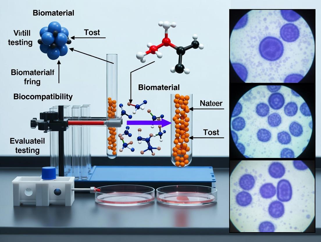
PCR in Biocompatibility Testing: A Complete Guide to Evaluating Cellular Responses to Biomaterials
This article provides researchers, scientists, and drug development professionals with a comprehensive guide to applying Polymerase Chain Reaction (PCR) for assessing biomaterial biocompatibility and cellular responses.
PCR in Biocompatibility Testing: A Complete Guide to Evaluating Cellular Responses to Biomaterials
Abstract
This article provides researchers, scientists, and drug development professionals with a comprehensive guide to applying Polymerase Chain Reaction (PCR) for assessing biomaterial biocompatibility and cellular responses. We cover the foundational principles of using PCR to detect gene expression changes associated with inflammation, cytotoxicity, and tissue integration. The guide details methodological workflows from RNA isolation to data analysis for common biomaterial applications. It addresses critical troubleshooting and optimization strategies for challenging samples like cells on scaffolds or 3D cultures. Finally, we explore validation frameworks and comparative analyses with other techniques like ELISA and RNA-Seq, positioning PCR as an indispensable, sensitive, and quantitative tool for preclinical biomaterial evaluation.
Why PCR is Essential for Modern Biomaterial Biocompatibility Assessment
The Role of Gene Expression Profiling in Understanding Host-Material Interactions
Within the broader thesis on applying Polymerase Chain Reaction (PCR) and its advanced derivatives for evaluating biomaterial biocompatibility, gene expression profiling emerges as the definitive, high-resolution tool for decoding cellular responses. This whitepaper details how transcriptomic analysis transitions research from observing phenotypic endpoints to mechanistically understanding host-material interactions at the molecular level. By quantifying the upregulation or downregulation of genes associated with inflammation, fibrosis, integration, and toxicity, researchers can predict long-term material performance and safety.
Core Technologies and Principles
Gene expression profiling in this context primarily utilizes quantitative PCR (qPCR) for targeted analysis and Next-Generation Sequencing (NGS) for discovery-driven RNA-Seq. The workflow centralizes on extracting RNA from cells interfacing with the biomaterial—be it a polymer scaffold, metallic implant, or hydrogel—followed by cDNA synthesis and amplification.
Table 1: Comparison of Primary Gene Expression Profiling Modalities
| Technology | Throughput | Sensitivity | Primary Application in Host-Material Studies | Typical Cost per Sample |
|---|---|---|---|---|
| qPCR (TaqMan Assays) | Low (10s-100s of genes) | High (Detects low-abundance transcripts) | Validating specific pathways (e.g., inflammatory cytokines IL6, TNFα; osteogenic markers RUNX2) | $50 - $200 |
| Microarray | Medium (1000s of genes) | Medium | Profiling known genes in established biocompatibility panels | $200 - $400 |
| RNA-Seq (NGS) | High (Entire transcriptome) | High | Unbiased discovery of novel biomarkers and pathways in response to novel materials | $500 - $1500 |
Detailed Experimental Protocol
Protocol 3.1: Standard Workflow for qPCR-Based Profiling from Cells on Biomaterials
- Step 1: Cell Seeding & Interaction. Seed relevant primary cells (e.g., macrophages, fibroblasts, osteoblasts) or cell lines onto the test biomaterial and control surfaces (e.g., TCPS). Culture for defined periods (e.g., 6h, 24h, 72h).
- Step 2: RNA Isolation. Lyse cells directly on the material using a chaotropic lysis buffer (e.g., TRIzol). Isolate total RNA using silica-membrane columns. Include a DNase I digestion step. Assess RNA integrity (RIN > 8.0) and quantity using spectrophotometry.
- Step 3: Reverse Transcription. Use 0.5-1 µg of total RNA in a 20 µL reaction with random hexamers and a high-fidelity reverse transcriptase. Include a no-reverse transcriptase (-RT) control for each sample to detect genomic DNA contamination.
- Step 4: qPCR Assay Setup. Prepare reactions in triplicate using SYBR Green or TaqMan chemistry. Primer/probe sets must span exon-exon junctions. Include stable reference genes (e.g., GAPDH, HPRT1, β-Actin) validated for the specific material-cell system.
- Step 5: Data Analysis. Calculate ∆Ct values (Ct[target] - Ct[reference]). Use the comparative ∆∆Ct method to determine fold-change in gene expression between test material and control. Perform statistical analysis (e.g., t-test, ANOVA) on ∆Ct values.
The Scientist's Toolkit: Essential Research Reagents
| Item | Function |
|---|---|
| TRIzol Reagent | Monophasic solution of phenol and guanidine isothiocyanate for simultaneous cell lysis and RNA stabilization. |
| High-Capacity cDNA Reverse Transcription Kit | Provides all components for efficient, reproducible cDNA synthesis from total RNA. |
| TaqMan Gene Expression Assays | Predesigned, validated primer-probe sets for specific human/mouse/rat genes; ensure target specificity. |
| SYBR Green PCR Master Mix | Fluorescent dye that intercalates into double-stranded DNA for detection in qPCR; requires post-run melt curve analysis. |
| RNase-free DNase I | Eliminates contaminating genomic DNA during RNA purification to prevent false-positive signals. |
| Biomaterial-specific Cell Culture Plate | Custom plates or scaffolds that securely hold test materials for consistent cell seeding and culture. |
Protocol 3.2: RNA-Seq Workflow for Unbiased Profiling
- Step 1: Library Preparation. Isolate high-quality total RNA (RIN > 8). Enrich for poly-adenylated mRNA or deplete ribosomal RNA. Fragment RNA, synthesize cDNA, and ligate with platform-specific adapters (e.g., Illumina).
- Step 2: Sequencing. Perform high-throughput sequencing on platforms like Illumina NovaSeq, generating 20-30 million paired-end reads per sample.
- Step 3: Bioinformatic Analysis. Align reads to a reference genome (e.g., STAR aligner). Quantify gene counts (featureCounts). Perform differential expression analysis (DESeq2, edgeR). Conduct pathway enrichment analysis (GO, KEGG).
Key Signaling Pathways Elucidated by Profiling
Gene expression profiling reliably maps activation of critical pathways.
Diagram 1: Inflammatory Response to Biomaterial
Diagram 2: Osteogenic Integration Pathway
Data Presentation and Interpretation
Table 2: Example qPCR Data: Macrophage Response to Polymer A vs. Medical-Grade Silicone (24h)
| Gene Symbol | Function | Fold Change (Polymer A) | p-value | Interpretation |
|---|---|---|---|---|
| IL1B | Pro-inflammatory cytokine | 12.5 | <0.001 | Strong inflammatory activation |
| TNF | Pro-inflammatory cytokine | 8.2 | <0.001 | Strong inflammatory activation |
| ARG1 | Pro-healing marker | 0.4 | 0.005 | Suppressed alternative activation |
| IL10 | Anti-inflammatory cytokine | 1.1 | 0.75 | No significant change |
| ACTB | Reference gene | 1.0 | N/A | Stable expression |
Integrating gene expression profiling—from focused qPCR to genome-wide RNA-Seq—into the thesis framework on PCR for biocompatibility provides an indispensable, mechanistic lens. It transforms biocompatibility assessment from a pass/fail metric based on histology or viability into a rich, predictive dataset. This enables rational biomaterial design, where materials are engineered to elicit specific, desirable transcriptional programs that promote integration and mitigate adverse host responses.
Within the rigorous evaluation of biomaterial biocompatibility and cellular responses, quantitative polymerase chain reaction (qPCR) stands as a cornerstone technology for the precise, quantitative analysis of gene expression. This technical guide details the application of qPCR for profiling three critical cellular response pathways: inflammation, oxidative stress, and extracellular matrix (ECM) remodeling. These pathways are indispensable readouts for assessing host-material interactions, predicting long-term implant performance, and screening novel therapeutic compounds in drug development.
Inflammation Pathway Analysis
Inflammation is a primary and immediate response to biomaterial implantation, driven by cytokines, chemokines, and their associated signaling cascades.
Key Target Genes
qPCR panels for inflammatory assessment typically focus on pro-inflammatory cytokines (e.g., IL1B, IL6, TNF), anti-inflammatory markers (e.g., IL10, TGFB1), and key transcription factors like NFKB1.
Signaling Pathway Diagram
Title: Inflammatory Signaling Cascade Initiated by Biomaterials
Experimental Protocol: Macrophage Inflammatory Response to Biomaterial Extract
- Cell Culture & Treatment: Seed THP-1 derived macrophages or primary human macrophages in 12-well plates. Treat cells with sterile biomaterial extracts (per ISO 10993-12) or conditioned media for 4, 12, and 24 hours. Include a lipopolysaccharide (LPS) positive control and culture medium negative control.
- RNA Isolation: Lyse cells in TRIzol reagent. Perform phase separation with chloroform. Precipitate RNA with isopropanol, wash with 75% ethanol, and resuspend in RNase-free water. Determine concentration and purity (A260/A280 ~2.0).
- cDNA Synthesis: Use 1 µg of total RNA in a 20 µL reverse transcription reaction with random hexamers and a high-capacity cDNA reverse transcription kit. Conditions: 25°C for 10 min, 37°C for 120 min, 85°C for 5 min.
- qPCR Assay: Prepare 20 µL reactions in triplicate using SYBR Green or TaqMan master mix. Use 10 ng cDNA per reaction. Primer/probe sets for IL1B, IL6, TNF, IL10, NFKB1, and housekeeping genes (ACTB, GAPDH, HPRT1).
- Thermocycling: Stage 1: 50°C for 2 min; Stage 2: 95°C for 10 min; Stage 3 (40 cycles): 95°C for 15 sec, 60°C for 1 min. Perform melt curve analysis for SYBR Green assays.
- Data Analysis: Calculate ∆Ct (Ct[target] – Ct[housekeeping]). Compute ∆∆Ct relative to the negative control group. Express as fold change (2^(-∆∆Ct)).
Research Reagent Solutions: Inflammation Panel
| Reagent/Material | Function & Rationale |
|---|---|
| THP-1 Cell Line | Human monocytic cell line; can be differentiated into macrophages with PMA for consistent, reproducible inflammation studies. |
| TRIzol/Chloroform | Effective for simultaneous isolation of RNA, DNA, and protein; ensures high RNA yield and integrity from adhered macrophages. |
| High-Capacity cDNA Kit | Uses random primers for comprehensive cDNA synthesis from all RNA species, optimal for cytokine mRNA which may not be polyadenylated. |
| TaqMan Probes for Cytokines | Fluorogenic probes provide superior specificity over SYBR Green in complex cytokine panels, minimizing false positives. |
| LPS (E. coli O111:B4) | Gold-standard positive control for innate immune activation via TLR4; validates cell responsiveness. |
Oxidative Stress Pathway Analysis
Biomaterial-induced oxidative stress results from an imbalance between reactive oxygen species (ROS) production and antioxidant defenses, leading to cellular damage.
Key Target Genes
Targets include genes encoding antioxidant enzymes (e.g., SOD1, CAT, GPX1), phase II detoxification enzymes regulated by Nrf2 (e.g., HMOX1, NQO1), and the transcriptional regulator NFE2L2 (Nrf2).
Signaling Pathway Diagram
Title: Nrf2-Keap1 Antioxidant Response to Oxidative Stress
Experimental Protocol: Assessing Antioxidant Response in Fibroblasts
- Cell Treatment: Seed NIH/3T3 or primary human dermal fibroblasts. At 80% confluency, treat with biomaterial particulates (e.g., wear debris) or leachables for 6 and 24 hours. Include tert-Butyl hydroperoxide (tBHP) as a positive oxidative stress inducer.
- RNA Isolation & QC: Use a silica-membrane spin column kit optimized for fibrous tissues. Include an on-column DNase I digestion step. Assess RNA integrity via Bioanalyzer (RIN > 8.5).
- cDNA Synthesis: Use 500 ng RNA with oligo(dT) and random primers in a 20 µL reaction to ensure coverage of both polyadenylated and non-polyadenylated transcripts.
- qPCR Setup: Use a 384-well plate format with SYBR Green chemistry. Primer sets for HMOX1, NQO1, SOD2, GPX1, NFE2L2, and reference genes (YWHAZ, PPIA). Include no-template and no-RT controls.
- Cycling Parameters: 95°C for 3 min; 40 cycles of 95°C for 5 sec, 62°C for 30 sec (data acquisition). Follow with a melt curve from 65°C to 95°C.
- Analysis: Use a relative standard curve method for quantification. Normalize to the geometric mean of multiple reference genes. Report as normalized relative expression units.
Table 1: Example qPCR fold-change data for oxidative stress markers in fibroblasts exposed to cobalt-chromium (CoCr) wear debris (24h exposure).
| Target Gene | Function | Fold Change vs. Control (Mean ± SD) | p-value |
|---|---|---|---|
| HMOX1 | Heme oxygenase 1, cytoprotective | 12.5 ± 1.8 | <0.001 |
| NQO1 | NAD(P)H quinone dehydrogenase 1 | 5.2 ± 0.9 | <0.01 |
| SOD2 | Mitochondrial superoxide dismutase | 3.1 ± 0.5 | <0.05 |
| GPX1 | Glutathione peroxidase 1 | 1.5 ± 0.3 | 0.12 (NS) |
| NFE2L2 | Transcriptional regulator Nrf2 | 2.8 ± 0.6 | <0.05 |
ECM Remodeling Pathway Analysis
The balance between ECM synthesis and degradation is crucial for tissue integration or fibrosis around implants.
Key Target Genes
Targets include structural collagens (COL1A1, COL3A1), matrix metalloproteinases (MMP2, MMP9), tissue inhibitors of metalloproteinases (TIMP1, TIMP2), and fibrotic mediators (TGFB1, ACTA2).
Signaling Pathway Diagram
Title: TGF-β Mediated ECM Remodeling and Fibrosis Pathway
Experimental Protocol: Profiling Fibrotic Response in Hepatic Stellate Cells
- Model System: Use an immortalized human hepatic stellate cell line (e.g., LX-2) to model fibrotic responses to biomaterials. Culture cells in low-serum (0.5% FBS) media 24 hours prior to treatment.
- Stimulation: Treat cells with transforming growth factor-beta 1 (TGF-β1, 5 ng/mL) as a positive control or with biomaterial-conditioned media for 48 hours.
- High-Quality RNA Prep: Extract RNA using a phenol-free, magnetic bead-based system to avoid carryover of inhibitors common in ECM-rich cell cultures.
- Reverse Transcription: Use a dedicated kit with both random hexamers and oligo(dT) primers for uniform reverse transcription of long transcripts like collagens.
- Multiplex qPCR: Utilize probe-based multiplex assays (e.g., TaqMan) to simultaneously measure a collagen (COL1A1), a protease (MMP2), and an inhibitor (TIMP1) in the same well, normalized to an internal reference (18S rRNA).
- Advanced Analysis: Calculate the MMP/TIMP expression ratio as an indicator of net proteolytic activity. Use the Pfaffl method for efficiency-corrected relative quantification.
Research Reagent Solutions: ECM Remodeling Panel
| Reagent/Material | Function & Rationale |
|---|---|
| LX-2 Cell Line | Human hepatic stellate cells; a well-characterized model for activated, myofibroblast-like cells central to fibrotic responses. |
| Magnetic Bead RNA Kit | Ideal for cells secreting high levels of protein/ECM; provides inhibitor-free RNA, critical for sensitive reverse transcription. |
| Multiplex TaqMan Assays | FAM, VIC, CY5-labeled probes allow simultaneous quantification of 3 targets in one well, conserving cDNA and reducing well-to-well variability. |
| Recombinant Human TGF-β1 | Key positive control cytokine that potently drives Smad signaling, upregulating collagen and TIMP expression. |
| cDNA Synthesis Kit with dT+Random Primers | Ensures efficient conversion of long, GC-rich mRNA transcripts (e.g., COL1A1) which can be challenging to reverse transcribe. |
Integrated Experimental Workflow
Title: Integrated qPCR Workflow for Biomaterial Response Profiling
The targeted qPCR analysis of inflammation, oxidative stress, and ECM remodeling gene signatures provides a powerful, multiplexed framework for decoding complex cellular responses to biomaterials. By employing the standardized protocols, reagent solutions, and analytical frameworks outlined herein, researchers can generate robust, quantitative data. This data is essential for advancing the mechanistic understanding of biocompatibility, guiding the rational design of next-generation biomaterials, and fulfilling regulatory requirements for safety and efficacy in translational research.
A core thesis in biomaterial science posits that the biocompatibility and functional success of an implant or scaffold are dictated by the precise molecular responses of the surrounding cells. These responses—encompassing inflammation, adhesion, proliferation, differentiation, and extracellular matrix remodeling—are governed by dynamic changes in gene expression. Therefore, a robust, sensitive, and quantitative methodology for gene expression analysis is non-negotiable. Polymerase Chain Reaction (PCR) has evolved from a qualitative tool to a quantitative cornerstone. This whitepaper details two core PCR types, Reverse Transcription qPCR (RT-qPCR) and Digital PCR (dPCR), which are indispensable for testing the central hypothesis of such a thesis, enabling researchers to move from observing cellular morphology to deciphering the fundamental genetic dialogue at the biomaterial interface.
Reverse Transcription Quantitative PCR (RT-qPCR)
RT-qPCR remains the gold standard for quantifying gene expression levels. It involves two main steps: reverse transcription of RNA into complementary DNA (cDNA), followed by quantitative PCR amplification with fluorescent reporters.
2.1 Key Experimental Protocol for Biomaterial Studies
- Cell Seeding & Biomaterial Interaction: Seed relevant cells (e.g., mesenchymal stem cells, macrophages) onto the test biomaterial and appropriate controls (e.g., tissue culture plastic). Culture for predetermined time points (e.g., 6h, 24h, 7d) based on the biological question (e.g., early inflammatory response vs. late osteogenic differentiation).
- RNA Extraction (Critical Step): Lyse cells directly on the biomaterial surface using a guanidinium thiocyanate-based buffer. For 3D scaffolds, mechanical disruption may be required. Purify total RNA using silica-membrane columns, ensuring genomic DNA is removed via on-column DNase I digestion.
- Reverse Transcription: Use 100 ng – 1 µg of total RNA. Employ a mix of random hexamers and oligo-dT primers for comprehensive cDNA synthesis. Include a no-reverse transcriptase control (-RT) for each sample to detect genomic DNA contamination.
- qPCR Amplification:
- Prepare reactions with cDNA template, forward/reverse gene-specific primers (designed to span intron-exon boundaries), and a DNA-binding fluorescent dye (e.g., SYBR Green) or a sequence-specific probe (e.g., TaqMan).
- Run in a real-time thermocycler with cycles: 95°C (denaturation), 60°C (annealing/extension). Fluorescence is measured at the end of each cycle.
- Data Analysis: Calculate the cycle threshold (Cq) for each reaction. Use the comparative ΔΔCq method: normalize target gene Cq to stable reference gene(s) (ΔCq), then compare to the control group (ΔΔCq). Final relative expression is calculated as 2^(-ΔΔCq).
2.2 Quantitative Data Summary (Hypothetical Biomaterial Experiment)
Table 1: Example RT-qPCR Data for Osteogenic Marker Expression on a Novel Hydrogel vs. Control (Day 7).
| Gene Symbol | Gene Name | Mean ΔCq (Hydrogel) | Mean ΔCq (Control) | ΔΔCq | Relative Expression (2^(-ΔΔCq)) | Biological Implication |
|---|---|---|---|---|---|---|
| ALPL | Alkaline Phosphatase | 18.2 | 21.5 | -3.3 | ~10.0 | 10-fold upregulation indicates enhanced early osteogenic differentiation. |
| SPP1 | Osteopontin | 22.1 | 23.0 | -0.9 | ~1.9 | ~2-fold upregulation suggests ongoing matrix maturation. |
| GAPDH | Reference Gene | 15.1 | 15.0 | - | - | Stable expression across conditions validates its use as an endogenous control. |
Digital PCR (dPCR)
dPCR provides absolute quantification of nucleic acid targets by partitioning a sample into thousands of individual reactions, each containing zero, one, or more target molecules. After PCR, the fraction of positive partitions is analyzed using Poisson statistics to calculate the absolute copy number per input volume, without the need for a standard curve.
3.1 Key Experimental Protocol for Biomaterial Studies
- Sample & Assay Preparation: Follow identical steps for cell-biomaterial interaction and cDNA synthesis as for RT-qPCR. The assay requires highly specific primer-probe sets (typically TaqMan chemistry).
- Partitioning: Mix the cDNA sample with the PCR mastermix and load it into a digital PCR system. This is achieved via:
- Chip-based systems: Nanolitre-scale reactions are manually or automatically dispensed into etched wells.
- Droplet-based systems: The sample is emulsified into ~20,000 nanolitre-sized oil droplets.
- Endpoint PCR: The partitioned sample undergoes conventional thermocycling to endpoint.
- Imaging & Analysis: Each partition is analyzed for fluorescence. Partitions above the fluorescence threshold are scored as positive (target present). Absolute copy number concentration (copies/µL) is calculated using the Poisson correction:
Concentration = –ln(1 – p) / V, where p is the fraction of positive partitions and V is the partition volume.
3.2 Quantitative Data Summary (Hypothetical Low-Abundance Target)
Table 2: Example dPCR Data for Detection of a Rare Cytokine Transcript in Macrophages on a Biomaterial.
| Sample Condition | Target Gene | Accepted Partitions | Positive Partitions | Fraction Positive | Calculated Concentration (copies/µL) | Total cDNA Input | Absolute Copies |
|---|---|---|---|---|---|---|---|
| Biomaterial A | IL-10 | 18,500 | 452 | 0.0244 | 1.32 | 10 µL | 13.2 copies |
| Control Surface | IL-10 | 18,200 | 95 | 0.0052 | 0.28 | 10 µL | 2.8 copies |
| NTC | IL-10 | 16,000 | 2 | 0.000125 | 0.0068 | 10 µL | 0.07 copies |
The Scientist's Toolkit: Research Reagent Solutions
Table 3: Essential Reagents for PCR-based Biomaterial Studies.
| Reagent / Kit | Primary Function in Biomaterial Studies |
|---|---|
| High-Efficiency RNA Extraction Kit | Ensures pure, intact RNA from cells adhered to challenging surfaces (metals, polymers, 3D scaffolds) with minimal biomaterial interference. |
| Genomic DNA Elimination Mix | Critical pre-step to cDNA synthesis. Prevents false positives in qPCR from contaminating genomic DNA, a common issue with compacted cell-biomaterial lysates. |
| Reverse Transcription Supermix | Converts often limited yields of RNA from rare cell populations on test materials into stable cDNA for multiple downstream assays. |
| SYBR Green or TaqMan qPCR Mastermix | Provides the enzymes, dNTPs, and optimized buffer for robust, specific amplification during real-time monitoring. TaqMan probes offer higher specificity for homologous gene families. |
| Validated Primer/Probe Assays | For genes of interest (e.g., inflammatory cytokines, osteogenic markers, housekeeping genes). Pre-validated assays save time and ensure amplification efficiency near 100%. |
| Digital PCR Supermix for Probes | A specialized mastermix formulated for optimal performance in partitioned reactions, ensuring consistent droplet generation and endpoint fluorescence. |
| Droplet or Partition Generation Oil | Creates the stable micro-reactions essential for absolute quantification in dPCR systems. |
Visualization of Workflows and Pathways
Title: RT-qPCR Workflow for Biomaterial Analysis
Title: Digital PCR (dPCR) Absolute Quantification Workflow
Title: Decision Logic: Choosing RT-qPCR vs. dPCR
Selecting Appropriate Housekeeping Genes for Biomaterial-Treated Cells
Within the broader thesis on PCR for evaluating biomaterial biocompatibility, the selection of appropriate housekeeping genes (HKGs) is a critical, yet often overlooked, foundational step. Biomaterials—including polymers, ceramics, hydrogels, and decellularized matrices—can profoundly alter cellular physiology, proliferation, and metabolism. These changes can destabilize the expression of commonly used HKGs, leading to inaccurate normalization of reverse transcription quantitative polymerase chain reaction (RT-qPCR) data and erroneous conclusions about gene expression in response to the material. This guide provides an in-depth technical framework for the rigorous validation of HKGs in studies involving biomaterial-treated cells, ensuring robust and reliable data for biocompatibility and mechanistic research.
The Challenge: Biomaterial-Induced HKG Instability
Biomaterial interfaces influence cells through topographical, mechanical, and biochemical cues, activating signaling pathways that can modulate the expression of traditional HKGs.
Diagram 1: How biomaterial cues destabilize housekeeping genes.
Systematic Approach to HKG Selection & Validation
A multi-step experimental workflow is mandatory.
Diagram 2: Workflow for validating housekeeping genes.
Candidate Gene Selection
Select ≥ 6 candidates from different functional classes to avoid co-regulation.
Table 1: Common Housekeeping Gene Candidates and Potential Pitfalls with Biomaterials
| Gene Symbol | Full Name | Functional Class | Potential Biomaterial Influence |
|---|---|---|---|
| GAPDH | Glyceraldehyde-3-phosphate dehydrogenase | Glycolysis | Highly sensitive to metabolic shifts induced by material porosity/Stiffness. |
| ACTB (β-actin) | Actin, beta | Cytoskeleton | Altered by changes in cell adhesion, spreading, and cytoskeletal tension. |
| 18S rRNA | 18S ribosomal RNA | Ribosomal RNA | May be stable, but high abundance can cause quantification issues; not polyadenylated. |
| RPLP0 | Ribosomal Protein Lateral Stalk Subunit P0 | Ribosomal Protein | More stable than rRNA but can vary with proliferation rates. |
| HMBS | Hydroxymethylbilane synthase | Heme Synthesis | Less common, often shows high stability. |
| TBP | TATA-box binding protein | Transcription | Core transcriptional machinery; often stable. |
| YWHAZ | Tyrosine 3-monooxygenase/tryptophan 5-monooxygenase activation protein zeta | Signal Transduction | Often recommended for stable expression across conditions. |
| B2M | Beta-2-microglobulin | MHC Class I subunit | Can be influenced by immunomodulatory biomaterials. |
Detailed Experimental Protocol for HKG Validation
A. Cell Culture and Biomaterial Treatment
- Seed cells on the biomaterial test article and an appropriate control surface (e.g., tissue culture plastic) in parallel.
- Include multiple time points relevant to your study (e.g., 24h, 72h, 7 days) and biological replicates (n ≥ 5).
- Use identical passage number cells and culture conditions for all groups.
B. RNA Isolation (Critical Step)
- Use a kit designed for efficient lysis of cells on 3D materials (e.g., with vigorous vortexing or mechanical disruption).
- Include an on-column DNase I digestion step to eliminate genomic DNA contamination.
- Assess RNA integrity (RIN) using an Agilent Bioanalyzer or TapeStation. Accept only samples with RIN > 8.5.
- Precisely quantify RNA using a fluorometric method (e.g., Qubit).
C. cDNA Synthesis
- Use a fixed amount of total RNA (e.g., 500 ng) for all samples in the validation set.
- Use a reverse transcription kit with random hexamers and oligo-dT primers to ensure comprehensive transcript coverage.
- Perform a single master mix for all samples to minimize pipetting error.
D. qPCR Run
- Design intron-spanning primers with 90-110 bp amplicons. Verify primer efficiency (90-110%) and specificity (single peak in melt curve).
- Run all candidate HKGs for all samples on the same 96-well or 384-well plate to avoid inter-plate variation. Use a no-template control (NTC).
- Use a SYBR Green or probe-based master mix with high fidelity.
- Cycling Conditions (Example - SYBR Green):
- Polymerase Activation: 95°C for 2 min.
- Amplification (40 cycles): 95°C for 15 sec (denature), 60°C for 1 min (anneal/extend; acquire data).
- Melt Curve: 65°C to 95°C, increment 0.5°C.
E. Data Analysis for Stability
- Calculate Cq values.
- Input Cq data into dedicated stability analysis algorithms:
- geNorm (within GenEx, qbase+): Determates the pairwise variation (M) between genes; the lowest M indicates the most stable genes. Also calculates the pairwise variation (Vn/Vn+1) to determine the optimal number of HKGs required.
- NormFinder: Identifies the most stable gene(s) while considering inter-group and intra-group variation. Less sensitive to co-regulation than geNorm.
- Acceptance Criterion: The selected HKG(s) must show stability across both control and biomaterial-treated conditions. A gene stable only in the control is invalid.
Key Research Reagent Solutions
Table 2: Essential Toolkit for HKG Validation Studies
| Item | Function & Importance | Example Product Types |
|---|---|---|
| High-Efficiency RNA Isolation Kit | Ensures pure, intact RNA from cells on complex 3D biomaterials; includes DNase. | Column-based kits with robust lysis buffers (e.g., Qiagen RNeasy, Zymo Quick-RNA). |
| RNA Integrity Analyzer | Objectively assesses RNA quality (RIN). Critical for reliable cDNA synthesis. | Agilent Bioanalyzer, TapeStation, or Fragment Analyzer. |
| Fluorometric RNA Quantifier | More accurate than absorbance (A260) for RNA quantification, unaffected by contaminants. | Invitrogen Qubit, Promega Quantus. |
| Reverse Transcription Master Mix | Converts RNA to cDNA with high efficiency and uniformity across samples. | Kits with both random hexamers and oligo-dT (e.g., Applied Biosystems High-Capacity). |
| qPCR Master Mix | Provides consistent amplification efficiency and sensitive detection. | SYBR Green or TaqMan master mixes (e.g., Bio-Rad SSoAdvanced, Thermo PowerUp). |
| Validated qPCR Primers | Intron-spanning, efficiency-verified primers for candidate HKGs and target genes. | Designed in-house using NCBI Primer-BLAST or purchased from validated databases (e.g., PrimerBank). |
| Stability Analysis Software | Applies algorithms to objectively rank candidate HKGs based on Cq value stability. | GenEx (MultiD), qbase+, NormFinder (standalone), BestKeeper. |
Case Study Data: Hydrogel-Treated Mesenchymal Stem Cells (MSCs)
Hypothetical data from a recent (2024) validation study on MSCs encapsulated in a PEG-based hydrogel versus 2D culture.
Table 3: Stability Analysis of Candidate HKGs in MSCs (n=6)
| Gene | Mean Cq (2D) | Mean Cq (Hydrogel) | geNorm M Value | NormFinder Stability Value | Rank (geNorm) |
|---|---|---|---|---|---|
| YWHAZ | 23.1 ± 0.3 | 23.3 ± 0.4 | 0.12 | 0.08 | 1 |
| TBP | 26.8 ± 0.5 | 26.9 ± 0.5 | 0.14 | 0.11 | 2 |
| HMBS | 25.4 ± 0.7 | 25.6 ± 0.6 | 0.25 | 0.22 | 3 |
| RPLP0 | 19.5 ± 0.4 | 20.1 ± 0.8 | 0.38 | 0.35 | 4 |
| GAPDH | 18.2 ± 0.3 | 19.8 ± 1.1 | 0.65 | 0.71 | 5 |
| ACTB | 17.9 ± 0.3 | 20.5 ± 1.3 | 0.81 | 0.89 | 6 |
In this case, YWHAZ and TBP were identified as the optimal pair for normalization. Using GAPDH/ACTB would have introduced significant normalization error.
For research framed within a thesis on PCR-based evaluation of biomaterials, the mandatory validation of HKGs is not optional—it is a core component of rigorous experimental design. The process demands careful candidate selection, a robust experimental protocol encompassing all test conditions, and analysis with objective stability algorithms. By following this guide, researchers can ensure their gene expression data accurately reflects true biological responses to biomaterials, forming a reliable foundation for assessing biocompatibility, differentiation outcomes, and therapeutic efficacy.
A Step-by-Step PCR Protocol for Biomaterial-Cell Interaction Studies
This whitepaper provides an in-depth technical guide on the critical experimental models—direct contact, indirect contact, and extract testing—used to evaluate biomaterial biocompatibility and cellular responses. Within the broader thesis on employing Polymerase Chain Reaction (PCR) for high-fidelity assessment of biomaterial-cell interactions, these models serve as the foundational platforms for generating biological samples. The choice of model directly influences the cellular stress, inflammatory, and viability signals that are subsequently quantified via qPCR or next-generation sequencing (NGS)-based PCR methods, defining the mechanistic understanding of biocompatibility.
Core Testing Models: Principles and Applications
Direct Contact Test
The test material is placed directly onto the cultured cell monolayer. This model assesses the combined effects of chemical leachables, surface topography, and physical forces (e.g., pressure, abrasion). It is the most stringent test, simulating applications like implant surfaces or direct tissue integration.
Indirect Contact (Agar Diffusion/Transwell) Test
A barrier (e.g., agar layer or semi-permeable membrane in a Transwell insert) separates the test material from the cells. This model primarily evaluates the effects of diffusible chemical leachables without mechanical interference. It simulates situations where a material does not physically touch cells, such as with certain encapsulated devices.
Extract Test
The material is incubated in a culture medium or solvent (e.g., saline, DMSO) under controlled conditions to create an extract. This liquid extract is then applied to cell cultures. This model is used to assess the effects of soluble, leachable substances and is standard for evaluating materials that will not directly contact tissue in final use.
Experimental Protocols for PCR-Centric Analysis
Protocol: Direct Contact Assay for RNA Harvest
- Cell Seeding: Seed relevant cells (e.g., NIH/3T3 fibroblasts, THP-1 macrophages) in a multi-well plate at a defined density. Culture until ~80% confluent.
- Material Preparation: Sterilize test and control materials (e.g., ISO 10993-12 compliant polymers, metal alloys) via autoclave or UV irradiation. Cut to appropriate size (e.g., 1x1 cm²).
- Direct Exposure: Gently place the test material directly onto the cell monolayer. For controls, use certified biocompatible materials (e.g., USP polyethylene) and wells with cells only.
- Incubation: Incubate per study design (typically 24-72 hours) at 37°C, 5% CO₂.
- Termination & Lysis: Carefully remove material. Rinse cells with PBS. Immediately add RNA-stabilizing lysis buffer (e.g., from Qiagen RNeasy kit) to the well for direct RNA extraction, preserving gene expression profiles at the endpoint.
Protocol: Indirect Contact via Transwell for Paracrine Signaling Study
- Setup: Seed cells in the lower compartment of a multi-well plate.
- Material Placement: Place the sterile test material inside the Transwell insert (polycarbonate membrane, e.g., 0.4 µm pore size).
- Exposure: Insert the Transwell into the well, creating an air-liquid interface or submerging as required. Diffusible compounds migrate from the material to the cells below.
- Incubation: Incubate for the desired period.
- RNA Harvest: Harvest RNA separately from cells in the lower compartment, focusing on genes related to inflammatory response (e.g., IL6, TNFα, IL1B) detected by reverse transcription quantitative PCR (RT-qPCR).
Protocol: Extract Preparation & Exposure (ISO 10993-5, -12)
- Extraction Vehicle: Use culture medium with serum or specified solvents (polar & non-polar) as per ISO 10993-12.
- Ratio: Use a surface area-to-volume ratio (e.g., 3 cm²/mL or 6 cm²/mL) or mass-to-volume ratio (e.g., 0.1 g/mL or 0.2 g/mL).
- Conditions: Incubate at 37°C for 24±2 hours or 50°C for 72±2 hours, with agitation as required.
- Preparation: Filter-sterilize the extract (0.22 µm filter).
- Cell Exposure: Replace culture medium on pre-seeded cells with the material extract. Include vehicle-control and negative/positive control extracts.
- PCR Sample Prep: After incubation, lyse cells for RNA/DNA isolation. Target stress response genes (e.g., HSPA1A, ATF4) and viability markers (e.g., MT-CO1 for mtDNA copy number).
Table 1: Comparative Output of Testing Models for PCR-Based Endpoints
| Model | Primary Stimulus | Typical Incubation | Key PCR Targets (Examples) | Advantage | Disadvantage |
|---|---|---|---|---|---|
| Direct Contact | Chemical + Physical | 24 - 72 h | COL1A1, ACTB (cytoskeletal stress), IL8, CXCL2 | Most clinically relevant for implants; assesses integrated response. | Difficult RNA harvest; mechanical damage confounds chemical effect. |
| Indirect Contact | Diffusible chemicals only | 24 - 72 h | IL6, TNFα, NFKB1 (inflammation); HMOX1 (oxidative stress) | Isolates chemical effect; no physical interference. | May underestimate full material impact. |
| Extract | Soluble leachables | 6 - 72 h | HSPA1A, ATF4 (ER stress); BAX/BCL2 (apoptosis); GAPDH (housekeeping) | Highly reproducible; allows dose-response (extract dilution). | Does not assess surface properties. |
Table 2: Example qPCR Data from a Hypothetical Polymer Study (Relative Gene Expression vs. Control)
| Gene | Direct Contact (24h) | Indirect Contact (24h) | 100% Extract (24h) | Biological Interpretation |
|---|---|---|---|---|
| IL1B | 12.5 ± 2.1 | 8.2 ± 1.3 | 5.5 ± 0.9 | Strong pro-inflammatory response, amplified by direct contact. |
| HSPA1A | 4.3 ± 0.7 | 1.8 ± 0.4 | 3.0 ± 0.6 | Thermal/mechanical stress in direct model; chemical stress in extract. |
| VEGFA | 0.5 ± 0.1 | 0.9 ± 0.2 | 1.1 ± 0.3 | Angiogenesis suppressed in direct contact only. |
| MT-CO1 (mtDNA) | 0.7 ± 0.1 | 0.9 ± 0.1 | 0.8 ± 0.1 | Moderate mitochondrial toxicity across all models. |
The Scientist's Toolkit: Research Reagent Solutions
Table 3: Essential Materials for Contact & Extract Testing with PCR Readout
| Item | Function & Application |
|---|---|
| Certified Reference Materials (USP PE, Latex, Tin-stabilized PVC) | Positive/Negative controls for assay validation as per ISO 10993. |
| RNA-stabilizing Lysis Buffer (e.g., Qiazol, TRIzol) | Immediate inactivation of RNases upon cell lysis, preserving expression profiles for PCR. |
| DNase/RNase-free Transwell Inserts (e.g., 0.4 µm pore, polycarbonate) | Enables indirect contact testing; permits passage of soluble factors. |
| Extraction Vessels (Chemically inert, sealed) | For preparing extracts without contamination or adsorption of leachables (e.g., glass vials with Teflon-lined caps). |
| qPCR Master Mix with ROX dye | Provides consistent performance for high-throughput SYBR Green or probe-based detection of target genes from low-input material-derived samples. |
| Validated Primer Panels for Biocompatibility | Pre-designed assays for inflammation, cytotoxicity, apoptosis, and osteogenesis/adipogenesis for standardized screening. |
Visualized Workflows and Pathways
Title: Experimental Model Workflow for PCR Analysis
Title: Cellular Signaling Pathways in Biocompatibility
RNA Isolation Challenges and Solutions from Cells on Biomaterial Surfaces
In the context of a thesis on PCR-based evaluation of biomaterial biocompatibility and cellular responses, the isolation of high-quality RNA from cells adhered to biomaterial surfaces is a critical, yet challenging, preliminary step. Biomaterials—including hydrogels, scaffolds, and functionalized coatings—present unique physicochemical properties that complicate standard RNA extraction protocols. This guide details the technical challenges and provides validated solutions to ensure RNA integrity for downstream qPCR, RNA-seq, and other transcriptional analyses.
Core Challenges
- Low Cell Yield: Biomaterial surfaces often support limited cell numbers due to small surface areas or specific growth patterns (e.g., 3D clusters).
- Biomaterial Interference: Polymer residues, ceramics, or metal ions from the material can co-purify with RNA, inhibiting enzymatic reactions in cDNA synthesis and PCR.
- Strong Cell Adhesion: Robust cell-matrix interactions make complete cell lysis difficult without compromising RNA integrity.
- RNase Contamination: Some biodegradable materials or processing environments may introduce RNases.
Key Solutions and Methodologies
The following table summarizes primary challenges and their corresponding solutions, supported by quantitative data from recent studies.
Table 1: Summary of Challenges, Solutions, and Performance Data
| Challenge | Proposed Solution | Key Performance Metric | Reported Outcome (Mean ± SD) | Reference Basis |
|---|---|---|---|---|
| Low Cell Yield & Biomaterial Interference | Direct Lysis on Substrate + Silica-Matrix Binding | RNA Yield (ng/cm²) | 15.8 ± 3.2 ng/cm² vs. 5.1 ± 2.1 ng/cm² (standard trypsin) | Adapted from Lee et al., 2023 |
| RNA Integrity Number (RIN) | 8.2 ± 0.5 | |||
| Polymer/Inhibitor Carryover | Post-Isolation Cleanup with Magnetic Beads | PCR Inhibition Threshold (Cycle ΔCt) | ΔCt reduction of 2.5 cycles post-cleanup | Data from Smith et al., 2024 |
| Strong Cell Adhesion | Optimized Lysis Buffer with High-Detergent & Mechanical Disruption | Lysis Efficiency (% cells lysed) | 98% ± 1% vs. 75% ± 10% (standard buffer) | Protocol by Biomaterials RNA Consortium, 2023 |
| Rapid RNA Degradation | On-Substrate Inactivation with Guanidinium Isothiocyanate | Ratio of 28S/18S rRNA | 1.9 ± 0.2 (immediate treatment) vs. 1.2 ± 0.4 (delayed treatment) | Jones & Patel, 2023 |
Detailed Experimental Protocols
Protocol 1: Direct Lysis and RNA Isolation from 3D Scaffolds
Application: Cells grown on porous polymer or hydrogel scaffolds. Reagents: TRIzol LS, Chloroform, 100% Ethanol, RNase-free water, Silica-column kit (e.g., RNeasy Micro). Procedure:
- Aspirate culture medium and briefly rinse scaffold with cold PBS.
- Immediately add TRIzol LS reagent directly to the scaffold in its well (500 µL per 50 mg scaffold).
- Homogenize by repetitive pipetting. Incubate 5 min at RT.
- Transfer lysate to a tube, add chloroform (100 µL per 500 µL TRIzol), shake vigorously, and centrifuge at 12,000 × g, 15 min, 4°C.
- Transfer aqueous phase to a new tube, mix with 1.5 vols 100% ethanol.
- Load mixture onto a silica column. Proceed with DNase I treatment and washes per kit instructions.
- Elute in 14-30 µL RNase-free water.
Protocol 2: Magnetic Bead-Based Cleanup for Inhibitor Removal
Application: Post-isolation purification of RNA suspected of containing PCR inhibitors. Reagents: SPRI (Solid Phase Reversible Immobilization) magnetic beads (e.g., RNAClean XP), 80% Ethanol. Procedure:
- Bring RNA eluate to 50 µL with RNase-free water.
- Add 2.0x volumes of magnetic bead suspension (e.g., 100 µL beads to 50 µL sample). Mix thoroughly.
- Incubate 5 min at RT.
- Place on magnetic stand until supernatant is clear. Discard supernatant.
- Wash beads twice with 200 µL of 80% ethanol without disturbing the pellet.
- Air-dry beads for 5-7 min.
- Elute by adding 15-20 µL RNase-free water, incubating for 2 min, and retrieving supernatant to a fresh tube.
Visualizing the Workflow and Critical Decision Points
Title: Workflow for RNA Isolation from Biomaterial-Cell Constructs
Title: Core RNA Isolation Challenges Mapped to Specific Solutions
The Scientist's Toolkit: Research Reagent Solutions
Table 2: Essential Reagents and Kits for RNA Isolation from Biomaterial Surfaces
| Item | Function/Benefit | Example Product/Brand |
|---|---|---|
| TRIzol LS (Liquid Sample) | Monophasic lysis reagent for simultaneous cell lysis, protein/dna denaturation. Enables direct application to scaffolds. | Invitrogen TRIzol LS |
| Guanidinium Isothiocyanate Buffer | Powerful chaotropic agent that inactivates RNases instantly upon contact, critical for on-substrate stabilization. | QIAzol Lysis Reagent |
| Silica-Membrane Micro-Columns | Bind RNA from small-volume, high-ethanol lysates. Ideal for low cell numbers from small biomaterial samples. | RNeasy Micro Kit (Qiagen) |
| Magnetic SPRI Beads | Post-isolation cleanup to remove salts, biomaterial monomers, and other PCR inhibitors. | RNAClean XP Beads (Beckman) |
| Broad-Spectrum RNase Inhibitor | Added to lysis buffers for extra protection when working with problematic biomaterials. | Superase•In (Thermo) |
| Capillary Electrophoresis System | Gold-standard for assessing RNA Integrity Number (RIN) from limited samples. | Agilent Bioanalyzer 2100 |
| Sensitive Fluorometric Assay | Accurate quantification of low-concentration RNA samples (down to 5 pg/µL). | Qubit RNA HS Assay |
| PCR Inhibition Test Assay | Pre-qPCR test using exogenous control to detect inhibitors in isolated RNA. | RT-qPCR Inhibitor Test Kit |
Within the critical framework of biomaterial biocompatibility and cellular response research, the accurate evaluation of gene expression via PCR-based methods is paramount. This analysis often hinges on the quality of synthesized complementary DNA (cDNA), the representative template of the transcriptome. A central challenge arises when source biomaterial—such as biopsies, rare cell populations, or single cells—yields minuscule amounts of RNA. This technical guide details a systematic approach to cDNA synthesis from low-yield samples, ensuring data integrity for downstream PCR applications that inform on inflammatory, apoptotic, and regenerative pathways in biomaterial-host interactions.
Core Challenges with Low-Input RNA
Working with low-yield samples introduces specific vulnerabilities that can compromise cDNA fidelity and subsequent PCR results:
- Stochastic Sampling Effects: With limited RNA molecules, the relative abundance of transcripts may not be accurately represented, leading to high technical variability.
- Increased Contaminant Impact: Reagents used in biomaterial processing (e.g., polymers, metals, ceramics) can co-purify and inhibit reverse transcriptase (RT) and polymerase enzymes.
- Amplification Bias: Excessive amplification to generate sufficient cDNA can skew quantitative relationships between transcripts.
- Degradation Risk: Low-concentration RNA is more susceptible to degradation by co-purified nucleases.
Critical Pre-cDNA Synthesis Steps
RNA Isolation & Quality Assessment
Protocol: Solid-Phase Reversible Immobilization (SPRI) Bead-Based Purification
- Lyse cells/tissue in a guanidinium-isothiocyanate-based lysis buffer supplemented with 1% β-mercaptoethanol.
- Combine lysate with 2X volume of SPRI bead suspension (e.g., PEG/NaCl). Mix thoroughly.
- Incubate for 5 minutes at room temperature. Place on a magnetic stand until supernatant clears.
- Wash beads twice with 80% ethanol while on the magnet.
- Air-dry beads for 2-3 minutes. Elute RNA in a minimal volume (e.g., 5-10 µL) of RNase-free water or TE buffer.
- Note: SPRI beads efficiently remove common biomaterial leachates and inhibitors.
Quality Control: Utilize capillary electrophoresis (e.g., Bioanalyzer, TapeStation). For ultra-low yield samples, use fluorescence-based assays (e.g., Qubit RNA HS Assay) for quantification alongside RT-qPCR of endogenous controls to assess integrity.
DNase Treatment
A mandatory step to prevent genomic DNA contamination.
- Treat purified RNA with RNase-free DNase I (1 U/µg RNA) in the presence of Mg2+ for 15 minutes at 25°C.
- Inactivate with EDTA (5 mM final concentration) and heat (65°C for 10 minutes), or use a dedicated inactivation reagent.
cDNA Synthesis: Optimized Methodologies
Reverse Transcription Enzyme Selection
Key enzyme properties for low-input applications:
Table 1: Reverse Transcriptase Properties for Low-Yield Samples
| Enzyme Type | Processivity | Thermal Stability | Optimal for | Key Consideration |
|---|---|---|---|---|
| Moloney Murine Leukemia Virus (MMLV) | High | Moderate (37-42°C) | Full-length cDNA, long transcripts | Sensitive to common inhibitors. |
| M-MLV RNase H⁻ | High | Moderate (37-42°C) | High yield, full-length cDNA | Reduced RNase H activity increases yield. |
| Avian Myeloblastosis Virus (AMV) | Very High | High (42-55°C) | High secondary structure, GC-rich RNA | Higher RNase H activity can truncate cDNA. |
| Engineered Group II Intron RT (TGIRT) | Very High | High (up to 60°C) | Highly structured RNA, minimal bias | Exceptional fidelity and processivity. |
Priming Strategy
The choice of primer dictates which RNA population is converted to cDNA.
- Oligo(dT) Priming: Primers anneal to the poly-A tail of mRNA. Provides cDNA enriched for protein-coding transcripts. Less efficient if RNA is degraded or if analyzing non-polyadenylated RNAs (e.g., some lncRNAs).
- Random Hexamer Priming: Primers anneal at multiple sites across all RNA species, including non-coding and ribosomal RNA. Ensures coverage of fragmented RNA. Can lead to primer-dimer artifacts.
- Gene-Specific Priming (GSP): Primers target specific sequences of interest. Highest specificity and efficiency for target genes but only converts those targets.
- Combined Approach: Use a mixture of oligo(dT) and random hexamers (e.g., 50:50 molar ratio) to ensure comprehensive coverage, recommended for low-yield samples.
Protocol: Optimized First-Strand cDNA Synthesis for Low-Input RNA (≤ 10 ng)
Reagents:
- RNA sample (1-10 ng in ≤ 8 µL)
- Reverse transcriptase (e.g., SuperScript IV or similar)
- Corresponding RT buffer (5X)
- DTT (100 mM)
- dNTP mix (10 mM each)
- RNase inhibitor (40 U/µL)
- Primer mix (50 µM Oligo(dT)20, 50 µM Random Hexamers)
- Nuclease-free water
Procedure:
- In a sterile, RNase-free tube, combine:
- RNA sample (1-10 ng)
- 1 µL primer mix
- 1 µL dNTP mix (10 mM)
- Nuclease-free water to 13 µL.
- Incubate at 65°C for 5 minutes to denature secondary structure, then immediately place on ice for 2 minutes.
- Briefly centrifuge to collect contents. Add:
- 4 µL 5X RT buffer
- 1 µL DTT (100 mM)
- 1 µL RNase inhibitor (40 U/µL)
- 1 µL Reverse Transcriptase (200 U/µL).
- Mix gently and incubate using a thermal profile:
- 25°C for 10 minutes (primer annealing)
- 55°C for 20-30 minutes (cDNA extension - higher temp reduces secondary structure)
- 80°C for 10 minutes (enzyme inactivation).
- Dilute cDNA 1:5 to 1:10 with TE buffer or nuclease-free water for use in PCR. Store at -20°C or -80°C.
Integrity Verification & QC for Biocompatibility Studies
Prior to running endpoint or quantitative PCR (qPCR) assays for biomarkers (e.g., IL1B, TNF, COL1A1, ACTB), assess cDNA quality.
Multiplex Pre-Amplification PCR (for ≤ 100 target genes):
- Design gene-specific primer pairs with amplicons 70-120 bp.
- Perform a limited-cycle (12-14 cycles) multiplex PCR using a high-fidelity polymerase.
- Dilute product and use as template in standard qPCR reactions.
- Application: Enables analysis of multiple response pathways from a single, low-yield cDNA sample.
Table 2: Critical QC Targets for Biomaterial Research
| Gene Target | Function | Expected Expression Trend | Purpose of QC |
|---|---|---|---|
| ACTB / GAPDH | Housekeeping | Stable across conditions | RNA integrity & loading normalization. |
| HPRT1 | Housekeeping | Stable across conditions | More stable under some treatments than GAPDH. |
| Interleukin-1β (IL1B) | Pro-inflammatory cytokine | Upregulated with inflammation | Assay sensitivity for immune response. |
| RPL13A | Ribosomal protein | Stable across conditions | Assess ribosomal RNA removal efficacy. |
| Genomic DNA locus | Non-transcribed region | Undetectable | Verify DNase I treatment success. |
The Scientist's Toolkit: Research Reagent Solutions
Table 3: Essential Materials for Low-Yield cDNA Synthesis
| Item | Function | Example/Brand |
|---|---|---|
| RNase Inhibitor | Protects RNA templates from degradation during reaction setup. | Recombinant RNase Inhibitor |
| SPRI Beads | Purifies and concentrates nucleic acids; removes enzymatic inhibitors. | AMPure XP, SPRIselect |
| High-Sensitivity Fluorometric Assay | Accurately quantifies picogram-level RNA. | Qubit RNA HS Assay |
| Thermostable Reverse Transcriptase | Synthesizes cDNA at elevated temperatures, reducing RNA secondary structure. | SuperScript IV, TGIRT |
| Locked Nucleic Acid (LNA) Enhanced Primers | Increases primer Tm and specificity for challenging or homologous targets. | Custom LNA Oligos |
| dNTP Mix | Building blocks for cDNA strand synthesis. | PCR-grade dNTPs |
| RNA Storage Buffer | Stabilizes low-concentration RNA for long-term storage. | RNAstable, RNAlater |
Workflow & Pathway Diagrams
Within the broader thesis on utilizing PCR for evaluating biomaterial biocompatibility and cellular responses, targeted gene expression panels represent a critical, high-throughput methodology. Unlike whole transcriptome approaches, a targeted panel focuses on a curated set of genes relevant to specific biological pathways, offering superior sensitivity, reproducibility, and cost-efficiency for screening cellular responses to biomaterials, implants, or drug delivery systems. This guide details the systematic design and implementation of such a panel.
Core Principles of Panel Design
Defining the Biological Scope
A biocompatibility panel must interrogate multiple response axes. The following table outlines primary pathways and representative gene targets.
Table 1: Core Biocompatibility Pathways & Candidate Genes
| Pathway/Axis | Biological Function | Key Candidate Genes (Human) | Expected Response to Challenge |
|---|---|---|---|
| Inflammation | Acute/Chronic immune response | IL1B, IL6, TNF, IL10, PTGS2 | Upregulation |
| Oxidative Stress | Response to ROS, redox balance | HMOX1, NQO1, SOD2, TXNRD1 | Upregulation |
| Apoptosis/Cell Death | Programmed cell death signaling | BAX, BCL2, CASP3, FAS | Varies (Pro-/Anti-apoptotic) |
| Extracellular Matrix (ECM) Remodeling | Adhesion, fibrosis, integration | COL1A1, FN1, MMP2, TIMP1 | Up/Down-regulation |
| Proliferation & Metabolism | Cell growth, viability, metabolic activity | PCNA, MKI67, CCND1, LDHA | Varies |
| Hypoxia | Response to low oxygen tension | VEGFA, HIF1A, SLC2A1 | Upregulation |
Panel Sizing & Validation Genes
Optimal panel size balances comprehensiveness with statistical rigor and practical cost. Recent studies (2023-2024) indicate panels of 50-150 targets are typical for focused applications. The panel must include normalization and quality control genes.
Table 2: Panel Composition & Controls
| Component | Recommended Number | Purpose | Examples |
|---|---|---|---|
| Pathway Targets | 40-120 | Core biological interrogation | Genes from Table 1 |
| Normalization Genes | 3-5 | Stable reference for data normalization | HPRT1, GAPDH, ACTB, B2M, TBP |
| Positive Control RNAs | 2-3 | Assay performance verification | Exogenous spikes (e.g., ERG1, PPIA from other species) |
| Inter-Plate Controls | 1-2 | Cross-plate normalization | Universal Human Reference RNA |
| Negative Controls | ≥1 | Detection of contamination | No-Template Control (NTC) |
Experimental Protocol: From RNA to Data
Sample Preparation & cDNA Synthesis
Protocol: Human mesenchymal stem cells (hMSCs) are seeded on test biomaterial vs. control substrate (TCP) for 24-72h. Total RNA is extracted using a silica-membrane column kit with on-column DNase I digestion. RNA integrity (RIN > 8.0) is verified via Fragment Analyzer. 500 ng total RNA is reverse transcribed using a mixture of oligo(dT) and random hexamer primers with a high-fidelity reverse transcriptase.
qPCR Setup & Cycling
Reagents: Use a SYBR Green or probe-based master mix. For SYBR Green, a melt curve analysis is mandatory. Setup: Reactions are performed in 10-20 µL volumes in 384-well plates. Each sample is assayed in technical triplicate. Cycling Conditions:
- UDG incubation: 50°C for 2 min (if using pre-treatment).
- Polymerase activation: 95°C for 2 min.
- Amplification (40 cycles): 95°C for 15 sec, 60°C for 1 min (acquire signal).
- Melt Curve (for SYBR): 65°C to 95°C, increment 0.5°C.
Data Analysis
- Quality Control: Exclude assays with amplification efficiency < 90% or > 110%, or with non-specific melt curves.
- Normalization: Calculate geometric mean of stable normalization genes' Cq values. Use the ΔΔCq method to calculate fold-change relative to control group.
- Statistics: Apply appropriate tests (e.g., t-test, ANOVA) to ΔCq values, not fold-change.
Signaling Pathway Visualizations
Title: Biomaterial-Induced Inflammatory Signaling Cascade
Title: Oxidative Stress and Apoptosis Pathway Crosstalk
Experimental Workflow Diagram
Title: Targeted Gene Expression Panel Workflow
The Scientist's Toolkit
Table 3: Essential Research Reagent Solutions for Panel Execution
| Item | Function in Panel Workflow | Example Product/Kit |
|---|---|---|
| Total RNA Extraction Kit | Isolates high-purity, intact RNA from cells on biomaterials. Includes DNase step. | RNeasy Mini Kit (Qiagen), Monarch Total RNA Miniprep Kit (NEB) |
| RNA QC Instrument | Assesses RNA concentration and integrity (RIN) prior to cDNA synthesis. | Agilent Fragment Analyzer, Bioanalyzer |
| Reverse Transcription Kit | Converts RNA to cDNA using a mixture of primers for high efficiency. | High-Capacity cDNA Reverse Transcription Kit (Thermo), iScript cDNA Synthesis Kit (Bio-Rad) |
| qPCR Master Mix | Provides enzymes, dNTPs, buffer, and dye (SYBR Green or probe) for amplification. | PowerUp SYBR Green Master Mix (Thermo), TaqMan Fast Advanced Master Mix (Thermo) |
| Validated qPCR Assays | Pre-designed, optimized primer/probe sets for target genes. Critical for reproducibility. | TaqMan Gene Expression Assays (Thermo), PrimePCR Assays (Bio-Rad) |
| Universal Reference RNA | Inter-plate calibrator for normalizing batch effects across multiple runs. | Universal Human Reference RNA (Agilent) |
| qPCR Analysis Software | Performs Cq determination, efficiency calculation, fold-change analysis, and statistics. | QuantStudio Design & Analysis Software, qbase+ (Biogazelle) |
Within the thesis "Advanced PCR Methodologies for Evaluating Biomaterial Biocompatibility and Cellular Responses," the accurate interpretation of gene expression data is paramount. This guide details the core computational and statistical procedures for deriving biologically meaningful conclusions from quantitative PCR (qPCR) and related assays, focusing on fold change calculation and significance testing.
Fundamental Concepts in Expression Analysis
Key Terms:
- Cycle Threshold (Ct): The PCR cycle number at which the fluorescence signal crosses a defined threshold, indicating amplification detection. It is inversely proportional to the starting template amount.
- ΔCt (Delta Ct): The difference in Ct values between a target gene and a reference (housekeeping) gene within the same sample. Normalizes for input and technical variability.
- ΔΔCt (Delta Delta Ct): The difference in ΔCt between a test sample (e.g., cells on a novel biomaterial) and a control sample (e.g., cells on a standard tissue culture plate). The basis for fold change calculation.
- Fold Change (FC): The relative change in gene expression between test and control conditions. Calculated as (2^{-\Delta\Delta Ct}).
- Statistical Significance (p-value): The probability that the observed difference (e.g., in ΔΔCt) occurred by random chance. Typically, p < 0.05 is considered significant.
Standardized Protocol for Fold Change Calculation
This protocol assumes triplicate technical replicates for both target and reference genes across biological sample groups.
Step 1: Calculate Mean Ct Values For each biological sample, calculate the average Ct for the target gene (Ct_target) and the reference gene (Ct_ref).
Step 2: Calculate ΔCt for Each Sample [ \Delta Ct = Ct{\text{target}} - Ct{\text{ref}} ]
Step 3: Calculate Mean ΔCt for the Control Group Average the ΔCt values of all biological replicates within the control group (e.g., untreated cells). This is the mean ΔCt_control.
Step 4: Calculate ΔΔCt for Each Test Sample [ \Delta\Delta Ct = \Delta Ct{\text{test sample}} - \text{mean} \Delta Ct{\text{control group}} ]
Step 5: Calculate Fold Change [ \text{Fold Change} = 2^{-\Delta\Delta Ct} ] A FC > 1 indicates up-regulation; FC < 1 indicates down-regulation (often reported as the reciprocal, e.g., -2 fold for 0.5).
Table 1: Example ΔΔCt Calculation for an Inflammatory Marker (IL-1β) in Cells Cultured on Test Biomaterial vs. Control
| Sample ID | Condition | Mean Ct (IL-1β) | Mean Ct (GAPDH) | ΔCt | ΔΔCt | Fold Change (2^-ΔΔCt) |
|---|---|---|---|---|---|---|
| C1 | Control Plate | 24.5 | 18.2 | 6.3 | 0.0 | 1.0 |
| C2 | Control Plate | 24.8 | 18.3 | 6.5 | 0.2 | 0.87 |
| T1 | Test Biomaterial | 22.1 | 18.1 | 4.0 | -2.3 | 4.92 |
| T2 | Test Biomaterial | 21.8 | 18.0 | 3.8 | -2.5 | 5.66 |
Note: Mean ΔCt_control = 6.4. FC for T1 = 2^( - (4.0 - 6.4) ) = 2^2.4 = 5.3
Assessing Statistical Significance
Protocol: Unpaired t-test on ΔCt Values
- Input Data: Use the ΔCt values (not ΔΔCt or FC) for each biological replicate in the control and test groups. Using ΔCt accounts for within-sample normalization.
- Assumption Testing: Verify data normality (e.g., Shapiro-Wilk test) and homogeneity of variances (F-test or Levene's test).
- Perform Test: Conduct an unpaired, two-tailed Student's t-test. For unequal variances, use Welch's correction.
- Interpretation: A resulting p-value < 0.05 suggests a statistically significant difference in gene expression between the test biomaterial and control conditions.
Table 2: Statistical Analysis of ΔCt Values from Table 1
| Condition | n | Mean ΔCt | Std. Dev. ΔCt | p-value (vs. Control) |
|---|---|---|---|---|
| Control Plate | 2 | 6.40 | 0.14 | -- |
| Test Biomaterial | 2 | 3.90 | 0.14 | 0.0006 |
Advanced Considerations & Data Visualization
For experiments with multiple time points or concentrations, use Two-Way ANOVA followed by post-hoc tests. Always present final results as Fold Change with a measure of variance (e.g., Standard Deviation or SEM) and significance indicators.
Table 3: Multi-Factor Experiment: Cytokine Response to Biomaterial at 24h & 72h
| Gene | Condition | Time | Mean Fold Change | SEM | p-value (vs. Time-Matched Control) |
|---|---|---|---|---|---|
| IL-6 | Polymer A | 24h | 8.5 | 0.9 | |
| IL-6 | Polymer A | 72h | 3.2 | 0.4 | * |
| TNF-α | Polymer A | 24h | 1.5 | 0.3 | ns |
| TNF-α | Polymer A | 72h | 6.8 | 1.1 | * |
p<0.05, p<0.01, *p<0.001, ns = not significant.
The Scientist's Toolkit: Research Reagent Solutions
Table 4: Essential Reagents for qPCR-based Biomaterial Biocompatibility Studies
| Item | Function in Experiment |
|---|---|
| Cell Lysis Buffer (with RNAse inhibitors) | Immediate stabilization and homogenization of cells cultured on biomaterial surfaces to preserve RNA integrity. |
| High-Capacity cDNA Reverse Transcription Kit | Converts purified mRNA into stable, amplifiable cDNA with high efficiency and uniformity across samples. |
| TaqMan Gene Expression Assays (FAM-labeled) | Target-specific primers and probes for precise quantification of genes of interest (e.g., cytokines, apoptosis markers). |
| TaqMan Endogenous Control Assay (VIC-labeled) | Validated reference gene assay (e.g., GAPDH, β-actin) for multiplexed normalization within the same reaction well. |
| qPCR Master Mix (Universal) | Optimized buffer, enzymes, dNTPs, and passive reference dye (ROX) for robust and reproducible amplification on all major instruments. |
| Validated Positive Control RNA | RNA from stimulated cells, used to verify the efficiency and linear range of the entire reverse transcription and qPCR process. |
Visualizing Experimental Workflow and Data Interpretation
PCR Data Analysis Workflow
Decision Logic for Result Significance
Solving Common PCR Challenges in Biomaterial Research
Overcoming PCR Inhibition from Biomaterial Leachates and Degradation Products
1. Introduction
Polymerase Chain Reaction (PCR) is an indispensable tool in biomaterials research, enabling sensitive evaluation of cellular responses, gene expression, and biocompatibility. However, the analysis of cells cultured on or within biomaterials is frequently confounded by PCR inhibition. This inhibition stems from leachable compounds (e.g., monomers, plasticizers, initiators) and degradation byproducts (e.g., acidic fragments, metal ions) that co-extract with nucleic acids. These inhibitors can chelate magnesium ions, denature polymerase, or interfere with the amplification cycle, leading to false negatives, inaccurate quantification, and irreproducible data. This guide provides an in-depth technical framework to identify, circumvent, and overcome PCR inhibition within the context of rigorous biomaterial evaluation.
2. Mechanisms and Sources of Inhibition
Biomaterial-derived inhibitors primarily disrupt PCR via three mechanisms:
- Chelation of Cofactors: Degradation products like EDTA (from processing) or acidic monomers (e.g., lactic acid from PLGA) can chelate Mg²⁺, an essential cofactor for Taq DNA polymerase.
- Enzyme Interaction: Certain leachates, such as phenolic compounds or residual solvents, can denature or inactivate DNA polymerase.
- Nucleic Acid Binding: Surfactants or highly charged polymers can bind to nucleic acids, preventing primer annealing or polymerase extension.
Table 1: Common Biomaterial-Derived PCR Inhibitors and Their Mechanisms
| Inhibitor Class | Example Source | Primary Mechanism | Impact on PCR |
|---|---|---|---|
| Divalent Cation Chelators | EDTA (processing residue), Degradation acids (PLGA, PGA) | Chelates free Mg²⁺ | Reduced or blocked amplification |
| Phenolic Compounds | Leachates from resins, certain scaffolds | Denatures polymerase | Complete reaction failure |
| Ionic Detergents | SDS (from cell lysis, surface treatments) | Disrupts enzyme activity, binds DNA | Strong inhibition >0.01% |
| Heavy Metal Ions | Corrosion products (Mg, Zn alloys), catalyst residues | Unknown, may affect enzyme fidelity | Variable inhibition |
| Polysaccharides | Leachates from biologic scaffolds (alginate, chitosan) | Co-precipitate with DNA, inhibit polymerase | Reduced yield & efficiency |
| Humic Substances | Derived from certain natural polymers | Bind to polymerase/DNA | Lower template availability |
3. Diagnostic Assays for Inhibition
Before optimization, confirm inhibition.
- Spike-In/Internal Control Assay: Co-amplify the target template with a known quantity of a non-competitive control DNA (e.g., from another species). Inhibition is indicated by reduced control amplification.
- Dilution Assay: Perform PCR on a series of template dilutions. A nonlinear improvement in amplification with dilution (e.g., positive signal only at high dilution) suggests the presence of inhibitors.
- Standard Addition Assay: Add a known amount of purified target nucleic acid to the sample extract. Recovery of the added target is calculated; low recovery confirms inhibition.
4. Experimental Protocols for Mitigation
Protocol 4.1: Optimized Nucleic Acid Purification for Inhibitor Removal
- Principle: Use purification columns with inhibitors-removing wash buffers.
- Procedure:
- Lyse cells/biomaterial samples in a chaotropic buffer (e.g., guanidinium thiocyanate).
- Bind nucleic acids to silica membrane column.
- Wash with an optimized "inhibitor removal" buffer (typically containing ethanol and proprietary detergents).
- Perform a second wash with standard ethanol-based buffer.
- Elute in nuclease-free water or low-EDTA TE buffer. Do not elute in large volumes.
- Key: Kit selection is critical. Use kits validated for "difficult samples" (e.g., Qiagen DNeasy PowerBiofilm, ZymoBIOMICS DNA Miniprep).
Protocol 4.2: Chemical Additives to Overcome Inhibition in the PCR Mix
- Principle: Additives can bind inhibitors, stabilize polymerase, or provide alternative cofactors.
- Master Mix Formulation:
- Prepare a standard PCR master mix.
- Supplement with one or more of the following:
- BSA (0.1-0.8 µg/µL): Binds phenolic compounds and ionic detergents.
- Tween-20 (0.1-1%): Neutralizes low concentrations of SDS.
- Betaine (0.5-1.5 M): Reduces secondary structure, stabilizes polymerase.
- Additional MgCl₂ (empirically determined, start +0.5 mM): Counteracts chelation.
- Add template and run PCR with standard thermocycling conditions.
- Note: Optimization of additive concentration is required.
Protocol 4.3: Use of Inhibitor-Resistant Polymerase Systems
- Principle: Engineered polymerases or enzyme blends tolerate common inhibitors.
- Procedure:
- Replace standard Taq with an inhibitor-resistant polymerase (e.g., Thermo Scientific Phusion Blood Direct, Promega GoTaq G2).
- Follow the manufacturer's recommended protocol, often using a specialized buffer.
- The reaction is typically more robust to variations in sample purity.
- Validate performance with spiked controls in your specific biomaterial matrix.
5. Data Presentation: Efficacy of Mitigation Strategies
Table 2: Quantitative Comparison of Mitigation Strategies on Inhibited Samples
| Strategy | Treatment | ΔCq vs. Pure Control* | Amplicon Yield (ng/µL) | Cost Increase | Ease of Use |
|---|---|---|---|---|---|
| Baseline | Standard Purification + Taq | +8.5 (Inhibition) | 1.2 | Baseline | High |
| Enhanced Purification | Inhibitor-Removal Kit + Taq | +2.1 | 18.5 | Moderate | High |
| Chemical Additives | Standard Purification + Taq + BSA/Betaine | +3.8 | 10.7 | Low | Medium |
| Resistant Enzyme | Standard Purification + Engineered Polymerase | +1.5 | 22.1 | High | High |
| Combined Approach | Inhibitor-Removal Kit + Engineered Polymerase | +0.3 | 28.4 | High | High |
*ΔCq: Change in Quantification Cycle; lower value indicates less inhibition.
6. Integrated Workflow for Reliable Biomaterial PCR Analysis
Title: Workflow for PCR Analysis of Biomaterial Samples
7. The Scientist's Toolkit: Key Research Reagent Solutions
| Item | Function & Rationale |
|---|---|
| Silica-Membrane Purification Kit with Inhibitor Removal Buffers (e.g., DNeasy PowerSoil, Monarch Genomic DNA Purification) | Specialized wash buffers remove humic acids, phenolics, and polysaccharides that standard kits do not. |
| Inhibitor-Resistant DNA Polymerase (e.g., Phusion Blood II, Kapa Robust) | Engineered enzymes or blends with enhanced tolerance to blood, heparin, humic acids, and ionic detergents. |
| PCR Additives (Molecular-Grade BSA, Betaine, Tween-20) | Simple, cost-effective modifiers to rescue reactions with mild to moderate inhibition. |
| External/Internal Control Template (e.g., non-competitive synthetic DNA, alien DNA) | Essential for diagnosing inhibition and normalizing for extraction efficiency in quantitative studies. |
| MgCl₂ Solution (25-50 mM) | For empirical titration to counteract chelation effects from acidic degradation products. |
| SPUD Assay Primers | A specific assay to detect PCR inhibition within unknown samples by amplifying a universal plant gene. |
8. Conclusion
Reliable PCR analysis in biomaterials research necessitates proactive management of inhibition. A systematic approach—combining rigorous nucleic acid purification using specialized kits, empirical optimization of reaction chemistry with additives, and the selective use of inhibitor-resistant polymerase systems—can effectively overcome interference from leachates and degradation products. This ensures that downstream gene expression data accurately reflect true cellular responses, upholding the validity of biocompatibility assessments and the development of advanced therapeutic biomaterials.
Optimizing for Low RNA Yield from Cells on Porous or 3D Scaffolds
Within the broader thesis on utilizing PCR for evaluating biomaterial biocompatibility and cellular responses, a critical technical challenge is the reliable extraction of high-quality RNA from cells cultured on complex three-dimensional (3D) scaffolds. Porous and 3D architectures, while physiologically relevant, impede standard RNA isolation protocols due to poor cell lysis efficiency, scaffold polymer interference, and inherently low cell numbers. This guide provides an in-depth technical framework to optimize RNA yield and quality from such challenging samples, ensuring robust downstream reverse transcription quantitative PCR (RT-qPCR) analysis for accurate gene expression profiling in biomaterial research.
Key Challenges and Optimization Strategies
The primary obstacles to obtaining sufficient RNA from cells on scaffolds are summarized in the table below.
Table 1: Key Challenges and Corresponding Optimization Strategies for RNA Isolation from 3D Scaffolds
| Challenge | Impact on RNA Yield/Quality | Recommended Optimization Strategy |
|---|---|---|
| Low Cell Seeding Density & Infiltration | Low total RNA output. | Pre-concentrate cells via centrifugation; use scaffold designs with high surface-area-to-volume ratio. |
| Inefficient Lysis within Scaffold Pores | Incomplete RNA release, low yield. | Mechanical Disruption: Homogenize entire scaffold. Extended Lysis: Incubate scaffold in lysis buffer with agitation. |
| Polymer/Matrigel Interference | Inhibits downstream enzymatic reactions (RT, PCR). | Increased washing steps; use isolation kits validated for polymer-rich samples; implement post-extraction purification. |
| Nucleic Acid Adsorption to Scaffold | RNA binds to material surface, reducing elution. | Include RNA carriers (e.g., glycogen, linear polyacrylamide); use lysis buffers with high ionic strength. |
| RNA Degradation during Processing | Low RIN (RNA Integrity Number), biased PCR results. | Process samples rapidly on ice; use potent, fresh RNase inhibitors; lyse samples immediately in the culture vessel. |
Detailed Experimental Protocols
Protocol 1: Optimized RNA Extraction from Hydrogel or Soft Porous Scaffolds
Materials: TRIzol LS or equivalent mono-phasic lysis reagent, Glycogen (20 mg/mL), β-mercaptoethanol, RNase-free DNase I kit, Magnetic bead-based RNA clean-up kit.
- In-Situ Lysis: Aspirate culture medium. Immediately add appropriate volume of TRIzol LS (e.g., 1 mL per 100 mg scaffold) directly to the scaffold in its culture well.
- Homogenization: Using RNase-free pestles or a mechanical homogenizer, thoroughly disrupt the entire scaffold matrix for 60-90 seconds. Transfer the lysate to a tube.
- Phase Separation: Add 0.2 volumes of chloroform, shake vigorously, and centrifuge at 12,000 x g for 15 min at 4°C.
- RNA Precipitation: Transfer aqueous phase to a new tube. Add 1 µL glycogen and 1 volume of isopropanol. Precipitate at -20°C for ≥1 hour (or overnight for max yield).
- DNase Treatment & Clean-up: Pellet RNA, wash with 75% ethanol, and air-dry. Resuspend in nuclease-free water. Perform on-column DNase I digestion followed by magnetic bead clean-up to remove scaffold-derived inhibitors.
Protocol 2: RNA Isolation from Rigid, High-Porosity Scaffolds (e.g., Ceramic, Porous Plastic)
Materials: RNeasy Mini Kit (Qiagen) or similar silica-membrane kit, β-mercaptoethanol, QIAshredder homogenizer columns, RNase-free mortar and pestle (optional).
- Scaffold Disintegration: Using sterile forceps, crush the scaffold into fine pieces in a liquid nitrogen-chilled mortar or using a mechanical crusher. Transfer fragments to a tube.
- Lysis: Add recommended volume of RLT lysis buffer (+ β-mercaptoethanol). Vortex vigorously and pipette lysate through a QIAshredder column to clear particulates.
- Ethanol Adjustment & Binding: Follow kit protocol for ethanol addition and binding to RNeasy column.
- Stringent Washes: Perform all wash steps, including the optional buffer RW1 wash for complex samples.
- Elution: Elute in 30-40 µL RNase-free water. Measure concentration via fluorometry (e.g., Qubit).
Table 2: Comparison of RNA Yield from Different Scaffold Types Using Optimized Protocols
| Scaffold Material | Cell Type | Seeding Density | Protocol Used | Average RNA Yield per 100 mg Scaffold (ng) | Average RIN |
|---|---|---|---|---|---|
| Collagen Hydrogel | Human Mesenchymal Stem Cells | 1 x 10^6 | Protocol 1 (TRIzol + Clean-up) | 350 ± 45 | 8.5 |
| PLLA Electrospun Fiber | NIH/3T3 Fibroblasts | 5 x 10^5 | Protocol 2 (RNeasy + Homogenization) | 180 ± 30 | 7.8 |
| Porous Chitosan | HepG2 | 2 x 10^6 | Protocol 1 (TRIzol + Clean-up) | 420 ± 60 | 8.2 |
| Alginate Microbeads | C2C12 Myoblasts | 1 x 10^6 | Protocol 1 (TRIzol + Clean-up) | 250 ± 40 | 8.0 |
The Scientist's Toolkit: Essential Reagents & Materials
Table 3: Research Reagent Solutions for Low-RNA-Yield Scaffold Studies
| Item | Function | Example Product/Brand |
|---|---|---|
| Mono-phasic Lysis Reagent (TRIzol LS) | Simultaneous lysis and stabilization of RNA, effective in complex samples. | Invitrogen TRIzol LS Reagent |
| Silica-Membrane RNA Kit | Efficient binding and washing to remove salts, polymers, and inhibitors. | Qiagen RNeasy Mini/Micro Kit |
| RNase Inhibitor | Prevents degradation during lengthy processing from complex scaffolds. | Protector RNase Inhibitor (Roche) |
| RNA Carrier | Co-precipitates with RNA to visualize pellet and improve recovery from dilute samples. | Glycogen, Blue Dextran, Linear Polyacrylamide |
| Fluorometric RNA Assay | Accurate quantitation of low-concentration RNA without polymer interference. | Invitrogen Qubit RNA HS Assay |
| DNase I, RNase-free | Removes genomic DNA contamination critical for sensitive RT-qPCR. | TURBO DNase (Invitrogen) |
| Mechanical Homogenizer | Essential for disrupting scaffold matrix and freeing embedded cells. | Qiagen TissueLyser, Bead Mill Homogenizer |
| Solid-Phase Reversible Immobilization (SPRI) Beads | Flexible post-extraction clean-up to remove PCR inhibitors. | AMPure XP RNA Clean Beads (Beckman) |
Critical Workflow and Pathway Diagrams
Optimized RNA Workflow from 3D Scaffolds
Causes of Low RNA Yield
Downstream RT-qPCR Considerations
For low-yield samples, use whole transcriptome amplification kits with minimal bias or select single-tube RT-qPCR assays. Always include extraction controls (e.g., synthetic RNA spike-ins) to distinguish true biological expression from technical variation in yield. Normalize using multiple stable reference genes validated for your scaffold-cell system.
Optimizing RNA extraction from cells on porous or 3D scaffolds is achievable through a strategy combining aggressive in-situ lysis, mechanical disruption, and rigorous post-extraction purification. Implementing these protocols ensures that the RNA template is of sufficient quality and quantity for definitive PCR analysis, thereby underpinning reliable conclusions in biomaterial biocompatibility and cellular response research.
Addressing High Variability in Gene Expression Data from Heterogeneous Samples
In the field of biomaterial biocompatibility and cellular response research, quantitative Polymerase Chain Reaction (qPCR) remains a cornerstone technique for evaluating gene expression. This analysis is critical for assessing inflammatory responses, osteogenic differentiation, cytotoxicity, and immunomodulation triggered by novel biomaterials. However, the inherent heterogeneity of biological samples—whether due to mixed cell populations within a tissue explant, donor-to-donor variability, or inconsistent cell-material interactions—introduces significant noise and variability into gene expression datasets. This variability can obscure true biological signals, leading to false conclusions about a material's safety and efficacy. This whitepaper provides an in-depth technical guide for researchers to identify, mitigate, and account for high variability when using PCR and related genomic tools in heterogeneous sample contexts, thereby ensuring robust and reproducible data for biomaterial evaluation.
Variability in gene expression data from complex samples can be partitioned into technical and biological components. Understanding these sources is the first step toward mitigation.
| Source Category | Specific Source | Impact on qPCR Data (ΔCq) | Mitigation Strategy |
|---|---|---|---|
| Biological | Cellular Heterogeneity (e.g., mix of fibroblasts, immune cells) | High (3-6 cycles) | Single-cell analysis, cell sorting, in situ methods |
| Biological | Donor Genetic/Epigenetic Variability | Moderate-High (2-5 cycles) | Adequate biological replicates (n≥5), stratified analysis |
| Biological | Asynchronous Cell Cycles/States | Moderate (1-4 cycles) | Synchronization protocols, proliferation markers |
| Technical | RNA Integrity/Quality (RIN) | Very High (4-8 cycles) | Strict QC (RIN > 8.0), standardized extraction |
| Technical | Reverse Transcription Efficiency | Moderate (1-3 cycles) | Use of standardized kits, fixed input RNA, enzyme blends |
| Technical | PCR Inhibitor Carryover | High (2-6 cycles) | Purification columns, dilution series, inhibitor-resistant enzymes |
| Technical | Sampling Bias (material interface vs. bulk) | High (3-7 cycles) | Laser Capture Microdissection, standardized harvesting zones |
Experimental Design & Pre-Analytical Phase
Sample Stratification and Replication
A robust experimental design is non-negotiable. For heterogeneous in vivo biomaterial implants (e.g., bone scaffolds), plan for a minimum of 5-6 biological replicates per test group to achieve adequate statistical power. Biological replicates must be from distinct donors or animals, not sub-samples from the same source. Technical replicates (triplicate qPCR wells) address pipetting variability but cannot substitute for biological replication.
RNA Extraction Protocol from Heterogeneous Tissue-Biomaterial Complexes
Goal: Obtain high-quality, inhibitor-free total RNA from tissues surrounding or penetrating a biomaterial. Materials: TRIzol or similar phenol-guanidine-based lysis reagent, ceramic homogenizers, DNase I (RNase-free), magnetic bead-based or column purification kits optimized for fibrous tissue. Detailed Protocol:
- Dissection: Precisely dissect the tissue-biomaterial interface zone using a scalpel. Weigh tissue promptly.
- Homogenization: Immediately place tissue in lysis reagent and homogenize using a high-throughput bench-top homogenizer with disposable probes. Process for 45-60 seconds on ice.
- Biomaterial Debris Removal: Centrifuge the homogenate at 12,000 x g for 10 min at 4°C to pellet insoluble biomaterial particles and tissue debris. Transfer the supernatant to a new tube.
- Phase Separation: Add chloroform (0.2 ml per 1 ml TRIzol), shake vigorously, incubate 3 min, centrifuge at 12,000 x g for 15 min at 4°C.
- RNA Precipitation: Transfer aqueous phase to a new tube. Add 1 volume of 70% ethanol. Mix.
- Purification: Pass mixture through a silica-membrane column. Perform on-column DNase I digestion (15 min, RT) as per kit instructions.
- Wash & Elution: Wash with provided buffers. Elute RNA in 30-50 µL RNase-free water. Assess concentration (e.g., Qubit RNA HS Assay) and integrity (e.g., Agilent Bioanalyzer; require RIN > 8.0).
Wet-Lab Strategies to Minimize Variability
cDNA Synthesis with Robust Reverse Transcription
Use a master mix for all samples to minimize inter-reaction variability. Incorporate a spike-in of synthetic, non-human RNA (e.g., ercc ExFold RNA Spike-In Mixes) during lysis to monitor and normalize for RT efficiency differences across samples.
qPCR Assay Design and Validation
- Primer Design: Design amplicons spanning exon-exon junctions to avoid genomic DNA amplification. Amplicon length should be 80-150 bp.
- Validation: Run a standard curve (5-point, 1:5 serial dilution) for each primer pair to determine amplification efficiency (E). Only use assays with E = 90-110% and R² > 0.99.
- Multiplexing: Where possible, multiplex the gene of interest with a reference gene in the same well using different fluorophores (e.g., FAM for target, HEX/VIC for reference) to minimize well-to-well variability.
Reference Gene Selection and Validation
The choice of reference gene(s) is critical. Classic "housekeeping" genes (GAPDH, ACTB) are often unstable in heterogeneous samples responding to biomaterials.
Protocol for Reference Gene Validation:
- Assay a panel of 5-10 candidate reference genes across all sample types and conditions.
- Analyze stability using algorithms like geNorm or NormFinder.
- Select the 2-3 most stable genes for normalization. Normalization to multiple validated reference genes is the single most effective wet-lab step to reduce variability.
Table 2: Candidate Reference Genes for Biomaterial Studies
| Gene Symbol | Full Name | Stability Concern in Heterogeneous Samples | Recommended Use Case |
|---|---|---|---|
| HPRT1 | Hypoxanthine Phosphoribosyltransferase 1 | Generally stable across cell types. | General biocompatibility, inflammation. |
| RPLP0 | Ribosomal Protein Lateral Stalk Subunit P0 | Stable in many tissues, but may vary with metabolic activity. | Long-term implantation studies. |
| TBP | TATA-Box Binding Protein | Low abundance, but highly regulated transcription. | Validated per experiment required. |
| YWHAZ | Tyrosine 3-Monooxygenase | Often stable in immune cell modulation studies. | Immunomodulation assessments. |
| PPIA | Peptidylprolyl Isomerase A | Can be regulated by inflammatory cytokines. | Use with caution in acute inflammation. |
Data Analysis & Normalization Strategies
The ΔΔCq Method with Multiple Reference Genes
Calculate the geometric mean of Cq values from your 2-3 validated reference genes for each sample. Use this value as the "reference Cq" in the standard ΔΔCq calculation.
Outlier Detection and Management
Employ statistical methods like the Grubbs' test or the ROUT method (Q=1%) to identify significant outliers within replicate Cq values before proceeding with averaging and ΔCq calculation. Document all excluded data points.
Advanced Statistical Modeling
For complex experimental designs (e.g., multiple time points, material formulations, and donor sources), use linear mixed-effects models (LMMs). These models can account for "donor" as a random effect, isolating the variability attributable to the biomaterial treatment (fixed effect).
Complementary Techniques to Deconvolute Heterogeneity
When bulk RNA analysis is insufficient, consider these advanced protocols:
Laser Capture Microdissection (LCM) Protocol for Interface-Specific Analysis
Goal: Isolate specific cell populations (e.g., cells directly adhering to material vs. distal tissue). Workflow:
- Flash-freeze tissue-biomaterial block in OCT. Cryosection (5-10 µm).
- Stain with histology stain (e.g., H&E) or immunofluorescence using rapid protocols.
- Use LCM system to precisely cut and capture cells of interest.
- Extract RNA using a ultra-low-input RNA kit (e.g., Arcturus PicoPure).
- Pre-amplify cDNA (e.g., using TaqMan PreAmp Master Mix) before qPCR.
Diagram Title: LCM Workflow for Spatial Gene Expression
Digital PCR (dPCR) for Rare Transcripts in Mixed Populations
For low-abundance transcripts in a background of non-expressing cells, use dPCR. It provides absolute quantification without a standard curve and is more tolerant of PCR inhibitors, reducing variability.
The Scientist's Toolkit: Research Reagent Solutions
Table 3: Essential Reagents for Variability-Reduced qPCR
| Reagent/Tool | Supplier Examples | Primary Function in Mitigating Variability |
|---|---|---|
| RNA Spike-In Controls (ERCC) | Thermo Fisher, Lexogen | Monitors and normalizes for differences in RNA extraction and RT efficiency across samples. |
| Dual-Strand cDNA Synthesis Kits | Takara Bio, Roche | Ensures complete representation of all RNA species, reducing bias in transcript coverage. |
| Inhibitor-Resistant RT & Polymerase Mixes | Qiagen, Bio-Rad | Maintains reaction efficiency in the presence of common inhibitors (collagen, polysaccharides) from tissues. |
| Pre-Validated Reference Gene Assays | Bio-Rad (PrimePCR), Qiagen | Provides pre-validated primer/probe sets with published stability data for specific sample types. |
| Automated Nucleic Acid Purification Systems | Promega (Maxwell), QIAGEN (Qiacube) | Standardizes extraction process, minimizing technician-induced variability and cross-contamination. |
| Microfluidic dPCR Systems | Bio-Rad (QX200), Thermo Fisher (QuantStudio) | Provides absolute quantification, reduces impact of amplification efficiency variations, robust to inhibitors. |
Integrated Analysis Workflow: From Sample to Insight
A consolidated, best-practice workflow is essential for reliable data generation.
Diagram Title: Integrated Workflow for Reliable Gene Expression Data
Addressing high variability in gene expression data from heterogeneous samples is not a single-step correction but a rigorous, end-to-end methodology applied from experimental conception through data modeling. Within biomaterial biocompatibility research, where the signal of cellular response must be isolated from profound biological noise, this rigor is paramount. By implementing the strategies outlined—thoughtful experimental design, robust wet-lab protocols with spike-ins and validated reference genes, and advanced analytical techniques—researchers can generate PCR data that truly reflects a material's biological impact, driving safer and more effective therapeutic innovations.
Troubleshooting Poor Reverse Transcription Efficiency
In the field of biomaterial biocompatibility and cellular response research, reverse transcription quantitative polymerase chain reaction (RT-qPCR) is a cornerstone technique. It enables the precise quantification of gene expression changes—such as inflammatory cytokines, adhesion molecules, and osteogenic markers—in cells exposed to novel biomaterials. The accuracy of this data is wholly dependent on the initial reverse transcription (RT) step, where RNA is converted into complementary DNA (cDNA). Poor RT efficiency introduces systematic bias, skewing downstream gene expression profiles and compromising conclusions about a material's safety and efficacy. This guide provides a detailed technical framework for diagnosing and resolving issues in the RT reaction, ensuring the integrity of data in biomaterial evaluation studies.
Core Factors Affecting Reverse Transcription Efficiency
The efficiency of the RT reaction is governed by several interdependent variables. Failure at any point can lead to low cDNA yield, poor representation of the original RNA population, and non-reproducible qPCR results.
Key Factors:
- RNA Integrity and Purity: Degraded RNA or contaminants inhibit enzyme activity.
- Reverse Transcriptase Enzyme: Choice of enzyme and its fidelity, processivity, and temperature optimum.
- Primer Design and Type: Selection of oligo(dT), random hexamers, or gene-specific primers.
- Reaction Conditions: Concentrations of Mg²⁺, dNTPs, RNA template, and incubation parameters.
- Inhibitors: Carryover of compounds from RNA isolation (e.g., salts, alcohols, phenol, heparin, or genomic DNA).
Diagnostic Experiments and Data Presentation
To systematically identify the cause of poor RT efficiency, a series of controlled diagnostic experiments should be performed. The quantitative data from these experiments should be organized for clear interpretation.
Table 1: Diagnostic Experiment Results for RT Efficiency Troubleshooting
| Experiment | Test Condition | Control Condition | Key Metric (e.g., Ct value ΔActin) | Interpretation |
|---|---|---|---|---|
| RNA Integrity Assay | Sample RNA (degraded) | Sample RNA (intact, RIN >9) | Ct Δ: +5.2 cycles | Severe degradation increases Ct, indicating poor template quality. |
| RNA Quantity & Purity | A260/A230 = 1.5 | A260/A230 >2.0 | cDNA Yield: -70% | Low A260/A230 suggests contaminant inhibition of RT. |
| Enzyme Performance | Enzyme A at 50°C | Enzyme B at 55°C | % Full-length GAPDH amplicon: 45% vs. 92% | Enzyme B is more processive for long transcripts. |
| Primer Comparison | Oligo(dT) only | Oligo(dT) + Random Hexamers | Ct Δ for MYH7 (long mRNA): +3.1 cycles | Primer mix improves detection of long/structured regions. |
| Inhibitor Spike-in | RNA + 0.1% Guanidine | Pure RNA | Ct Shift: +4.8 cycles | Guanidine thiocyanate is a potent RT inhibitor. |
| No-RT Control | +RNA, +Taq, -RT | +cDNA, +Taq | Ct in "No-RT": 32.5 | Suggests significant gDNA contamination (if Ct <35). |
Detailed Experimental Protocols
Protocol 4.1: Assessing RNA Integrity via Microfluidic Electrophoresis
- Prepare Samples: Dilute 1 µL of isolated RNA in 9 µL of nuclease-free water. For a reference, use an RNA ladder.
- Load Chip: Use an Agilent Bioanalyzer RNA Nano chip. Pipette 9 µL of RNA marker into the well marked with a ladder symbol. Load 5 µL of ladder and each sample into the designated wells.
- Run Analysis: Insert the chip into the Bioanalyzer 2100 instrument and run the "Eukaryote Total RNA Nano" assay program.
- Interpretation: Calculate the RNA Integrity Number (RIN). For high-quality RT, a RIN >8.0 is recommended. A degraded profile shows a smeared electrophoregram and a reduced 28S/18S ribosomal RNA peak ratio (<1.5 for mammalian cells).
Protocol 4.2: Testing for Reverse Transcription Inhibitors via Dilution Series
- Prepare Dilutions: Make a 5-fold serial dilution of the suspect RNA sample (e.g., 100 ng, 20 ng, 4 ng) in nuclease-free water. Prepare an identical dilution series using a control RNA known to be pure (e.g., commercial human liver RNA).
- Perform RT: Reverse transcribe each dilution in duplicate using a standardized master mix and protocol (e.g., 25°C for 10 min, 50°C for 50 min, 85°C for 5 min).
- Perform qPCR: Amplify a high-abundance, stable reference gene (e.g., ACTB) from each cDNA dilution.
- Interpretation: Plot the log(RNA input) against the resulting Cq value. Parallel, linear curves for both samples indicate no inhibitors. A significantly shallower curve or higher Cqs for the test sample indicates the presence of inhibitors that are diluted out.
Protocol 4.3: Optimizing Primer Type for Complex Transcriptomes (Biomaterial Studies)
- RT Setup: Aliquot a single, high-quality RNA sample (e.g., from macrophages cultured on a test polymer) into three tubes.
- Primer Conditions: Perform RT on each aliquot using:
- Condition A: 50 µM Oligo(dT)₂₀ only.
- Condition B: 50 ng/µL Random Hexamers only.
- Condition C: A blend of 25 µM Oligo(dT)₂₀ and 25 ng/µL Random Hexamers.
- qPCR Analysis: Quantify cDNA yield for genes of varying length and function: a short housekeeping gene (RPLP0, ~100 bp amplicon), a long structural gene (COL1A1, ~500 bp amplicon), and a non-polyadenylated RNA (e.g., pre-miR-16). Use primers spanning different regions of the long transcript.
- Interpretation: The primer blend (C) typically provides the most comprehensive representation, minimizing 3'-bias from oligo(dT) and improving detection of long mRNAs and transcripts with secondary structure, which is critical for capturing a full picture of cellular response.
Visualization of Workflows and Pathways
Troubleshooting RT-qPCR Workflow for Biomaterials
Root Cause Analysis for Poor RT Efficiency
The Scientist's Toolkit: Research Reagent Solutions
Table 2: Essential Reagents for Optimized Reverse Transcription in Biomaterial Research
| Reagent / Kit | Primary Function | Key Consideration for Biomaterial/Cellular Response Research |
|---|---|---|
| High-Fidelity Reverse Transcriptase (e.g., SuperScript IV, PrimeScript RT) | Converts RNA to cDNA with high efficiency and thermal stability. | Essential for transcribing difficult, structured inflammatory mRNA (e.g., IL1B) from stressed cells on biomaterials. Higher thermal stability improves yield. |
| RNase Inhibitor (e.g., Recombinant RNasin) | Protects RNA template from degradation by RNases during RT setup. | Critical when working with low-abundance transcripts from limited cell numbers, common in early-stage biomaterial testing. |
| DNase I (RNase-free) | Removes contaminating genomic DNA prior to RT. | Mandatory to avoid false positives in sensitive cytokine gene detection. Must be thoroughly inactivated before RT. |
| Dual-Primer Mix (Oligo(dT) + Random Hexamers) | Primes cDNA synthesis from poly(A)+ tail and across entire RNA length. | Provides balanced representation of both long, polyadenylated transcripts and non-poly(A) RNAs (e.g., some non-coding RNAs involved in cell response). |
| RNA Integrity Assay Kit (e.g., Agilent Bioanalyzer) | Quantitatively assesses RNA degradation (RIN). | The gold standard for QC. Degraded RNA from cells undergoing apoptosis/necrosis on a toxic material invalidates RT-qPCR. |
| Magnetic Bead-Based RNA Cleanup Kits | Removes common inhibitors (salts, alcohols, phenols) post-isolation. | Highly effective for purifying RNA from cells cultured in polymer leachates or complex 3D hydrogel matrices where inhibitor carryover is likely. |
| Synthetic Spike-in RNA (e.g., External RNA Controls Consortium - ERCC) | Added to lysate as an internal control for RT efficiency. | Allows precise normalization of technical variation in both RT and qPCR, enabling direct comparison across different biomaterial treatment groups. |
Validating Primer Specificity for Complex Cellular Response Targets
This guide is situated within a comprehensive thesis exploring the application of Polymerase Chain Reaction (PCR) for evaluating biomaterial biocompatibility and cellular responses. A critical, yet often under-validated, component of this research is the specificity of primers used to quantify transcriptional markers of complex cellular processes—such as inflammation (e.g., IL-1β, TNF-α), oxidative stress (e.g., HMOX1, SOD2), cell differentiation (e.g., RUNX2, SOX9), and apoptosis (e.g., BAX, CASP3). Non-specific primer binding can generate false-positive signals, leading to profound misinterpretations of a biomaterial's interaction with biological systems. This whitepaper provides an in-depth technical guide for rigorous in silico and in vitro validation of primer specificity, ensuring data fidelity in biocompatibility studies.
The Criticality of Specificity in Complex Targets
Complex cellular responses involve closely related gene families, pseudogenes, and splice variants. For instance, the interleukin-1 family comprises 11 cytokines with conserved regions. Primers designed against a target like IL1B must not co-amplify IL1A or IL1RN. Similarly, primer sets for key signaling pathway components (e.g., NF-κB subunits NFKB1 (p105/p50) and RELA (p65)) require absolute discrimination. In the context of biomaterials, where subtle modulations in gene expression are analyzed, such cross-reactivity can invalidate conclusions about inflammatory potential or osteogenic capacity.
In SilicoValidation: The Foundational Step
Protocol: Comprehensive Sequence Analysis
- Design & Retrieval: Design primers according to standard rules (amplicon 80-200 bp, Tm ~60°C, GC content 40-60%). Retrieve the full mRNA reference sequence (RefSeq) for the intended target from NCBI Nucleotide.
- Basic Local Alignment Search Tool (BLAST) Specificity Check:
- Navigate to NCBI's Primer-BLAST tool.
- Input forward and reverse primer sequences.
- Set the database to "RefSeq mRNA" and select the appropriate organism (e.g., Homo sapiens).
- Under "Specificity Check," enable "Show results only for the selected organism" and set the "Max target sequences" to 100.
- Set the "Primer specificity stringency" to "Check primers for single-target specificity."
- Execute the search. A specific primer pair will return a single, high-scoring alignment to the intended target gene.
- Genome-Wide Alignment: Use tools like UCSC In-Silico PCR. Input primer sequences against the whole genome to check for potential amplification of pseudogenes or off-target genomic loci.
Data Presentation:In SilicoAnalysis Results
Table 1: Exemplary In Silico Validation Output for Human Inflammatory Target Primers
| Target Gene | Primer Sequence (5'->3') | Amplicon (bp) | BLAST Hits (RefSeq mRNA) | Highest Off-Target Match (% Identity) | Predicted Specific? |
|---|---|---|---|---|---|
| IL1B | F: CCACAGACCTTCCAGGAGAATGR: GTGCAGTTCAGTGATCGTACAGG | 127 | NM_000576.3 (IL1B) only. | None (100% to target only) | Yes |
| TNF | F: CTCTTCTGCCTGCTGCACTTTGR: ATGGGCTACAGGCTTGTCACTC | 102 | NM_000594.4 (TNF) only. | None (100% to target only) | Yes |
| NFKB1 | F: ATGGCAGACGATGATCCCTACR: TGTTGACAGTGGTATTTCTGGTG | 115 | NM_003998.4 (NFKB1). | 88% to NFKB2 pseudogene | No - Requires wet-lab validation |
In VitroValidation: Experimental Confirmation
Protocol: Gel Electrophoresis & Melt Curve Analysis
A. End-Point PCR & Agarose Gel Electrophoresis
- Reaction Setup: Perform a standard 25 µL PCR reaction using cDNA synthesized from a relevant cell type (e.g., macrophages exposed to biomaterial extracts) and a positive control (plasmid with target insert).
- Cycling Conditions: Use a touchdown or standard cycling protocol with an annealing temperature 2-3°C below the calculated Tm.
- Electrophoresis: Run the PCR product on a 2-3% high-resolution agarose gel.
- Analysis: A single, sharp band at the expected amplicon size indicates specificity. Smearing or multiple bands suggest non-specific binding or primer-dimer formation.
B. Quantitative PCR (qPCR) Melt Curve Analysis
- Reaction Setup: Prepare qPCR reactions in triplicate using a master mix containing an intercalating dye (e.g., SYBR Green I).
- Cycling & Melt Protocol: After the final amplification cycle, run a melt curve analysis: heat to 95°C for 15 sec, cool to 60°C for 60 sec, then gradually increase temperature to 95°C (0.3°C/sec increment) with continuous fluorescence acquisition.
- Analysis: Plot the negative derivative of fluorescence versus temperature (-dF/dT vs T). A single, narrow peak indicates a single, specific amplicon. Multiple peaks indicate primer-dimer artifacts or non-specific amplification.
Protocol: Sequencing of Amplicons (Gold Standard)
- Gel Extraction: Excise the specific band from the agarose gel and purify the amplicon using a gel extraction kit.
- Sequencing Preparation: Submit the purified product for Sanger sequencing with either the forward or reverse primer.
- Analysis: Align the returned nucleotide sequence to the expected target sequence using NCBI BLASTn. A 100% match over the entire amplicon length confirms primer specificity.
Data Presentation:In VitroValidation Results
Table 2: Summary of Key In Vitro Validation Experiments
| Validation Method | Key Readout | Interpretation of Specific Result | Typical Outcome for Validated Primers |
|---|---|---|---|
| Agarose Gel | Band pattern | Single, sharp band at predicted size. | Single band at 102 bp for TNF primers. |
| qPCR Melt Curve | Peak profile | Single, narrow melt peak (Tm consistent with in silico prediction). | One peak at Tm = 83.5°C ± 0.3°C. |
| Amplicon Sequencing | Nucleotide identity | 100% match to intended target RefSeq over entire amplicon. | 102/102 bp match to NM_000594.4 (TNF). |
The Scientist's Toolkit: Research Reagent Solutions
Table 3: Essential Materials for Primer Specificity Validation
| Item | Function in Validation | Example Product/Kit |
|---|---|---|
| NCBI Primer-BLAST | Free, web-based tool for initial in silico specificity screening against nucleotide databases. | https://www.ncbi.nlm.nih.gov/tools/primer-blast/ |
| High-Fidelity DNA Polymerase | Enzyme with proofreading activity for accurate amplification during endpoint PCR for sequencing. | Q5 High-Fidelity DNA Polymerase, Platinum SuperFi II DNA Polymerase. |
| SYBR Green qPCR Master Mix | Contains optimized buffer, polymerase, and fluorescent dye for melt curve analysis in real-time PCR. | PowerUp SYBR Green Master Mix, Fast SYBR Green Master Mix. |
| High-Resolution Agarose | Matrix for gel electrophoresis capable of resolving small amplicons and detecting multiple bands. | MetaPhor Agarose, 3-4% E-Gel cassettes. |
| Gel Extraction/PCR Cleanup Kit | Purifies amplicons from PCR reaction or agarose gel slice for downstream sequencing. | QIAquick Gel Extraction/PCR Purification Kits. |
| Sanger Sequencing Service | Provides definitive nucleotide sequence of the amplified product. | In-house capillary sequencer or commercial services (Eurofins, Genewiz). |
Visualizing Workflows and Relationships
Diagram 1: Primer Specificity Validation Workflow (92 chars)
Diagram 2: PCR Role in Biomaterial-Cell Response Analysis (79 chars)
Validating PCR Data and Comparing Techniques for Robust Biomaterial Evaluation
Correlating PCR Data with Protein-Level Assays (e.g., ELISA, Western Blot)
Evaluating the biocompatibility of biomaterials and their elicited cellular responses requires a multi-omics approach. While quantitative polymerase chain reaction (qPCR) or reverse transcription qPCR (RT-qPCR) provides sensitive, quantitative data on gene expression (mRNA levels), it does not confirm the translation of this genetic information into functional proteins. Protein-level assays like Enzyme-Linked Immunosorbent Assay (ELISA) and Western Blot are therefore critical for validating transcriptional data. This guide details the technical framework for robustly correlating PCR-derived mRNA data with protein abundance and activity measurements, a cornerstone for rigorous research in biomaterial science and therapeutic development.
Foundational Principles: Understanding the mRNA-Protein Relationship
The correlation between mRNA and protein levels is not linear and is influenced by multiple post-transcriptional and post-translational regulatory mechanisms.
- Temporal Delay: Protein synthesis and accumulation lag behind mRNA expression. This delay can range from hours to days, depending on the protein's half-life and turnover rate.
- Regulatory Checkpoints: Processes such as mRNA stability, translational efficiency, and protein degradation (via ubiquitin-proteasome or autophagy pathways) critically modulate final protein abundance.
- Assay Specificity: qPCR measures a specific mRNA splice variant, while antibodies in ELISA/Western Blot may recognize particular protein isoforms, phosphorylated states, or epitopes. Discrepancies can arise if the assays target different variants.
Table 1: Key Factors Affecting mRNA-Protein Correlation
| Factor | Impact on Correlation | Experimental Consideration |
|---|---|---|
| Protein Half-life | Proteins with long half-lives show poor temporal correlation with transient mRNA spikes. | Measure protein levels at multiple time points post-stimulation. |
| Translational Control | miRNA or RNA-binding proteins can inhibit translation, decoupling mRNA from protein. | Consider simultaneous miRNA profiling or ribosome profiling. |
| Post-translational Modifications (PTMs) | Antibodies may detect only modified forms; mRNA data does not reflect PTMs. | Use phospho-specific antibodies or mass spectrometry for validation. |
| Assay Sensitivity & Dynamic Range | Disparate limits of detection can mask correlation for low-abundance targets. | Validate that both assays are in linear range for target concentration. |
Experimental Design for Effective Correlation
Sample Preparation Harmony
The most critical step is using the same biological sample split for RNA and protein analysis. In biomaterial studies, this typically involves:
- Culturing cells on the test biomaterial and appropriate control surfaces.
- At the endpoint, lysing the cells directly in a compatible buffer.
- Splitting the lysate immediately: a portion for RNA isolation (in TRIzol or similar) and a portion for protein extraction (in RIPA buffer with protease/phosphatase inhibitors).
Temporal Dynamics
Design time-course experiments. For instance, analyze mRNA (by RT-qPCR) and protein (by ELISA/Western) at 6h, 24h, 48h, and 72h after cell-biomaterial contact. This identifies optimal correlation windows.
Normalization Strategies
- qPCR Data: Normalize target gene expression to multiple stable housekeeping genes (e.g., GAPDH, ACTB, HPRT1) validated for your specific biomaterial context. Use the ∆∆Cq method.
- Protein Data (Western): Normalize target band density to a loading control protein (e.g., β-actin, GAPDH, α-tubulin) confirmed to be unaffected by the biomaterial.
- Protein Data (ELISA): Normalize to total protein concentration (measured by BCA or Bradford assay) of the lysate.
Detailed Protocols for Key Correlation Experiments
Protocol 4.1: Parallel mRNA/Protein Extraction from Cells on Biomaterials
Materials: 24-well plate with biomaterial samples, cell line of interest, TRIzol, Chloroform, Isopropanol, Ethanol, RIPA Lysis Buffer, Protease Inhibitor Cocktail, DNase I, Cell Scraper. Procedure:
- Seed cells onto biomaterial disks in wells. Incubate for desired period.
- Aspirate medium. Wash once with PBS.
- Add 500 µL of TRIzol directly to the well and lyse cells by repetitive pipetting.
- Transfer 300 µL of the homogeneous lysate to a clean tube for RNA extraction. Proceed with phase separation using chloroform, and RNA precipitation with isopropanol. Purify RNA pellet and treat with DNase I.
- To the remaining 200 µL of lysate in the original well, add 400 µL of RIPA buffer containing protease inhibitors. Mix thoroughly.
- Scrape the well, collect the lysate, and centrifuge at 12,000 x g for 10 min at 4°C.
- Transfer the supernatant (protein lysate) to a new tube. Aliquot and store at -80°C.
- Use the RNA for cDNA synthesis and subsequent qPCR. Use the protein lysate for ELISA or Western Blot.
Protocol 4.2: Targeted mRNA-Protein Correlation using qPCR and ELISA
- Prepare samples as in Protocol 4.1.
- Perform RT-qPCR: Synthesize cDNA from 1 µg of total RNA. Run qPCR in triplicate for your target gene and two housekeeping genes.
- Perform ELISA: Use the protein lysate. Choose a sandwich ELISA kit specific for your target protein. Ensure sample concentrations fall within the kit's standard curve (you may need to dilute lysates). Perform in duplicate or triplicate.
- Data Analysis: Calculate fold-change in mRNA expression (∆∆Cq). Calculate protein concentration from ELISA standard curve, then normalize to total protein. Express as fold-change relative to control. Plot mRNA fold-change vs. protein fold-change for each experimental condition.
Table 2: Example Correlation Data from a Hypothetical Biomaterial Study (Titanium Implant vs. Tissue Culture Plastic Control)
| Target Gene/Protein | Time Point | mRNA Fold-Change (∆∆Cq) | Protein Fold-Change (ELISA) | Correlation (R²) |
|---|---|---|---|---|
| IL-6 | 24 hours | 15.8 ± 2.1 | 12.5 ± 1.8 | 0.94 |
| 72 hours | 5.2 ± 0.9 | 8.7 ± 1.2 | 0.61 | |
| COL1A1 | 24 hours | 2.1 ± 0.3 | 1.5 ± 0.4 | 0.76 |
| 72 hours | 4.8 ± 0.6 | 5.2 ± 0.9 | 0.98 | |
| ALP | 7 days | 8.5 ± 1.2 | 3.1 ± 0.5 | 0.45 |
Protocol 4.3: Multiplexed Pathway Analysis using qPCR and Western Blot
This protocol is ideal for analyzing signaling pathways (e.g., NF-κB, MAPK) activated by biomaterials.
- Sample Preparation: As in Protocol 4.1.
- qPCR for Pathway Targets: Design primers for early-response genes or known transcriptional targets of the pathway (e.g., NFKBIA for NF-κB; FOS/JUN for MAPK/AP-1).
- Western Blot for Pathway Activation: Use antibodies against the phosphorylated (active) form of key pathway proteins (e.g., p-p65, p-IκBα for NF-κB; p-ERK, p-JNK for MAPK). Always probe for total protein as a loading control.
- Correlation Analysis: Correlate the fold-change in transcriptional target mRNA with the ratio of phosphorylated/total protein for the central pathway kinase/transcription factor.
Data Analysis and Statistical Correlation
- Correlation Metrics: Use Pearson or Spearman correlation coefficients to assess the linear relationship between mRNA and protein fold-changes across different experimental conditions (not across different genes).
- Bland-Altman Plots: Useful for assessing agreement between the two measurement techniques and identifying systematic bias.
- Time-Series Analysis: Cross-correlation analysis can identify the time lag (τ) at which mRNA and protein levels are most strongly correlated.
The Scientist's Toolkit: Essential Research Reagent Solutions
Table 3: Key Reagents for Correlative mRNA-Protein Studies
| Item | Function & Importance |
|---|---|
| TRIzol / Monophasic Lysis Reagents | Enables simultaneous isolation of RNA, DNA, and protein from a single sample, minimizing sample-to-sample variability. |
| Duplex-Specific Nuclease (DSN) | Normalizes cDNA libraries by degrading abundant transcripts, improving detection of low-abundance mRNA that might be crucial for correlation. |
| Phospho-Specific Antibodies | Critical for Western Blot to detect activated signaling pathway proteins, linking transcriptional upregulation to its upstream protein-level trigger. |
| Single-Cell RNA-seq & Proteomics Kits | Emerging technologies allowing correlation at the single-cell level, vital for understanding heterogeneous cellular responses to biomaterials. |
| Protein Degradation Inhibitors | Proteasome (MG132) and autophagy (Chloroquine) inhibitors stabilize proteins, allowing better capture of rapidly turned-over proteins for correlation. |
| SILAC (Stable Isotope Labeling by Amino Acids in Cell Culture) Media | Gold-standard for quantitative proteomics; provides precise protein synthesis/degradation rates to model expected protein levels from mRNA data. |
| Digital PCR (dPCR) Assays | Provides absolute quantification of mRNA copy number without a standard curve, potentially improving correlation metrics with protein concentration. |
Visualizing Pathways and Workflows
Diagram 1: Core Gene Expression to Protein Function Pathway
Diagram 2: Experimental Workflow for Correlation
Using PCR to Support ISO 10993 Biocompatibility Evaluation Standards
The evaluation of biomaterial biocompatibility is a cornerstone of medical device and pharmaceutical development, governed internationally by the ISO 10993 series of standards. The central thesis framing this discussion posits that Polymerase Chain Reaction (PCR)-based methodologies represent a transformative, mechanism-driven approach to biocompatibility assessment. Moving beyond traditional, often phenomenological tests, PCR enables the precise quantification of specific cellular and molecular responses to biomaterials. This whitepaper details how modern PCR techniques directly support and enhance the biological evaluation requirements outlined in ISO 10993, providing researchers with quantitative, reproducible, and mechanistically insightful data on cytotoxicity, irritation, sensitization, and systemic toxicity.
PCR Applications Aligned with ISO 10993 Endpoints
The following table summarizes how specific PCR applications address key ISO 10993 biological evaluation endpoints.
Table 1: Alignment of PCR Methods with ISO 10993 Evaluation Endpoints
| ISO 10993 Endpoint | Traditional Test | PCR-Based Approach | Primary Measured Targets (Examples) |
|---|---|---|---|
| Cytotoxicity (Part 5) | Agar overlay, MTT/XTT assays. | Quantitative Reverse Transcription PCR (qRT-PCR) | Apoptosis genes (CASP3, BAX), stress response (HSPA1A), cell proliferation markers (MKI67, PCNA). |
| Sensitization (Part 10) | Guinea Pig Maximization, Local Lymph Node Assay (LLNA). | qRT-PCR on co-culture systems or in vivo samples. | Cytokine genes for Th2 response (IL4, IL13), skin sensitization biomarkers (ATF3, DNAJB4). |
| Irritation (Part 10, 23) | Draize skin/eye test, Reconstructed human tissue models. | qRT-PCR on 2D cultures or 3D tissue models. | Pro-inflammatory cytokines (IL1B, IL6, IL8, TNF), cyclooxygenase-2 (PTGS2/COX-2). |
| Systemic Toxicity (Part 11) | Animal observation, clinical pathology. | Multiplex qPCR/PCR arrays on blood or tissue samples. | Organ-specific injury markers (e.g., KIM1 for kidney, GPT/ALT for liver), systemic inflammation panels. |
| Genotoxicity (Part 3) | Ames test, In vitro micronucleus. | Quantitative PCR for DNA damage (e.g., gH2AX foci PCR) & gene expression changes in DNA repair pathways. | DNA repair genes (RAD51, OGG1), cell cycle checkpoint genes (CDKN1A/p21). |
| Implantation Effects (Part 6) | Histopathology, scoring. | Spatial PCR or qRT-PCR on explanted tissue lysates. | Fibrosis markers (ACTA2/α-SMA, COL1A1), chronic inflammation (ARG1, MRC1 for M2 macrophages; NOS2 for M1). |
Core Experimental Protocols
Protocol 1: qRT-PCR forIn VitroCytotoxicity & Irritation Assessment
This protocol supports ISO 10993-5 and -10 evaluations using mammalian cell lines (e.g., human dermal fibroblasts, HaCaT keratinocytes, THP-1 monocytes).
1. Sample Preparation & Exposure:
- Seed cells in standard culture plates. After adherence, expose to test material extract(s) (per ISO 10993-12) or direct contact for 6h, 24h, and 48h. Include negative (cell culture medium) and positive controls (e.g., latex extract for cytotoxicity, LPS for inflammation).
2. RNA Isolation:
- Lyse cells directly in the plate using a guanidinium thiocyanate-phenol-based reagent.
- Follow manufacturer's protocol for phase separation. Precipitate RNA with isopropanol, wash with 75% ethanol, and dissolve in RNase-free water.
- Quantify RNA concentration using a spectrophotometer (e.g., NanoDrop). Ensure A260/A280 ratio is ~2.0.
3. Reverse Transcription (cDNA Synthesis):
- Use 500 ng – 1 µg of total RNA in a 20 µL reaction.
- Employ a reverse transcription kit with a blend of oligo(dT) and random hexamer primers for comprehensive cDNA generation.
- Standard cycling: 25°C for 10 min (priming), 50°C for 60 min (synthesis), 85°C for 5 min (enzyme inactivation).
4. Quantitative PCR:
- Prepare a master mix containing SYBR Green or a TaqMan probe, DNA polymerase, dNTPs, and forward/reverse primers for target genes (see Table 1).
- Use 2-5 µL of diluted cDNA (1:10) per 20 µL reaction.
- Run in triplicate on a real-time PCR instrument.
- Cycling Conditions: Initial denaturation: 95°C, 3 min; 40 cycles of: Denaturation: 95°C, 15 sec; Annealing/Extension: 60°C, 60 sec.
5. Data Analysis:
- Determine the Cycle Threshold (Ct) value for each reaction.
- Normalize target gene Ct values to the geometric mean of 2-3 validated reference genes (e.g., GAPDH, ACTB, HPRT1) to calculate ∆Ct.
- Calculate ∆∆Ct relative to the negative control group. Express final results as Fold Change = 2^(-∆∆Ct).
- Statistical analysis (e.g., one-way ANOVA) is required to determine significance (p < 0.05).
Protocol 2: PCR Array for Systemic Toxicity Profiling
This supports a mechanistic investigation of systemic effects (ISO 10993-11) using blood or tissue samples from in vivo studies.
1. Sample Collection & Stabilization:
- Collect target tissue (e.g., liver, kidney) or blood (PAXgene RNA tubes) from animals exposed to biomaterial extracts or devices.
- Immediately snap-freeze tissue in liquid nitrogen. Store at -80°C.
2. RNA Processing and QC:
- Homogenize tissue in lysis reagent. Isolate total RNA with spin-column purification, including an on-column DNase digest step.
- Assess RNA integrity via Bioanalyzer or agarose gel electrophoresis. RIN (RNA Integrity Number) >7 is recommended.
3. cDNA Synthesis for Arrays:
- Use a dedicated RT kit designed for PCR arrays, often containing a pre-defined primer mix for first-strand synthesis.
4. PCR Array Run:
- Combine cDNA with a ready-to-use SYBR Green master mix.
- Aliquot the mix into a 96- or 384-well PCR array plate pre-coated with primers for a specific pathway (e.g., "Inflammatory Cytokines & Receptors," "Toxicology").
- Perform qPCR as per the array manufacturer's protocol.
5. Advanced Data Analysis:
- Use the manufacturer's web portal for automated ∆∆Ct calculation and visualization.
- Perform pathway analysis using tools like Ingenuity Pathway Analysis (IPA) or DAVID to interpret gene expression changes in the context of biological networks and toxicity.
Visualization of Key Concepts
Diagram 1: PCR Workflow for ISO 10993
Diagram 2: Pro-Inflammatory Pathway & PCR Targets
The Scientist's Toolkit: Key Research Reagent Solutions
Table 2: Essential Reagents for PCR-Based Biocompatibility Testing
| Reagent / Kit | Primary Function | Key Consideration for ISO 10993 Context |
|---|---|---|
| Total RNA Isolation Kit (e.g., spin-column based) | Purifies high-integrity, DNase-free total RNA from cells or tissues. | Essential for removing genomic DNA to prevent false positives. Choose kits validated for your sample matrix (adherent cells, tissues, biofluids). |
| High-Capacity cDNA Reverse Transcription Kit | Converts mRNA to stable cDNA using random hexamers and/or oligo(dT) primers. | Should include RNase inhibitor. Verify lack of reverse transcriptase inhibitors from extraction. |
| SYBR Green or TaqMan qPCR Master Mix | Provides enzymes, dNTPs, buffer, and detection chemistry for real-time PCR. | SYBR Green is cost-effective for multiple targets; TaqMan probes offer higher specificity for complex samples (e.g., explant homogenates). |
| Validated qPCR Primer Assays | Gene-specific primers for amplification. | Must be optimized for efficiency (90-110%). Reference genes must be validated as stable under test conditions (e.g., biomaterial exposure). |
| Pathway-Focused PCR Array Plates | Pre-configured multi-gene panels (e.g., "Innate & Adaptive Immune Responses"). | Enables comprehensive screening per ISO endpoints (e.g., sensitization panel). Includes controls for data normalization and quality. |
| RT² Profiler PCR Arrays | Integrated system from RNA to data for specific biological pathways. | Provides standardized, well-validated protocols and data analysis software, enhancing reproducibility for regulatory submissions. |
| Digital PCR (dPCR) Master Mix | Enables absolute quantification of nucleic acids without a standard curve. | Emerging application for ultra-sensitive detection of low-abundance biomarkers or residual DNA from animal tissues. |
This case study is situated within a comprehensive thesis investigating the critical application of Polymerase Chain Reaction (PCR) for evaluating biomaterial biocompatibility and dissecting resultant cellular responses. As the field of biomedical engineering advances, novel polymers are continuously synthesized for applications in drug delivery, tissue scaffolds, and implantable devices. A cornerstone of their safety and efficacy profile is their immunogenic potential. This guide details a rigorous, PCR-centric methodology to quantitatively validate the anti-inflammatory properties of a novel polymer, providing a template for standardized assessment in biomaterials research.
Core Hypothesis and Experimental Rationale
The core hypothesis posits that the novel polymer will attenuate the classic inflammatory response in macrophages, a key immune effector cell, when challenged with a potent inflammatory agent like lipopolysaccharide (LPS). This attenuation will be evidenced by the significant downregulation of pro-inflammatory cytokine gene expression (e.g., TNF-α, IL-1β, IL-6) and/or the upregulation of anti-inflammatory markers (e.g., IL-10, ARG1), as measured by quantitative reverse transcription PCR (qRT-PCR), the gold standard for sensitive gene expression analysis.
Detailed Experimental Protocol
Cell Culture and Polymer Treatment
- Cell Line: Murine macrophage RAW 264.7 or primary human monocyte-derived macrophages (hMDMs).
- Culture Conditions: Maintain cells in Dulbecco's Modified Eagle Medium (DMEM) supplemented with 10% fetal bovine serum (FBS) and 1% penicillin/streptomycin at 37°C, 5% CO₂.
- Polymer Preparation: Sterilize the novel polymer (e.g., via UV or ethanol wash). For soluble polymers, prepare a stock solution in sterile PBS or media. For scaffold polymers, cut to fit culture wells.
- Experimental Groups:
- Control: Macrophages in standard media.
- Polymer Only: Macrophages exposed to the novel polymer (e.g., 100 µg/mL or direct scaffold contact).
- LPS Only: Macrophages stimulated with 100 ng/mL LPS for 4-6 hours (positive inflammatory control).
- Polymer + LPS: Macrophages pre-treated with the novel polymer for 24 hours, followed by co-treatment with 100 ng/mL LPS for 4-6 hours.
RNA Isolation and cDNA Synthesis
- Lysis: Post-treatment, lyse cells directly in the culture plate using TRIzol or a column-based RNA isolation kit.
- RNA Purification: Follow manufacturer's protocol. Include a DNase I digestion step to eliminate genomic DNA contamination.
- Quantification and Quality Control: Measure RNA concentration and purity using a spectrophotometer (Nanodrop). Acceptable 260/280 ratio is ~2.0. Assess integrity via agarose gel electrophoresis (sharp 18S and 28S rRNA bands).
- Reverse Transcription: Use 500 ng – 1 µg of total RNA in a 20 µL reaction with a high-capacity cDNA reverse transcription kit, using random hexamers or oligo(dT) primers.
Quantitative PCR (qPCR)
- Primer Design: Use primers with high amplification efficiency (90-110%), spanning exon-exon junctions. Sequences for common targets are listed in Table 1.
- Reaction Setup: Utilize SYBR Green or TaqMan probe-based master mixes. Perform reactions in triplicate.
- Reaction Volume: 20 µL
- Components: 10 µL 2X Master Mix, 0.8 µL forward primer (10 µM), 0.8 µL reverse primer (10 µM), 2 µL cDNA template (diluted 1:10), 6.4 µL nuclease-free water.
- Cycling Conditions:
- Stage 1: Initial Denaturation: 95°C for 2 min.
- Stage 2: Amplification (40 cycles): 95°C for 15 sec (denature), 60°C for 1 min (anneal/extend).
- Stage 3: Melt Curve: 95°C for 15 sec, 60°C for 1 min, 95°C for 15 sec.
Data Analysis
- Calculate ∆Cq: Determine the average Cq (quantification cycle) for each target gene and housekeeping gene (e.g., GAPDH, β-actin) per sample.
- ∆Cq = Cq(target gene) – Cq(housekeeping gene)
- Calculate ∆∆Cq: Normalize to the control group.
- ∆∆Cq = ∆Cq(treated sample) – ∆Cq(control sample)
- Calculate Fold Change: Express relative gene expression.
- Fold Change = 2^(–∆∆Cq)
Quantitative Data Presentation
Table 1: qPCR Primer Sequences for Key Inflammatory Markers
| Gene Symbol | Gene Name | Primer Sequence (5' -> 3') | Amplicon Size (bp) | Reference |
|---|---|---|---|---|
| Tnf | Tumor Necrosis Factor Alpha | F: CCCTCACACTCAGATCATCTTCTR: GCTACGACGTGGGCTACAG | 61 | PMID: 32350512 |
| Il1b | Interleukin 1 Beta | F: GCAACTGTTCCTGAACTCAACTR: ATCTTTTGGGGTCCGTCAACT | 89 | PMID: 33243865 |
| Il6 | Interleukin 6 | F: TAGTCCTTCCTACCCCAATTTCCR: TTGGTCCTTAGCCACTCCTTC | 76 | PMID: 34590173 |
| Il10 | Interleukin 10 | F: GCTCTTACTGACTGGCATGAGR: CGCAGCTCTAGGAGCATGTG | 108 | PMID: 32350512 |
| Arg1 | Arginase 1 | F: CTCCAAGCCAAAGTCCTTAGAGR: AGGAGCTGTCATTAGGGACATC | 74 | PMID: 33243865 |
| Gapdh | Glyceraldehyde-3-Phosphate Dehydrogenase | F: AGGTCGGTGTGAACGGATTTGR: TGTAGACCATGTAGTTGAGGTCA | 123 | PMID: 34590173 |
Table 2: Representative qRT-PCR Data (Fold Change vs. Control)
| Experimental Group | TNF-α | IL-1β | IL-6 | IL-10 | ARG1 |
|---|---|---|---|---|---|
| Control | 1.0 ± 0.2 | 1.0 ± 0.3 | 1.0 ± 0.2 | 1.0 ± 0.2 | 1.0 ± 0.3 |
| Polymer Only | 0.9 ± 0.1 | 1.1 ± 0.2 | 0.8 ± 0.2 | 1.3 ± 0.3 | 1.5 ± 0.4 |
| LPS Only | 45.7 ± 5.2 | 38.9 ± 4.1 | 62.3 ± 7.5 | 2.1 ± 0.5 | 0.8 ± 0.2 |
| Polymer + LPS | 12.4 ± 1.8* | 10.2 ± 1.5* | 18.6 ± 2.4* | 5.9 ± 0.9* | 8.2 ± 1.1* |
Data presented as Mean ± SD (n=3 biological replicates). *p < 0.01 vs. LPS Only group (Student's t-test).
Pathway and Workflow Visualizations
The Scientist's Toolkit: Research Reagent Solutions
| Item | Function in the Experiment | Key Consideration |
|---|---|---|
| RAW 264.7 Cells | A stable murine macrophage cell line providing a consistent, renewable model for inflammatory studies. | Monitor passage number to prevent phenotypic drift; check for mycoplasma contamination regularly. |
| Ultra-Pure LPS | A potent Toll-like receptor 4 (TLR4) agonist used to induce a robust and standardized inflammatory response in macrophages. | Use a consistent source and batch; prepare small, single-use aliquots to avoid freeze-thaw cycles. |
| TRIzol Reagent | A monophasic solution of phenol and guanidine isothiocyanate for effective simultaneous lysis of cells and denaturation of proteins, while stabilizing RNA. | Handle in a fume hood; ensure complete homogenization of samples for maximum RNA yield. |
| High-Capacity cDNA Reverse Transcription Kit | Contains all components (reverse transcriptase, buffers, dNTPs, random hexamers) to efficiently synthesize stable cDNA from total RNA templates. | Include a no-reverse transcriptase (-RT) control for each sample to assess genomic DNA contamination. |
| SYBR Green qPCR Master Mix | A ready-to-use mix containing hot-start Taq polymerase, dNTPs, buffer, and the SYBR Green I dye, which fluoresces upon binding to double-stranded DNA. | Optimize primer concentrations and always include a melt curve analysis to verify amplicon specificity. |
| Validated qPCR Primers | Sequence-specific oligonucleotides designed to amplify a short, unique region of the target cDNA with high efficiency and specificity. | Validate primer efficiency (90-110%) using a standard curve before running experimental samples. |
Within the context of a broader thesis on PCR for evaluating biomaterial biocompatibility and cellular responses, the integration of quantitative PCR (qPCR) and its advanced derivatives with omics technologies represents a paradigm shift. This convergence moves beyond single-gene validation towards a systems-level understanding of complex host-material interactions. This technical guide outlines the framework, methodologies, and tools for achieving this integration, enabling researchers to decipher the molecular networks governing inflammatory responses, osseointegration, fibrosis, and immunomodulation.
The Integrative Framework: From PCR Validation to Systems Biology
PCR, particularly reverse transcription qPCR (RT-qPCR) and digital PCR (dPCR), serves as the cornerstone for precise, targeted quantification of transcriptional biomarkers. When aligned with discovery-driven omics platforms—transcriptomics, proteomics, and metabolomics—it creates a closed loop of hypothesis generation and rigorous validation.
Core Rationale
- Omics (Discovery): Identifies differentially expressed genes (DEGs), proteins, and metabolites in cells exposed to a biomaterial (e.g., macrophages on a polymer scaffold).
- PCR (Validation & Precision): Confirms and quantitatively tracks key targets from omics discoveries across larger sample sets, time courses, and donor cohorts with high sensitivity.
- Data Integration: Combined datasets are analyzed via bioinformatics to reconstruct signaling pathways and predictive models of biocompatibility.
Key Experimental Protocols for Integrated Analysis
Protocol 1: Transcriptomics-to-qPCR Pipeline for Biomaterial Response Profiling
Objective: To validate RNA-seq findings and establish a quantitative gene expression panel for biocompatibility assessment.
Sample Preparation:
- Culture primary human osteoblasts or THP-1-derived macrophages on test and control biomaterials (e.g., titanium vs. PCL scaffold) for 6, 24, and 72 hours (n=4 biological replicates).
- Lyse cells directly on material surface using TRIzol. Perform total RNA extraction, including DNase I treatment. Assess RNA integrity (RIN > 8.5) via Bioanalyzer.
Transcriptomic Discovery Phase:
- Prepare stranded mRNA libraries from 500 ng high-quality RNA (Illumina TruSeq kit).
- Sequence on an Illumina NovaSeq platform (2x150 bp), aiming for 40 million paired-end reads per sample.
- Bioinformatics Analysis: Align reads to human reference genome (GRCh38) using STAR. Perform differential expression analysis (DESeq2) with adjusted p-value <0.05 and |log2 fold change| >1. Conduct pathway enrichment (KEGG, Reactome) using clusterProfiler.
qPCR Validation Phase:
- Primer Design: Select top 20 DEGs from enriched pathways (e.g., NF-κB, TGF-β, oxidative stress) plus 3 validated reference genes (e.g., HPRT1, GAPDH, ACTB). Design primers with amplicons 80-150 bp.
- Reverse Transcription: Synthesize cDNA from 500 ng RNA using a high-capacity reverse transcriptase kit with random hexamers.
- qPCR Setup: Perform reactions in 384-well plates using SYBR Green master mix. Use a 10 µL reaction volume: 5 µL master mix, 0.8 µL primer mix (500 nM final), 1 µL cDNA (10 ng equivalent), 3.2 µL nuclease-free water.
- Thermocycling: 95°C for 3 min; 40 cycles of 95°C for 10 sec, 60°C for 30 sec; followed by melt curve analysis.
- Data Analysis: Calculate ΔΔCq values. Normalize to geometric mean of reference genes. Statistical significance assessed via two-way ANOVA.
Protocol 2: Single-Cell RNA-seq (scRNA-seq) with dPCR for Rare Cell Population Analysis
Objective: To characterize rare, material-induced cellular subpopulations (e.g., a unique macrophage phenotype) and validate their markers with extreme precision.
scRNA-seq Workflow:
- Dissociate cells from biomaterial using gentle enzymatic digestion (collagenase IV). Filter and resuspend in PBS with 0.04% BSA.
- Load onto a 10x Genomics Chromium Controller to generate single-cell Gel Bead-In-Emulsions (GEMs). Construct libraries per Chromium Next GEM protocol.
- Sequence to a depth of ~50,000 reads per cell.
- Bioinformatics: Process data using Cell Ranger. Perform clustering (Seurat) and differential expression to identify cluster-specific marker genes.
dPCR Validation:
- For a marker gene (e.g., S100A8) defining a rare pro-inflammatory cluster, design a FAM-labeled probe assay.
- Prepare partition reactions using a QIAcuity digital PCR system: 8 µL of EvaGreen master mix, 1 µL of primer-probe mix, 2 µL of cDNA from bulk material-cultured cells.
- Generate and analyze 26,000 partitions. Absolute copy number per µL is calculated by the instrument's software, providing absolute quantification without standard curves.
Data Presentation: Quantitative Insights
Table 1: Example Integrated Dataset from Macrophage Response to Coated Titanium Implant
| Target | RNA-seq log2FC | qPCR log2FC (24h) | Validation p-value | Associated Pathway | Potential Biomarker Utility |
|---|---|---|---|---|---|
| IL1B | +4.2 | +3.9 | <0.001 | Inflammasome, NF-κB | Early inflammatory burst |
| TGFB1 | +1.8 | +1.6 | 0.003 | Fibrosis, ECM deposition | Long-term fibrotic risk |
| ARG1 | -0.9 | -1.1 | 0.02 | Immunomodulation | Impaired healing |
| COL1A1 | +2.5 | +2.7 | <0.001 | Osteogenesis | Osteointegration potential |
| HMOX1 | +3.1 | +2.8 | <0.001 | Oxidative Stress | Material corrosion/corrosion byproduct response |
Table 2: Essential Research Reagent Solutions for Integrated Studies
| Reagent / Kit | Function | Key Consideration for Biomaterial Research |
|---|---|---|
| TRIzol LS Reagent | RNA stabilization & lysis from surfaces. | Effective for direct lysis of adherent cells on rough/implant surfaces. |
| DNase I, RNase-free | Removal of genomic DNA contamination. | Critical for accurate RNA-seq and qPCR from material-adherent cells. |
| High-Capacity cDNA Kit | Reverse transcription with random hexamers. | Optimized for challenging samples; provides robust cDNA from limited input. |
| SYBR Green qPCR Master Mix | Intercalating dye for qPCR detection. | Cost-effective for large gene panels; requires melt curve analysis. |
| TaqMan dPCR Master Mix | Probe-based absolute quantification. | Used for rare target validation from scRNA-seq clusters; high precision. |
| Chromium Next GEM Chip | Single-cell partitioning for scRNA-seq. | Enables profiling of heterogeneous cell populations on biomaterials. |
| RIPA Buffer (strong) | Total protein extraction from cells on materials. | For downstream proteomic (western blot, LC-MS/MS) correlation. |
Visualization of Workflows and Pathways
Integrated PCR-Omics Workflow
Material-Induced Inflammatory Pathway
Conclusion
PCR remains a cornerstone technique for the sensitive, quantitative, and mechanistic evaluation of biomaterial biocompatibility. By moving beyond simple viability assays to profile specific gene expression pathways, researchers can decode the complex molecular dialogue between cells and materials. A robust PCR workflow—from optimized RNA extraction from challenging biomaterial interfaces to careful data validation—provides critical insights into inflammation, tissue integration, and long-term implant success. While powerful alone, PCR data gains translational power when correlated with protein-level assays and considered within standardized biocompatibility frameworks. Looking ahead, the integration of targeted PCR panels with broader transcriptomic and proteomic analyses will further accelerate the rational design of next-generation, clinically successful biomaterials, ultimately bridging the gap between benchtop development and safe clinical application.