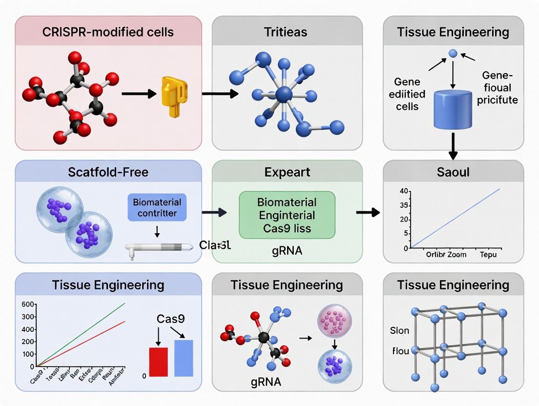
Engineering the Future: How CRISPR-Modified Cells Enable Scaffold-Free Tissue Regeneration
This article provides a comprehensive exploration of scaffold-free tissue engineering powered by CRISPR-Cas9 genome editing.
Engineering the Future: How CRISPR-Modified Cells Enable Scaffold-Free Tissue Regeneration
Abstract
This article provides a comprehensive exploration of scaffold-free tissue engineering powered by CRISPR-Cas9 genome editing. Targeted at researchers and drug development professionals, it details the foundational principles of using CRISPR to enhance cell aggregation and self-organization. The scope covers key methodologies for generating organoids and tissue spheroids, addresses common challenges in homogeneity and scaling, and validates the approach against traditional scaffold-based methods. By synthesizing current research, the article highlights the transformative potential of this convergent technology for creating physiologically relevant tissue models, accelerating drug discovery, and advancing regenerative medicine.
The Convergence of CRISPR and Self-Assembly: Foundations of Scaffold-Free Tissue Engineering
Scaffold-Free Tissue Engineering (SFTE) is an approach that focuses on guiding cells to self-assemble into functional 3D tissues without the use of exogenous biomaterial scaffolds. This paradigm shift leverages innate cellular processes like self-organization, self-sorting, and cell-cell adhesion to form complex structures, often termed organoids, spheroids, or tissue sheets. When integrated with CRISPR-modified cells, SFTE provides a powerful platform for generating genetically precise tissue models for research, drug screening, and therapeutic development. This content is framed within a thesis exploring the synergistic potential of CRISPR genome editing and SFTE to create next-generation, physiologically relevant human tissue constructs.
Core Principles of Scaffold-Free Engineering
The fundamental principles differentiating SFTE from scaffold-based methods are:
- Self-Assembly & Self-Organization: Cells are instructed to secrete and organize their own extracellular matrix (ECM), mimicking natural tissue development.
- Emergent Complexity: Through cell-cell signaling and interactions, simple cell aggregates evolve into structured tissues with distinct regions.
- High Cell Density: Constructs begin with high cell density, leading to rapid tissue formation and enhanced cell-cell communication.
- Minimal Exogenous Material: Avoids complications associated with synthetic or decellularized scaffolds, such as batch variability, immune rejection, degradation byproducts, and potential distortion of cell phenotype.
Advantages Over Traditional Scaffold-Based Methods
The following table quantifies key comparative advantages.
Table 1: Quantitative Comparison of Scaffold-Free vs. Traditional Tissue Engineering
| Parameter | Traditional (Scaffold-Based) | Scaffold-Free (Self-Assembly) | Advantage & Implication |
|---|---|---|---|
| ECM Composition & Remodeling | Pre-defined by scaffold material. | Dynamic, cell-secreted, tissue-specific. | SFTE offers more physiologically relevant ECM, crucial for signaling and mechanics. |
| Cell Density at Initiation | Typically low (< 5x10^6 cells/mL). | Very high (spheroids > 1x10^7 cells/mL). | SFTE accelerates tissue maturation and function. |
| Diffusion Limitations | Can be significant, causing necrotic cores in thick scaffolds. | Present in large spheroids (>500 μm), but can be mitigated via vascularization strategies or bioprinting. | Traditional methods may require complex porosity engineering. |
| Throughput for Drug Screening | Lower, due to scaffold handling. | High (e.g., >1000 spheroids/plate in ULA plates). | SFTE is superior for high-content screening applications. |
| Immunogenicity Risk | High (from scaffold material/residues). | Very Low (purely cellular/autologous ECM). | SFTE is favorable for clinical implantation. |
| CRISPR Delivery & Analysis Efficiency | Can be hindered by scaffold barriers. | High; direct cell access improves editing and clonal analysis. | SFTE synergizes with CRISPR for generating isogenic tissue models. |
| Maturation Timeline | Variable, often slower. | Rapid initial aggregation (24-72 hrs). | SFTE enables faster model generation. |
Application Notes & Protocols
Application Note 1: Generating CRISPR-Edited Cardiac Spheroids for Toxicity Screening
Aim: To create uniform human iPSC-derived cardiomyocyte (iPSC-CM) spheroids with a CRISPR-introduced mutation in a cardiotoxicity-associated gene (e.g., HERG) for drug safety pharmacology.
Protocol:
- CRISPR Editing: Electroporate iPSCs with ribonucleoprotein (RNP) complexes targeting the gene of interest. Isolate and validate clonal lines via sequencing and functional assays.
- Differentiation: Differentiate isogenic wild-type and mutant iPSC lines into cardiomyocytes using established small-molecule protocols (e.g., via modulation of Wnt signaling).
- Spheroid Formation:
- Harvest iPSC-CMs at day 10-12 of differentiation.
- Count and resuspend cells in maintenance medium supplemented with 10 µM Y-27632 (ROCK inhibitor).
- Seed cells into ultra-low attachment (ULA) 96-well round-bottom plates at 10,000 cells/well in 150 µL.
- Centrifuge plates at 300 x g for 3 minutes to aggregate cells at the well bottom.
- Incubate at 37°C, 5% CO₂. Compact spheroids form within 24-48 hours.
- Maturation & Assay: Culture spheroids for 7-14 days, changing 50% medium every 2 days. Assess contractility (video microscopy), viability (ATP assay), and gene expression (qPCR) in response to drug compounds.
Application Note 2: Self-Assembling CRISPR-Modified Hepatic Organoids
Aim: To develop a 3D human liver organoid model from CRISPR-edited primary hepatocytes or adult stem cells for studying metabolic disease.
Protocol:
- Cell Preparation: Isolate primary human hepatocytes or liver progenitor cells. Transduce with lentivirus containing CRISPR/Cas9 and gRNA constructs to knock out a metabolic enzyme (e.g., CYP3A4). Use puromycin selection.
- Matrix-Free Organoid Culture:
- Prepare a single-cell suspension of edited cells.
- Mix cells with unedited hepatic stellate cells (HSCs) and liver endothelial cells (LECs) at a 70:15:15 ratio in advanced DMEM/F12 medium.
- Supplement medium with: 1x B-27, 1x N-2, 10 mM HEPES, 1% GlutaMAX, 10% (v/v) R-spondin-1 conditioned medium, 50 ng/mL EGF, 10 nM Gastrin, 1 µM A-83-01 (TGF-β inhibitor), 10 µM Forskolin, 25 ng/mL HGF, and 10 µM Y-27632.
- Seed 10,000 cells/well in 20 µL droplets of medium onto the lid of a culture dish (inverted hanging drop method). Incubate for 3 days, allowing aggregation.
- Embedding & Expansion: Carefully transfer formed organoids to a ULA 24-well plate with fresh medium (without Y-27632). Culture for 14+ days, passaging every 7-10 days via mechanical dissociation.
- Functional Validation: Assess albumin/urea secretion, CYP450 activity, and bile canaliculi formation (using CLF dye) compared to wild-type organoids.
The Scientist's Toolkit
Table 2: Key Research Reagent Solutions for CRISPR-Enabled SFTE
| Item | Function in SFTE | Example Product/Catalog # |
|---|---|---|
| Ultra-Low Attachment (ULA) Plates | Prevents cell adhesion, forcing 3D aggregation via gravity. | Corning Costar Spheroid Microplates |
| Hydrogel-Based Hanging Drop Plates | Enables high-throughput spheroid formation in defined volumes. | 3D Biomatrix Perfecta3D Hanging Drop Plates |
| ROCK Inhibitor (Y-27632) | Enhances single-cell survival post-dissociation, critical for aggregate formation. | STEMCELL Technologies, #72304 |
| Synthetic ECM (for supportive gels) | Optional soft, inert hydrogel (e.g., PEG) for organoid support without bioactive signaling. | BioLamina's LN-based hydrogels |
| CRISPR RNP Complexes | For high-efficiency, transient editing with minimal off-target effects in primary/Stern cells. | Synthego or IDT's engineered Cas9 and synthetic gRNAs |
| Clonal Isolation Medium | For selecting and expanding CRISPR-edited single-cell clones prior to 3D culture. | With appropriate antibiotics or fluorescent reporters. |
| Metabolic Assay Kits (3D-optimized) | Assess viability/function in dense 3D structures (e.g., ATP, Albumin, Urea). | Promega CellTiter-Glo 3D |
| Live-Cell Imaging Dyes | For long-term tracking of cell viability, apoptosis, or specific organelles in 3D cultures. | Invitrogen CellTracker dyes |
Visualized Pathways and Workflows
Title: CRISPR-Enhanced Scaffold-Free Tissue Engineering Workflow
Title: Key Signaling Pathways Driving SFTE Self-Assembly
Within scaffold-free tissue engineering, generating functional, self-organizing tissues requires cells with precisely engineered behaviors. CRISPR-Cas9 has become the foundational tool for introducing targeted genetic modifications to direct cellular processes such as proliferation, differentiation, matrix production, and intercellular communication. This protocol set details methods for enhancing chondrogenic and osteogenic behavior in human mesenchymal stem cells (hMSCs), a common cell source, for musculoskeletal tissue engineering applications.
Key Application Areas:
- Knockout of Differentiation Inhibitors: Inactivating genes like HES1 (Notch signaling) or SOX9 inhibitors to potentiate chondrogenesis.
- Knock-in of Master Regulators: Integrating inducible RUNX2 or SOX9 expression cassettes for controlled osteogenic or chondrogenic differentiation.
- Editing of ECM Components: Modifying genes for collagen type II (COL2A1) to enhance cartilage matrix quality.
- Reporter Cell Line Generation: Introducing fluorescent tags (e.g., GFP under ACAN promoter) to non-destructively monitor differentiation in 3D organoids.
Key Research Reagent Solutions
| Reagent / Material | Function in CRISPR Protocol |
|---|---|
| RNP Complex Components | Highly specific, transient editing with reduced off-target effects. |
| S. pyogenes Cas9 Nuclease | The endonuclease that creates double-strand breaks at the target DNA site. |
| Synthetic sgRNA (chemically modified) | Guides Cas9 to the specific genomic locus; chemical modifications enhance stability. |
| Delivery Vehicles | |
| Neon Transfection System (Thermo Fisher) | Electroporation system optimized for high-efficiency delivery of RNP into hMSCs. |
| Lipofectamine CRISPRMAX (Thermo Fisher) | Lipid-based transfection reagent for RNP delivery, alternative to electroporation. |
| Validation & Analysis | |
| T7 Endonuclease I / Surveyor Assay | Detects insertions/deletions (indels) at the target site by cleaving heteroduplex DNA. |
| DNeasy Blood & Tissue Kit (Qiagen) | Reliable genomic DNA isolation for downstream sequence analysis. |
| Sanger Sequencing & TIDE Analysis | Quantitative decomposition of sequencing traces to determine editing efficiency. |
| Cell Culture & Differentiation | |
| Human Mesenchymal Stem Cells (hMSCs) | Primary cell model for skeletal tissue engineering. |
| StemMACS MSC Expansion Media | Defined, xeno-free media for maintaining hMSC pluripotency. |
| Chondrogenic/Osteogenic Differentiation Media | Media containing TGF-β3/BMP-6 or ascorbate/β-glycerophosphate/Dex to direct differentiation. |
Detailed Protocol: CRISPR-Cas9 MediatedHES1Knockout in hMSCs to Enhance Chondrogenesis
Aim: To disrupt the HES1 gene in hMSCs via non-homologous end joining (NHEJ), reducing Notch-mediated inhibition of chondrogenic differentiation.
Pre-Experimental Design & sgRNA Preparation
- sgRNA Design: Using a validated tool (e.g., CRISPick, IDT), design two sgRNAs targeting early exons of the human HES1 gene (NCBI: NG_029777.1). Include a negative control (non-targeting) sgRNA.
- Example target sequence (sgRNA1): 5'-GACACCGGACAAACCAAAGG-3' (PAM: TGG)
- Order: Procure chemically modified synthetic sgRNAs (2'-O-methyl analogs, 3' phosphorothioate bonds) and Alt-R S.p. HiFi Cas9 nuclease.
Day 1: RNP Complex Formation & hMSC Electroporation
- RNP Complex Assembly: For a single reaction, combine:
- 3 µL of 10 µM Cas9 Nuclease
- 3 µL of 10 µM HES1 sgRNA (or control sgRNA)
- 4 µL of Nuclease-Free Duplex Buffer
- Incubate at room temperature for 10-20 minutes.
- Cell Preparation: Harvest and count low-passage (P3-P5) hMSCs. For the Neon system, resuspend 2e5 cells in 10 µL Resuspension Buffer R.
- Electroporation: Mix 10 µL cell suspension with the 10 µL RNP complex. Aspirate into a Neon tip. Electroporate (Neon 100 µL kit) at 1100V, 20ms, 2 pulses. Immediately transfer cells to pre-warmed, antibiotic-free complete medium in a 12-well plate.
Day 2-4: Recovery & Expansion
- Refresh culture medium 24 hours post-electroporation.
- Allow cells to recover and proliferate for 72 hours before analysis or differentiation induction.
Day 5: Genomic Editing Efficiency Validation
- Genomic DNA (gDNA) Extraction: Use the DNeasy Kit to isolate gDNA from a portion of the edited and control cell populations.
- PCR Amplification: Amplify a ~500-800bp region surrounding the HES1 target site using high-fidelity polymerase.
- T7 Endonuclease I Assay:
- Hybridize PCR products: 95°C for 5 min, ramp to 85°C at -2°C/s, then to 25°C at -0.1°C/s.
- Digest heteroduplex DNA with T7E1 enzyme for 30 min at 37°C.
- Run products on a 2% agarose gel. Cleaved bands indicate presence of indels.
- Quantitative Analysis (TIDE): Purify the other half of the PCR product and submit for Sanger sequencing. Analyze the sequencing trace data using the TIDE web tool to calculate precise indel percentages.
Day 6-28: Functional Assessment via 3D Chondrogenic Differentiation
- Pellet Culture: Harvest edited and control hMSCs. Form micromass pellets (2.5e5 cells/pellet) by centrifugation in V-bottom plates.
- Chondrogenic Induction: Maintain pellets in serum-free chondrogenic medium (high-glucose DMEM, 1x ITS, 50 µg/mL ascorbate, 40 µg/mL proline, 100 nM dexamethasone, 10 ng/mL TGF-β3) for 28 days. Change medium every 2-3 days.
- Analysis: At day 28, assess:
- Histology: Alcian Blue staining for sulfated glycosaminoglycans (GAGs).
- Biochemistry: DMMB assay for quantitative GAG content, normalized to DNA.
- Gene Expression: qRT-PCR for SOX9, ACAN, COL2A1 (relative to HES1-edited cells should show significant upregulation).
Table 1: Typical Editing Efficiency & Outcomes for HES1 Knockout in hMSCs
| Metric | Control (Non-targeting sgRNA) | HES1-Targeted RNP (Pooled sgRNAs) | Measurement Method |
|---|---|---|---|
| Transfection Viability | 92% ± 3% | 85% ± 5% | Live/Dead assay, 24h post-electroporation |
| Indel Efficiency | 0.5% ± 0.2% | 78% ± 6% | TIDE analysis (Day 5) |
| HES1 Protein Knockdown | 100% ± 8% | 15% ± 7% | Western Blot (Day 7) |
| Chondrogenic GAG/DNA | 12 µg/µg ± 2 | 28 µg/µg ± 3* | DMMB assay (Day 28) |
| COL2A1 Expression | 1.0 ± 0.3 (fold-change) | 8.5 ± 1.2 (fold-change)* | qRT-PCR (Day 28) |
*Indicates statistically significant increase (p < 0.01) vs. control.
Table 2: Comparison of Delivery Methods for RNP in hMSCs
| Method | Editing Efficiency (%) | Cell Viability (%) | Throughput | Relative Cost |
|---|---|---|---|---|
| Neon Electroporation | 75-85 | 80-90 | Medium | High |
| Lipofectamine CRISPRMAX | 60-75 | >95 | High | Medium |
| Nucleofection (4D-Nucleofector) | 70-80 | 75-85 | Medium | Highest |
Diagrams & Workflows
CRISPR-hMSC Engineering Workflow
HES1 Knockout Enhances Chondrogenesis
Application Notes
Within the paradigm of scaffold-free tissue engineering, the generation of robust, self-organizing tissues hinges on the autonomous ability of cells to produce, organize, and respond to their microenvironment. CRISPR-mediated genetic modification of progenitor or somatic cells presents a precise strategy to enhance these intrinsic capabilities. This document outlines key applications targeting adhesion molecules, signaling pathways, and extracellular matrix (ECM) production to engineer tissues with improved structural integrity, morphogenetic potential, and functionality.
1. Editing Cell Adhesion Molecules (CAMs) Enhancing homotypic and heterotypic cell adhesion is critical for the cohesion of scaffold-free organoids or tissue spheroids. CRISPR knockout of inhibitory cadherins (e.g., substituting endogenous cadherins with E-cadherin for stronger epithelial cohesion) or knock-in of engineered integrin subunits can direct cell sorting and enhance binding to specific, endogenously secreted ECM components, improving tissue compaction and reducing necrotic cores.
2. Modulating Morphogenetic Signaling Pathways Precise control of developmental pathways can guide self-organization. CRISPR can be used to:
- Hyperactivate Pathways: Introduce constitutively active mutants of β-catenin (Wnt pathway) or SMADs (TGF-β/BMP pathway) to promote proliferation and specific lineage commitment.
- Create Synthetic Switches: Insert drug-inducible gene expression systems (e.g., doxycycline-inducible CRISPRa) to temporally control pathway activation, enabling staged morphogenesis.
3. Augmenting and Customizing ECM Production The quality and composition of the cell-secreted matrix define the tissue's mechanical properties and biochemical niche. CRISPR strategies include:
- Knock-in of Engineered ECM Genes: Replace wild-type collagen (e.g., COL1A1) alleles with variants resistant to pathological cleavage or incorporating fluorescent tags (e.g., HALO tag) for live imaging of matrix deposition.
- Knockout of ECM Degraders: Delete genes for matrix metalloproteinases (e.g., MMP1) or their activators to reduce catabolism, promoting net matrix accumulation.
- Co-expression of Modifying Enzymes: Knock-in LOXL2 or other cross-linking enzymes under strong promoters to enhance ECM stiffness and stability.
4. Synthetic Biology Circuits for Self-Regulation Advanced constructs can link ECM production to physiological cues. For example, a CRISPR-based gene circuit where a hypoxia-response element drives expression of VEGF and a modified, pro-angiogenic fibronectin isoform, creating a positive feedback loop for vascularized tissue formation.
Protocols
Protocol 1: CRISPR-Cas9 Knock-in for an Engineered E-Cadherin Fusion Protein
Objective: Integrate a sequence encoding E-cadherin fused to a fluorescent protein (mNeonGreen) and a self-cleaving P2A peptide followed by a puromycin resistance gene into the CDH1 safe-harbor locus in human mesenchymal stem cells (hMSCs) to enhance spheroid cohesion.
Materials:
- Cells: hMSCs (passage 3-5)
- Nucleofection System: Lonza 4D-Nucleofector
- CRISPR Components: Alt-R S.p. Cas9 Nuclease V3, synthetic gRNA targeting CDH1 safe-harbor locus, ssODN homology-directed repair (HDR) template.
- HDR Template: 120-nt ssODN with homology arms (60nt each) flanking the insertion sequence:
[PAM-disrupted sequence]-mNeonGreen-P2A-PuroR-[PAM-disrupted sequence]. - Culture Media: Growth medium + 1µM HDR Enhancer (e.g., Alt-R HDR Enhancer). Puromycin selection medium.
Procedure:
- Design & Complex Formation: Resuspend Alt-R Cas9 ribonucleoprotein (RNP) with gRNA at 3:1 molar ratio. Incubate 10 min at RT. Add 1µg of ssODN HDR template.
- Nucleofection: Harvest 2x10^5 hMSCs. Use SE Cell Line 4D-Nucleofector X Kit and program CA-137. Resuspend cell pellet in RNP+ssODN mix, transfer to cuvette, and nucleofect.
- Recovery & Selection: Immediately transfer cells to pre-warmed medium with HDR Enhancer. At 48h post-nucleofection, replace medium with puromycin (1.5 µg/mL). Maintain selection for 7 days.
- Validation: Isolate clones. Confirm integration via PCR (junction amplification) and Sanger sequencing. Assess fusion protein expression via fluorescence microscopy and western blot for E-cadherin. Perform spheroid cohesion assay (see Table 1).
Protocol 2: Inducible Activation of the Wnt/β-Catenin Pathway via CRISPRa
Objective: Establish a stable cell line with doxycycline-inducible expression of a CRISPR activation (CRISPRa) system targeting the promoter of AXIN2, a negative regulator of Wnt signaling, to transiently hyperactivate the pathway.
Materials:
- Plasmids: lenti-dCas9-VPR (CRISPRa), lenti-sgRNA (targeting AXIN2 promoter), Tet-On 3G transactivator plasmid.
- Lentiviral Packaging: psPAX2, pMD2.G, HEK293T cells.
- Inducer: Doxycycline hyclate.
- Reporter: TOPFlash luciferase reporter plasmid.
Procedure:
- Virus Production: Co-transfect HEK293T cells with packaging plasmids and each lentiviral vector using PEI transfection reagent. Harvest virus-containing supernatant at 48h and 72h.
- Cell Line Generation: Infect target cells (e.g., iPSCs) sequentially with Tet-On 3G, then lenti-dCas9-VPR viruses. Select with appropriate antibiotics (G418, blasticidin). Finally, transduce with lenti-sgRNA(AXIN2) and select with puromycin.
- Induction & Validation: Add doxycycline (1 µg/mL) to culture medium for 48h.
- qRT-PCR: Harvest RNA, analyze expression of AXIN2 (initial target), MYC, and CCND1 (downstream targets).
- Reporter Assay: Co-transfect stable line with TOPFlash and Renilla control plasmids pre- and post-induction. Measure luciferase activity (see Table 1).
- Functional Assay: Induce cells pre-aggregation into spheroids; assess changes in size and morphology over 5 days.
Table 1: Representative Experimental Outcomes from Genetic Modifications
| Genetic Target | Modification Type | Assay | Control Value | Modified Value | Key Outcome |
|---|---|---|---|---|---|
| E-Cadherin (CDH1) | Knock-in fusion protein | Spheroid Compaction (Diameter at 72h) | 450 ± 35 µm | 320 ± 28 µm | 29% increase in compaction |
| Integrin α5 (ITGA5) | Overexpression (KI) | Cell-Matrix Adhesion (Absorbance 570nm) | 0.45 ± 0.05 | 0.78 ± 0.07 | 73% increase in adhesion to fibronectin |
| Wnt Pathway (AXIN2 promo.) | CRISPRa activation | TOPFlash Luciferase Activity (Fold Change) | 1.0 ± 0.2 | 8.5 ± 1.3 | ~8.5x pathway activation |
| Collagen I (COL1A1) | Knock-in of tagged variant | ECM Deposition (Fluor. Intensity) | 100 ± 12 AU | 255 ± 30 AU | 155% increase in detectable matrix |
| MMP1 (MMP1) | CRISPR-Cas9 Knockout | Collagen Degradation (µg/mL) | 12.4 ± 1.8 | 4.1 ± 0.9 | 67% reduction in degradation |
Research Reagent Solutions Toolkit
Table 2: Essential Reagents for CRISPR-Enhanced Tissue Engineering
| Reagent / Material | Supplier Examples | Function in Context |
|---|---|---|
| Alt-R S.p. Cas9 Nuclease V3 | Integrated DNA Technologies (IDT) | High-fidelity Cas9 protein for RNP complex formation, reducing off-target effects. |
| Synthetic crRNA & tracrRNA | IDT, Sigma-Aldrich | For customizable gRNA assembly with Cas9 protein. Allows rapid screening of multiple targets. |
| HDR Enhancer V2 | IDT | Small molecule that improves HDR efficiency, critical for precise knock-in experiments. |
| Lonza 4D-Nucleofector System | Lonza | High-efficiency delivery of RNP complexes into hard-to-transfect primary cells like hMSCs. |
| Tet-On 3G Inducible System | Takara Bio, Clontech | Enables precise, dose-temporal control of dCas9-effector (CRISPRa/i) expression. |
| Lenti-X Concentrator | Takara Bio | For simple, high-titer lentivirus concentration to transduce sensitive stem cells. |
| Geltrex or Cultrex BME | Thermo Fisher, R&D Systems | Defined basement membrane extract for supporting 3D culture of modified cells pre-self-assembly. |
| CellTiter-Glo 3D | Promega | Luminescent assay for viability measurement within dense 3D spheroids/organoids. |
Pathway & Workflow Diagrams
Title: CRISPR Editing Strategies for Tissue Engineering
Title: Key Pathways Edited via CRISPR for Morphogenesis
Title: Generic Workflow for Creating CRISPR-Edited Tissues
Within the broader thesis on CRISPR-modified cells for scaffold-free tissue engineering, this document details the application notes and protocols for harnessing cell self-organization. The core principle is that genetically edited stem or progenitor cells, when provided with appropriate biochemical and biophysical cues, can undergo morphogenesis to form complex, functional 3D structures—organoids or tissuoids—without exogenous scaffolds. This process recapitulates key aspects of embryonic development, including cell sorting, differential adhesion, and patterned signaling.
Key Signaling Pathways in Self-Organization
Self-organization in edited cells is governed by evolutionarily conserved signaling pathways. CRISPR/Cas9 is used to introduce reporters, actuators, or knockout alleles into these pathways to study and control morphogenesis.
Title: Core Signaling Pathways Guiding 3D Self-Organization
Quantitative Data on Self-Organizing Structures
Table 1: Characterization Metrics for CRISPR-Edited Self-Organizing Structures
| Organoid Type | Starting Cell # | Time to 3D Structure (Days) | Avg. Diameter (µm) | Key CRISPR Edit for Guidance | Reporter Gene Used |
|---|---|---|---|---|---|
| Cerebral Cortical | 10,000 | 20-30 | 400-500 | PAX6 KO / Reporter | SOX2-GFP |
| Intestinal | 500 | 7-14 | 200-300 | LGR5 Reporter / LGR5 OE | LGR5-tdTomato |
| Kidney | 5,000 | 15-25 | 300-400 | SIX2 Reporter / WNT7B OE | SIX2-H2B-CFP |
| Hepatic | 2,500 | 10-20 | 250-350 | HNF4α Reporter / ATF5 KO | ALB-Luciferase |
Table 2: Comparison of Scaffold-Free vs. Scaffold-Based Methods
| Parameter | Scaffold-Free Self-Organization | Conventional Scaffold-Based |
|---|---|---|
| ECM Composition | Endogenous, cell-derived | Exogenous (e.g., Matrigel, collagen) |
| Structural Fidelity | High (self-patterning) | Medium (dependent on scaffold design) |
| Protocol Complexity | High (requires precise cell prep) | Medium |
| Throughput for Screening | Medium to High (96/384-well plates) | Low to Medium |
| Cost per Unit | Low (cells & media only) | High (scaffold materials) |
Detailed Experimental Protocols
Protocol 4.1: CRISPR Editing of hPSCs for Self-Organization Studies
Aim: Generate a stable, homozygous reporter knock-in in human pluripotent stem cells (hPSCs) to trace a specific lineage during self-organization. Materials: See "Scientist's Toolkit" below. Procedure:
- Design & Cloning: Design gRNAs targeting the safe-harbor locus AAVS1 or a specific gene's STOP codon. Clone gRNA and donor template (containing your reporter, e.g., GFP-P2A-puromycin, flanked by ~800bp homology arms) into a CRISPR/Cas9 plasmid.
- hPSC Transfection: Culture hPSCs in mTeSR Plus on Matrigel-coated plates. At 70% confluence, dissociate to single cells. Transfect 1x10^6 cells with 5 µg of CRISPR plasmid using a nucleofection system (Program B-016). Immediately plate in mTeSR Plus with 10 µM ROCK inhibitor (Y-27632).
- Selection & Cloning: After 48 hours, begin puromycin selection (0.5 µg/mL) for 5-7 days. Surviving colonies are manually picked and expanded in 96-well plates.
- Genotyping & Validation: Extract genomic DNA from clones. Confirm correct integration via junction PCR (using one primer inside the inserted reporter and one outside the homology arm) and sequencing. Validate reporter expression via fluorescence microscopy and flow cytometry.
- Banking: Expand validated clones, cryopreserve in aliquots.
Protocol 4.2: 3D Aggregation and Directed Differentiation for Cerebral Organoid Formation
Aim: Differentiate CRISPR-edited hPSCs into self-organized cerebral organoids. Materials: AggreWell400 plates, neural induction medium (NIM), cerebral organoid differentiation medium (CDM), orbital shaker. Procedure:
- Embryoid Body (EB) Formation: Harvest edited hPSCs as single cells. Count and resuspend at 1.2x10^6 cells/mL in mTeSR Plus with ROCK inhibitor. Add 1 mL cell suspension per well of a AggreWell400 plate (centrifuged per manufacturer's instructions to seed cells into microwells). Centrifuge at 100 x g for 3 min. Incubate (37°C, 5% CO2) for 24h to form uniform EBs.
- Neural Induction: At Day 1, transfer EBs (using a wide-bore pipette tip) to a low-adherence 6-well plate containing NIM. Culture for 6 days, changing medium every other day.
- Matrix Embedding & Neuroepithelial Budding: At Day 7, individually transfer EBs to Matrigel droplets (~15 µL per EB) in a culture dish. Incubate for 30 min at 37°C to polymerize. Overlay with CDM. Over the next 5-10 days, neuroepithelial buds will form.
- Long-Term Maturation: Carefully release Matrigel-embedded organoids and transfer to a low-adherence 125 mL flask containing CDM. Place on an orbital shaker at 60 rpm. Culture for up to 90 days, changing medium twice weekly. Monitor morphology and reporter expression.
- Analysis: Fix for immunostaining (e.g., PAX6, TUJ1, CTIP2) or dissociate for single-cell RNA-seq.
Title: Workflow for Generating CRISPR-Edited Cerebral Organoids
The Scientist's Toolkit: Research Reagent Solutions
Table 3: Essential Materials for Self-Organization Research with Edited Cells
| Item | Function & Rationale | Example Product/Catalog # |
|---|---|---|
| CRISPR/Cas9 System | Precise genome editing to introduce reporters, lineage tags, or modify morphogen receptors. | TrueCut Cas9 Protein v2; Synthetic gRNA. |
| Stem Cell Culture Medium | Maintains pluripotency of hPSCs prior to differentiation. | mTeSR Plus (Stemcell Technologies #100-0276). |
| Low-Adhesion Plates | Prevents cell attachment, forcing 3D aggregation and growth. | Corning Costar Ultra-Low Attachment plates. |
| AggreWell Plates | Microwell plates for forming uniformly sized embryoid bodies, critical for reproducibility. | AggreWell400 (Stemcell Technologies #34415). |
| Basement Membrane Matrix | Provides a complex, physiological 3D environment to support polarized budding (for organoids). | Corning Matrigel Growth Factor Reduced. |
| Rho-Kinase (ROCK) Inhibitor | Improves survival of single hPSCs and newly formed aggregates post-dissociation. | Y-27632 (Tocris #1254). |
| Small Molecule Inducers | Directs differentiation toward specific lineages (e.g., Wnt agonists, BMP inhibitors). | CHIR99021 (GSK-3 inhibitor), LDN-193189 (BMP inhibitor). |
| Orbital Shaker | Provides gentle agitation in suspension culture, improving nutrient/waste exchange for larger organoids. | Thermo Scientific MaxQ 4000. |
| Live-Cell Reporter Dyes | Tracks cell viability, apoptosis, or calcium flux in real-time within 3D structures. | CellTracker CM-Dil, Calcein AM, Fluo-4 AM. |
Application Notes
Within the thesis framework of CRISPR-modified cells for scaffold-free tissue engineering, selecting the optimal primary cell source is critical. Each source presents unique advantages and challenges for genetic modification and subsequent self-assembly into functional tissues.
- Induced Pluripotent Stem Cells (iPSCs): Ideal for generating patient-specific tissues and studying developmental pathways. CRISPR/Cas9 editing is highly efficient in iPSCs, allowing for precise disease modeling, reporter line generation, or correction of genetic defects prior to differentiation. Their unlimited expansion potential supports complex genome editing workflows. However, undefined differentiation and potential teratoma risk post-transplantation require rigorous quality control.
- Mesenchymal Stem/Stromal Cells (MSCs): Valued for their immunomodulatory properties, trophic factor secretion, and relative ease of isolation. CRISPR editing in MSCs is challenging due to low transfection/transduction efficiency and limited proliferative capacity, but is used to enhance paracrine functions or direct differentiation. Their inherent ability to condense into spheroids makes them a prime candidate for scaffold-free engineering of bone, cartilage, and adipose tissues.
- Differentiated Somatic Cells (e.g., fibroblasts, chondrocytes, keratinocytes): Provide a mature, stable phenotype for engineering tissues like skin, cartilage, or myocardium. CRISPR editing enables the introduction of therapeutic genes or the knockout of pathological pathways. While more genetically stable, they often have limited proliferative capacity and may dedifferentiate in culture, posing challenges for generating sufficient edited cell numbers for tissue fabrication.
Table 1: Comparative Analysis of Primary Cell Sources for CRISPR-Modified Tissue Engineering
| Feature | iPSCs | MSCs (Bone Marrow) | Differentiated Somatic Cells (Dermal Fibroblasts) |
|---|---|---|---|
| Proliferative Capacity | Unlimited (theoretically) | 30-40 population doublings before senescence | 50-70 population doublings (varies by donor age) |
| CRISPR Editing Efficiency | High (>80% for indel formation) | Low to Moderate (10-40% depending on delivery) | Moderate (20-60% for fibroblasts) |
| Key Editing Applications | Disease modeling, gene correction, developmental studies | Enhancing secretion, controlling differentiation, immunoengineering | Direct therapeutic protein expression, knockout of fibrotic pathways |
| Time to Edited Tissue | Long (weeks for editing, clonal selection, differentiation) | Moderate (days for editing/expansion, direct use or brief differentiation) | Short (days for editing/expansion, direct use in assemblies) |
| Tumorigenic Risk | High (requires pure differentiated population) | Very Low | None |
| Ideal Tissue Engineering Target | Patient-specific neural, cardiac, or hepatic organoids | Bone, cartilage, stromal-vascular assemblies | Dermal substitutes, fibrotic disease models |
Protocols
Protocol 1: CRISPR/Cas9 Knock-in of a Fluorescent Reporter in Human iPSCs for Lineage Tracing
Objective: To generate a clonal iPSC line with an endogenously tagged gene of interest (e.g., OCT4) via HDR for monitoring differentiation in tissue assemblies.
Materials: See "Research Reagent Solutions" below.
Workflow:
- Design & Preparation: Design sgRNAs targeting near the STOP codon of the target gene using an online design tool. Order sgRNA as crRNA and synthesize ssODN donor template with ~60bp homology arms, P2A sequence, and fluorescent protein (e.g., eGFP) sequence.
- Ribonucleoprotein (RNP) Complex Formation: Combine 6 µg of purified Cas9 protein, 200 pmol of sgRNA (crRNA:tracrRNA duplex), and 1 nmol of ssODN donor in nucleofection buffer. Incubate at 25°C for 10 min.
- iPSC Nucleofection: Dissociate a confluent well of a 6-well iPSC culture into single cells using Accutase. Pellet 1x10^6 cells, resuspend in RNP complex, and nucleofect using program B-016 on a 4D-Nucleofector.
- Recovery & Selection: Immediately transfer cells to pre-warmed mTeSR Plus with 10 µM Y-27632 (ROCKi). After 72 hours, apply appropriate antibiotic selection (e.g., Puromycin, 0.5 µg/mL) for 5-7 days.
- Clonal Isolation & Validation: Pick >30 single colonies, expand, and screen by PCR and Sanger sequencing for correct 5’ and 3’ junction integration. Confirm mono-allelic/bi-allelic editing by sequencing and fluorescent microscopy.
Protocol 2: CRISPRa-Mediated Enhancement of Paracrine Factor in MSCs for Spheroid Formation
Objective: To activate the transcription of VEGFA in bone marrow-derived MSCs using dCas9-VPR to enhance angiogenic potential in engineered spheroids.
Materials: See "Research Reagent Solutions" below.
Workflow:
- sgRNA Cloning: Design and clone 3 sgRNAs targeting the VEGFA promoter into a lentiviral guide vector (e.g., lenti-sgRNA(MS2)_zeo).
- Lentiviral Production: Co-transfect Lenti-dCas9-VPR, psPAX2, and pMD2.G (packaging plasmids) with the sgRNA transfer vector into HEK293T cells using PEI transfection reagent. Harvest virus at 48h and 72h.
- Transduction of MSCs: At passage 3-4, transduce 1x10^5 MSCs with viral supernatant containing 8 µg/mL polybrene via spinfection (1000g, 90 min, 32°C). Refresh complete media (α-MEM + 10% FBS).
- Selection & Expansion: Apply Zeocin (200 µg/mL) and Puromycin (1 µg/mL) for 7 days to select double-positive cells. Expand pooled population.
- Spheroid Formation & Assay: Harvest edited MSCs and plate 2x10^4 cells per well in a 96-well ultra-low attachment plate. Centrifuge at 300g for 3 min to aggregate. Culture for 3 days. Assay spheroids for VEGFA secretion via ELISA and conditioned media for endothelial tube formation.
The Scientist's Toolkit: Research Reagent Solutions
| Item | Function in Protocol | Example Product/Catalog # |
|---|---|---|
| Synthego crRNA | High-purity synthetic sgRNA component for RNP complex formation. | Synthego CRISPR RNA, Gene-specific crRNA |
| Alt-R S.p. Cas9 Nuclease | Recombinant, high-activity Cas9 protein for RNP delivery. | IDT, Cat# 1081058 |
| Neon Transfection System | Electroporation device for high-efficiency delivery into iPSCs. | Thermo Fisher, Neon Transfection System |
| mTeSR Plus Medium | Defined, feeder-free maintenance medium for human iPSCs. | STEMCELL Technologies, Cat# 100-0276 |
| Lenti-dCas9-VPR | Lentiviral vector for constitutive expression of CRISPR activation machinery. | Addgene, Cat# 63800 |
| psPAX2 & pMD2.G | 3rd generation lentiviral packaging plasmids for virus production. | Addgene, Cat# 12260 & #12259 |
| Polybrene | Cationic polymer that enhances viral transduction efficiency. | Sigma-Aldrich, Cat# TR-1003-G |
| Ultra-Low Attachment (ULA) Plate | Prevents cell adhesion, forcing aggregation into spheroids. | Corning, Cat# 7007 |
| Human VEGFA ELISA Kit | Quantifies secreted VEGF protein from edited MSC spheroids. | R&D Systems, Cat# DVE00 |
Application Notes
The integration of CRISPR-Cas9 genome editing with scaffold-free tissue engineering represents a paradigm shift in regenerative medicine. From 2023-2024, research has moved beyond simple 2D knockout studies to the precise, multi-gene engineering of progenitor cells for the directed self-assembly of complex, functional tissues. Key advancements include the generation of immunologically inert universal donor cells, the knockout of senescence pathways to enhance proliferative capacity in engineered tissues, and the precise regulation of morphogenetic signaling cascades to control tissue patterning. Proof-of-concept models now demonstrate the feasibility of creating vascularized cardiac micro-tissues, stratified epidermis, and osteogenic bone spheroids entirely from CRISPR-modified cells without exogenous scaffolds. These models rely on the ability to edit genes governing cell-cell adhesion, extracellular matrix production, and paracrine signaling, enabling cells to act as their own biofactories and structural guides.
Protocols
Protocol 1: Generation of a Universal Donor iPSC Line for Tissue Engineering
Objective: Create an immunocompatible induced pluripotent stem cell (iPSC) line via knockout of HLA class I/II genes and overexpression of CD47. Materials: Human iPSCs, CRISPR-Cas9 ribonucleoprotein (RNP) complexes (targeting B2M, CIITA), AAVS1-safe harbor CD47 overexpression donor template, Nucleofector Kit. Procedure:
- Culture human iPSCs in mTeSR Plus medium on Matrigel-coated plates.
- Prepare two RNP complexes: Cas9 protein complexed with sgRNAs targeting exon 1 of B2M and exon 3 of CIITA.
- Harvest 1x10^6 iPSCs using Accutase. Pellet and resuspend in Nucleofector solution with RNPs (5 µg each) and 2 µg of ssODN donor template.
- Electroporate using program B-016.
- Plate cells on vitronectin-coated plates in recovery medium with 10 µM ROCK inhibitor.
- After 72 hours, apply puromycin (0.5 µg/mL) selection for 5 days.
- Isolate single-cell clones and validate via flow cytometry (loss of HLA-ABC/DR, gain of CD47) and Sanger sequencing of target loci.
Protocol 2: Directed Self-Assembly of CRISPR-Engineered Cardiac Micro-Tissues
Objective: Form beating, vascularized cardiac spheroids from iPSC-derived cardiomyocytes (iPSC-CMs) and endothelial cells (ECs) engineered for enhanced adhesion. Materials: CRISPR-edited iPSC-CMs (NKX2-5eGFP reporter), CRISPR-edited iPSC-ECs (CDH5-mCherry, PECAM1 overexpressing), AggreWell400 plates. Procedure:
- Differentiate the universal donor iPSC line (from Protocol 1) into cardiomyocytes and endothelial cells using established small molecule protocols.
- Prior to differentiation, engineer the iPSCs to overexpress PECAM1 (CD31) in the AAVS1 locus to enhance endothelial barrier function and homophilic adhesion.
- Harvest differentiated iPSC-CMs and iPSC-ECs using gentle cell dissociation reagent.
- Mix cells at a 70:30 (CM:EC) ratio. Prepare a suspension of 1.2x10^6 cells in 4 mL of cardiac spheroid medium.
- Seed 1000 cells per microwell (400 µm) in an AggreWell plate. Centrifuge at 100 x g for 3 min.
- Culture for 96 hours, allowing spheroid formation. Change medium every 48 hours.
- Transfer spheroids to a low-adhesion 96-well plate for long-term culture (>14 days). Assess contractility via video analysis and vascular network formation via confocal microscopy (mCherry signal).
Data Tables
Table 1: Key Landmark Studies (2023-2024) in CRISPR/Cell-Based Tissue Engineering
| Study Focus (First Author, Journal) | Cell Type Edited | CRISPR Target(s) | Key Quantitative Outcome |
|---|---|---|---|
| Immune-evasive tissue spheroids (Lee, Nat. Biomed. Eng. 2023) | Human iPSCs | B2M, CIITA, CD47 (KI) | >90% reduction in NK cell-mediated killing; Spheroid survival in humanized mice: 85% at 28 days vs. 15% in controls. |
| Senescence-resistant osteogenic constructs (Vargas, Sci. Adv. 2024) | Human MSCs | p16INK4a (CDKN2A KO) | 2.5-fold increase in cell proliferation in 3D aggregates; 3-fold increase in mineralized matrix deposition at day 21. |
| Pre-patterned neural organoids (Schmidt, Cell Stem Cell 2023) | Human iPSCs | HES1 (oscillatory reporter KI), ASCL1 (enhancer deletion) | Precise control of neurogenic domains; 75% correlation between in silico pattern prediction and experimental GFP expression. |
| Self-vascularizing cardiac patches (Gao, Circulation 2024) | iPSC-CMs & iPSC-ECs | VEGF-A (doxycycline-inducible KI in AAVS1) | On-demand VEGF expression induced capillary density increase from 50 to 210 capillaries/mm² within engineered tissue. |
Table 2: Performance Metrics of Proof-of-Concept Models
| Tissue Model | Key Edited Feature | Functional Metric | Result (Mean ± SD) | Measurement Method |
|---|---|---|---|---|
| Epidermal Equivalents | FLG (Filaggrin) KO & COL7A1 KI | Barrier Function (TEWL)* | 15.2 ± 3.1 g/m²/h (Edited) vs. 8.5 ± 1.2 (Wild-type) | Tewameter |
| Hepatic Spheroids | CYP3A4 (Overexpression) | Metabolic Activity (Testosterone 6β-hydroxylation) | 45.2 ± 5.6 pmol/min/mg protein | LC-MS/MS |
| Myobundles | MSTN (Myostatin) KO | Contractile Force (Twitch Force) | 1.8 ± 0.3 mN (Edited) vs. 1.1 ± 0.2 mN (Control) | Force transducer |
| Cartilage Micromasses | IL1R1 KO | Inflammation Resistance (IL-6 secretion post IL-1β challenge) | 75% reduction compared to control | ELISA |
TEWL: Transepidermal Water Loss (lower is better for barrier; edited model shows a *compromised barrier as intended for disease modeling).
Diagrams
CRISPR-Engineered Tissue Generation Workflow
IL1R1 KO Blocks Inflammatory Signaling
The Scientist's Toolkit: Key Research Reagent Solutions
| Reagent / Material | Vendor Examples | Function in CRISPR/Scaffold-Free Engineering |
|---|---|---|
| CRISPR-Cas9 RNP Complex | Synthego, IDT | Deliver pre-complexed Cas9 and sgRNA for high-efficiency, transient editing with reduced off-target effects. |
| AAVS1 Safe Harbor Donor Template | VectorBuilder, Takara | Provides a standardized DNA template for consistent, safe knock-in of transgenes (e.g., CD47, reporters). |
| AggreWell Plates | STEMCELL Technologies | Microwell plates for the reproducible formation of uniform, size-controlled cell aggregates (spheroids, organoids). |
| Low-Adhesion U-bottom Plates | Corning, Greiner Bio-One | Enable long-term suspension culture of 3D tissue spheroids without attachment and flattening. |
| Extracellular Matrix (ECM) Hydrogels (for Maturation) | Cultrex BME, Matrigel | Used sparingly not as a scaffold, but as an overlay to provide apical-basal polarity cues to engineered tissues. |
| Live-Cell Analysis Imagers (e.g., Incucyte) | Sartorius | Non-invasive, kinetic tracking of spheroid growth, contractility (cardiac), and fluorescent reporter expression. |
| Tunable Reseeding Plates (e.g., Elplasia) | Corning | Allow for sequential addition of different cell types to build complex, layered tissue structures over time. |
From Design to Dish: Methodologies for Building CRISPR-Engineered Tissues
Application Notes
This protocol details the generation of scaffold-free, three-dimensional tissue constructs using CRISPR-Cas9 engineered human cells. Within the broader thesis of CRISPR-modified cells for scaffold-free tissue engineering, this workflow is pivotal for creating advanced in vitro models that recapitulate native tissue function and pathology. This approach is essential for high-fidelity disease modeling, drug screening, and regenerative medicine applications, offering a more physiologically relevant alternative to 2D cultures and scaffold-dependent methods. The process integrates precision genome editing with self-organization principles of cells to form complex tissues.
Protocols
sgRNA Design and Vector Construction
Objective: To design and clone sequence-specific sgRNAs targeting genes of interest (GOIs) for knock-out (KO) or knock-in (KI).
- sgRNA Design: Identify the target genomic locus. Using current design tools (e.g., CRISPOR, ChopChop), select a 20-nucleotide guide sequence directly 5' of a Protospacer Adjacent Motif (PAM; NGG for SpCas9). Prioritize guides with high on-target and low off-target scores.
- Oligo Annealing: Synthesize complementary oligonucleotides for the chosen guide sequence with appropriate overhangs for your chosen cloning system (e.g., BsaI sites for Golden Gate assembly into a backbone like pSpCas9(BB)-2A-Puro (PX459)).
- Cloning: Anneal oligos and ligate into the digested plasmid backbone using T4 DNA ligase.
- Transformation: Transform the ligation product into competent E. coli, plate on selective agar, and incubate overnight.
- Validation: Pick colonies, perform colony PCR and Sanger sequencing to confirm correct sgRNA insertion.
Cell Line Engineering and Clonal Selection
Objective: To generate a stable, clonal population of cells harboring the desired genetic modification.
- Cell Culture: Maintain human primary or immortalized cells (e.g., mesenchymal stem cells, fibroblasts) in appropriate medium.
- Transfection/Transduction: Deliver the constructed sgRNA/Cas9 plasmid (for KO) or plasmid plus donor template (for KI) via nucleofection or lentiviral transduction, optimized for your cell type.
- Selection and Expansion: Apply selection (e.g., puromycin, 1-2 µg/mL) 48 hours post-transfection for 3-5 days. Allow surviving cells to recover and expand.
- Clonal Isolation: Seed cells at low density in a 10-cm dish or by serial dilution into 96-well plates to obtain single-cell-derived colonies. Expand clones for 2-3 weeks.
- Genotyping: Harvest genomic DNA from each clone. Analyze by PCR followed by T7 Endonuclease I assay or Tracking of Indels by Decomposition (TIDE) analysis for KO. For KI, perform PCR spanning the homology arms and sequence to verify precise integration.
Formation of Scaffold-Free Spheroids
Objective: To aggregate edited cells into three-dimensional spheroids, the foundational units for tissue fusion.
- Harvesting: Trypsinize validated clonal cells and count.
- Aggregation: Seed 5,000-10,000 cells per well in a 96-well ultra-low attachment (ULA) round-bottom plate. The ULA coating prevents cell adhesion, forcing aggregation.
- Centrifugation: Centrifuge the plate at 300 x g for 3 minutes to pellet cells together at the bottom of each well.
- Incubation: Culture the plate undisturbed at 37°C, 5% CO₂ for 48-72 hours. Compact, spherical aggregates will form.
Maturation into a Macroscopic Tissue Construct
Objective: To fuse individual spheroids into a larger, cohesive, and functional tissue construct.
- Mold Preparation: Use a non-adhesive agarose or PDMS mold with a desired shape (e.g., a ring or channel).
- Spheroid Transfer: Using a wide-bore pipette tip, carefully transfer ~50-100 mature spheroids into the mold cavity.
- Fusion Culture: Carefully submerge the mold in culture medium. Over 5-10 days, spheroids will fuse into a single, continuous construct due to cell migration and extracellular matrix deposition.
- Maturation: Maintain the construct in culture for up to 4 weeks, with medium changes every 2-3 days. Optionally apply mechanical stimulation (e.g., cyclic stretching) or use a bioreactor to enhance tissue maturation and function.
Data Presentation
Table 1: Key Quantitative Parameters for Workflow Steps
| Workflow Step | Key Parameter | Typical Value/Range | Purpose/Notes |
|---|---|---|---|
| sgRNA Design | On-target Score | >60 (tool-dependent) | Predicts cleavage efficiency. |
| Off-target Count | Aim for 0 high-quality hits | Minimizes unintended edits. | |
| Cell Transfection | Transfection Efficiency | 40-80% (cell-type dependent) | Measured via control fluorescent plasmid. |
| Selection Duration | 3-5 days | Eliminates non-transfected cells. | |
| Spheroid Formation | Cells per Spheroid | 5,000 - 10,000 | Determines initial spheroid size. |
| Spheroid Diameter (Day 3) | 300 - 500 µm | Indicator of compaction health. | |
| Tissue Maturation | Spheroids per Construct | 50 - 100 | Determines final construct size. |
| Fusion Time (Initial) | 24 - 72 hours | Time for spheroids to adhere. | |
| Maturation Period | 14 - 28 days | For ECM production and functional maturation. |
Visualizations
Title: CRISPR Tissue Engineering Workflow
Title: From Gene Edit to Tissue Phenotype
The Scientist's Toolkit
Table 2: Essential Research Reagent Solutions for CRISPR Tissue Engineering
| Item | Function in Workflow |
|---|---|
| High-Fidelity Cas9 Expression Plasmid (e.g., pX459) | All-in-one vector expressing SpCas9, sgRNA, and a selection marker (puromycin resistance) for efficient knock-out workflows. |
| Chemically Competent E. coli (High-Efficiency) | For rapid and efficient amplification of constructed sgRNA plasmids after cloning. |
| Cell-Type Specific Nucleofection Kit / Lentiviral System | Critical for delivering CRISPR machinery into hard-to-transfect primary or stem cells. |
| Ultra-Low Attachment (ULA) Round-Bottom Plates | Enforces scaffold-free, spontaneous cell aggregation to form uniform, single spheroids per well. |
| Recombinant ECM Proteins (e.g., Collagen I, Fibronectin) | May be used to functionalize fusion molds or in post-fusion assays to assess construct integration capabilities. |
| Live/Dead Viability/Cytotoxicity Kit | Standardized fluorescence-based assay (Calcein AM/EthD-1) to assess 3D construct viability throughout maturation. |
| T7 Endonuclease I / Surveyor Mutation Detection Kit | Enables rapid detection of CRISPR-induced indels in pooled or clonal populations without sequencing. |
| Wide-Bore or Low-Adhesion Pipette Tips | Essential for transferring intact spheroids without causing mechanical disruption or shearing. |
Application Notes
The selection of a CRISPR-Cas9 delivery method for primary cells is a critical determinant of success in scaffold-free tissue engineering research. Primary cells, being non-immortalized and often delicate, present unique challenges. The chosen strategy must balance editing efficiency, cell viability, and the suitability of the resulting modified cells for forming coherent, functional tissues without exogenous scaffolds.
Lentiviral Vectors provide stable genomic integration and sustained Cas9/gRNA expression, enabling high editing efficiencies in hard-to-transfect primary cells (e.g., mesenchymal stem cells). This is advantageous for long-term tissue development studies. However, insertional mutagenesis risks, immunogenicity, and potential for prolonged Cas9 expression leading to off-target effects limit their use in therapeutic applications. Size constraints (~8kb) can also complicate delivery of larger Cas variants.
Electroporation (including nucleofection) is a physical method for delivering CRISPR ribonucleoproteins (RNPs) or plasmids directly into the cytoplasm/nucleus. It is highly efficient for many immune cells (T-cells, NK cells) and some stem cells. RNP delivery offers rapid, transient activity, minimizing off-target events. The primary drawback is significant cellular stress, leading to variable viability that can compromise the number of cells available for 3D tissue assembly.
Nanoparticles (e.g., lipid nanoparticles, polymeric nanoparticles) are emerging as versatile, non-viral carriers. They protect CRISPR payloads (RNPs, mRNA, plasmid DNA) and facilitate endosomal escape. They are highly customizable, can be targeted to specific cell types, and typically evoke lower immune responses than viruses. While often less efficient than viral methods, they offer an excellent balance of efficiency, safety, and scalability for generating edited primary cell populations destined for tissue constructs.
The optimal strategy depends on the primary cell type, desired editing outcome (knockout, knock-in), and the downstream requirement for the cells to self-organize into functional tissues. A combination of high-efficiency RNP electroporation for initial editing followed by nanoparticle-mediated delivery for in-situ modifications in forming tissues is a forward-looking approach.
Protocols
Protocol 1: Lentiviral Transduction of Human Mesenchymal Stem Cells (hMSCs) for Stable Gene Knockout
Objective: Generate a stable, homogeneous population of gene-edited hMSCs for long-term chondrogenic tissue formation studies.
Materials:
- Primary hMSCs (passage 2-4)
- Lentiviral particles (VSV-G pseudotyped) encoding SpCas9 and target-specific gRNA
- Polybrene (8 µg/mL final concentration)
- Complete MSC growth medium (α-MEM, 10% FBS, 1% GlutaMAX)
- Puromycin or appropriate selection antibiotic
- 6-well tissue culture plates
- Centrifuge
Procedure:
- Day 0: Plate 2 x 10^5 hMSCs per well in a 6-well plate in 2 mL complete growth medium. Incubate overnight (37°C, 5% CO2).
- Day 1: Prepare viral transduction mixture. Replace medium with 1 mL fresh medium containing polybrene (8 µg/mL). Add lentiviral particles at a pre-optimized Multiplicity of Infection (MOI, typically 5-20). Include a no-virus control well.
- Spinoculation: Centrifuge the plate at 800 x g for 30 minutes at 32°C. Then, return plate to incubator.
- Day 2: (24 hours post-transduction) Carefully remove medium containing virus and replace with 2 mL fresh complete growth medium.
- Day 3: Begin antibiotic selection. Replace medium with complete growth medium containing the appropriate selective agent (e.g., 1-2 µg/mL puromycin). Maintain selection for 5-7 days, changing medium every 2-3 days, until all cells in the control well are dead.
- Day 10+: Expand surviving, transduced cell pool. Validate knockout efficiency via genomic DNA PCR, T7E1 assay, or next-generation sequencing. Differentiate edited cells towards target lineage (e.g., chondrocytes) to assess functional impact in 3D pellet culture.
Protocol 2: Electroporation of Primary Human T Cells with CRISPR-Cas9 RNP
Objective: Achieve high-efficiency knockout of a target gene (e.g., PD-1) in primary T cells for immunomodulatory tissue engineering applications.
Materials:
- Isolated primary human CD3+ T cells
- Recombinant SpCas9 protein
- Synthetic target-specific crRNA and tracrRNA (or pre-complexed sgRNA)
- Electroporation buffer (commercial, e.g., P3 buffer)
- Electroporation cuvettes (2 mm gap) and electroporator (e.g., Lonza 4D-Nucleofector)
- Pre-warmed complete T-cell medium (RPMI-1640, 10% FBS, IL-2)
- 24-well tissue culture plate
Procedure:
- RNP Complex Formation: Resuspend purified Cas9 protein at 10 µM in sterile duplex buffer. Anneal crRNA and tracrRNA (or use sgRNA) at 10 µM. Mix Cas9 protein and gRNA at a 1:1.2 molar ratio (e.g., 5 µL Cas9 + 6 µL gRNA). Incubate at room temperature for 10-20 minutes to form RNP complexes.
- Cell Preparation: Isolate and activate T cells as per standard protocol. Harvest 1-2 x 10^6 cells per condition. Centrifuge and resuspend cells in 20 µL of pre-warmed electroporation buffer.
- Electroporation: Combine 20 µL cell suspension with 11 µL RNP complex. Transfer entire mixture into a 2 mm cuvette. Electroporate using the appropriate pre-optimized program (e.g., EO-115 for human T cells). Immediately add 80 µL of pre-warmed medium to the cuvette.
- Recovery: Gently transfer cells from the cuvette to a well of a 24-well plate containing 1 mL pre-warmed complete T-cell medium supplemented with IL-2 (e.g., 200 U/mL).
- Analysis: Assess cell viability at 24h using trypan blue exclusion. Expand cells. Assess editing efficiency at the genomic level 72-96 hours post-electroporation via flow cytometry (if a surface protein is targeted) or T7E1 assay.
Protocol 3: Lipid Nanoparticle (LNP) Mediated Delivery of Cas9 mRNA and sgRNA to Primary Hepatocytes
Objective: Transient, high-efficiency editing of primary hepatocytes for constructing metabolically active liver tissue models.
Materials:
- Primary mouse or human hepatocytes
- Cas9 mRNA (chemically modified)
- Target-specific sgRNA
- Ionizable cationic lipid-based LNP formulation reagents (e.g., DLin-MC3-DMA, DSPC, Cholesterol, PEG-lipid)
- Microfluidic mixer
- Hepatocyte maintenance medium
- 96-well or 24-well collagen-coated plates
Procedure:
- LNP Formulation: Prepare an aqueous phase containing Cas9 mRNA and sgRNA at a specific mass ratio (e.g., 3:1) in citrate buffer (pH 4.0). Prepare a lipid phase in ethanol containing the ionizable lipid, phospholipid, cholesterol, and PEG-lipid at precise molar ratios. Using a microfluidic mixer, rapidly mix the aqueous and lipid phases at a controlled flow rate ratio (typically 3:1 aqueous:ethanol) to form LNPs via self-assembly.
- LNP Processing: Dialyze or buffer-exchange the formed LNP suspension into PBS (pH 7.4) to remove ethanol. Concentrate if necessary. Filter-sterilize (0.22 µm). Characterize particle size (Z-average ~80-100 nm) and encapsulation efficiency (>90%).
- Cell Transfection: Plate primary hepatocytes on collagen-coated plates in maintenance medium. Allow cells to attach for 4-6 hours. Dilute LNPs in serum-free medium. Add LNP solution to cells at a final mRNA concentration of 0.1-0.5 µg/well in a 96-well plate. Incubate for 4-6 hours, then replace with fresh complete maintenance medium.
- Analysis: Monitor cell health. Harvest cells 48-72 hours post-transfection for analysis. Assess editing efficiency via next-generation sequencing of the target locus and confirm protein knockout via western blot (if antibody available).
Data Tables
Table 1: Comparative Overview of CRISPR Delivery Strategies for Primary Cells
| Parameter | Lentivirus | Electroporation (RNP) | Nanoparticles (LNP-mRNA) |
|---|---|---|---|
| Typical Editing Efficiency | 70-95% (stable cells) | 50-90% | 40-80% |
| Cell Viability (Post-Delivery) | High (>80%) | Variable (30-70%) | Moderate to High (60-85%) |
| Payload Type | DNA (integrated) | Protein/RNA Complex (RNP) | mRNA/gRNA or RNP |
| Kinetics of Action | Slow, sustained (days-weeks) | Rapid, transient (<48-72h) | Moderate, transient (days) |
| Risk of Off-Target Effects | Higher (prolonged expression) | Lowest (transient) | Low (transient) |
| Immunogenicity | High | Low | Low to Moderate |
| Scalability for Tissue Engineering | Moderate | Low (for large cell numbers) | High |
| Ideal Primary Cell Types | Stem cells (MSCs, HSCs), Fibroblasts | Immune cells (T, NK), iPSCs | Hepatocytes, Endothelial cells, some Stem cells |
Table 2: Key Performance Metrics from Recent Studies (2023-2024)
| Study (Cell Type) | Delivery Method | Target Gene | Efficiency (% Indel) | Viability | Key Outcome for Tissue Engineering |
|---|---|---|---|---|---|
| hMSCs (Cell Stem Cell, 2023) | Lentivirus (all-in-one) | BMP2 | 88% | 92% | Enhanced osteogenic differentiation in 3D spheroids. |
| Primary Human T Cells (Nat. Comm., 2024) | Electroporation (RNP) | TRAC | 95% | 65% | Generated universal CAR-T cells for tumor-infiltrating tissue models. |
| Primary Hepatocytes (Sci. Adv., 2023) | Biodegradable LNPs (mRNA) | PCSK9 | 75% | 78% | Created edited hepatocytes with improved metabolic function for liver assembloids. |
| Human iPSCs (Meth. Prot., 2024) | Electroporation (RNP) | AAVS1 (KI) | 45% (KI) | 55% | Safe-harbor knock-in for tissue-specific reporter expression. |
Diagrams
The Scientist's Toolkit: Research Reagent Solutions
| Item | Function in CRISPR Delivery to Primary Cells |
|---|---|
| Lentiviral Packaging Plasmids (psPAX2, pMD2.G) | Second/third-generation systems for producing replication-incompetent lentiviral particles with high biosafety. |
| Polybrene (Hexadimethrine Bromide) | A cationic polymer that reduces charge repulsion between viral particles and cell membrane, enhancing transduction efficiency. |
| Recombinant Cas9 Protein (NLS-tagged) | High-purity, ready-to-complex protein for RNP formation with synthetic gRNA. Enables rapid, DNA-free editing via electroporation. |
| Chemically Modified sgRNA | Synthetic single-guide RNA with phosphorothioate bonds and 2'-O-methyl modifications; increases stability and reduces immune response. |
| Ionizable Cationic Lipid (e.g., DLin-MC3-DMA) | Key component of LNPs; protonates in acidic endosomes, facilitating endosomal escape of CRISPR payload into the cytoplasm. |
| Cell-Type Specific Nucleofector Kit | Optimized buffers and pre-set electroporation programs for specific primary cell types (e.g., Human T Cell Kit). |
| IL-2 (Interleukin-2) | Essential cytokine for the survival and proliferation of primary T cells post-electroporation. |
| Puromycin Dihydrochloride | Common selection antibiotic for cells transduced with lentiviral vectors containing a puromycin resistance gene. |
| T7 Endonuclease I (T7E1) | Enzyme for detecting small insertions/deletions (indels) at the target site via surveyor nuclease assay. |
| Collagen I, Rat Tail | Substrate for coating culture vessels to promote attachment and maintenance of primary cells like hepatocytes and MSCs. |
Application Notes
The integration of CRISPR-Cas9 genome editing with scaffold-free tissue engineering represents a paradigm shift in constructing physiologically relevant tissue models. These techniques enable the fabrication of 3D structures using only cells and their secreted extracellular matrix (ECM), eliminating potential biocompatibility and immunogenicity concerns associated with exogenous scaffolds. CRISPR-modified cells allow for precise interrogation of gene function in tissue morphogenesis, disease modeling, and drug response within a 3D context. Hanging drop and aggregation techniques are ideal for medium-throughput generation of uniform spheroids for fundamental studies, while 3D bioprinting offers architectural control for creating complex, macroscale tissues. The synergy of these fabrication methods with edited cells accelerates research in developmental biology, cancer metastasis, and high-content drug screening.
Table 1: Comparison of Core Fabrication Techniques for CRISPR-Edited Cells
| Parameter | Hanging Drop | Aggregation (Liquid Overlay) | Extrusion Bioprinting |
|---|---|---|---|
| Typical Spheroid/Construct Size | 200 - 500 µm | 200 - 1000 µm | 1 mm - several cm |
| Throughput | Medium (96-384 spheroids/plate) | High (hundreds to thousands) | Low to Medium (build time-dependent) |
| Uniformity | High (low coefficient of variation) | Moderate (requires optimization) | High (computer-controlled) |
| Architectural Control | Low (simple spheroids) | Low (simple spheroids/aggregates) | High (complex 3D structures) |
| Cell Density | High (starting density controlled) | Adjustable | High (bioink-dependent) |
| Cost per Unit | Low | Very Low | High (printer & bioink) |
| Compatibility with CRISPR Workflow | Post-editing aggregation | Post-editing aggregation | Can print pre-edited cells or bioinks containing editing vectors |
| Key Application | Developmental studies, medium-throughput screening | Large-scale spheroid production, co-culture models | Vascularized tissues, multi-tissue interfaces, implantable constructs |
Table 2: Common CRISPR Edit Types & Their Impact on 3D Fabrication
| Edit Type | Purpose in Scaffold-Free Engineering | Optimal Fabrication Technique(s) | Key Readout Metrics |
|---|---|---|---|
| Gene Knockout (e.g., ECM protein) | Study ECM role in self-assembly & mechanics | Hanging Drop, Aggregation | Spheroid compaction rate, stiffness (AFM), histology |
| Fluorescent Reporter Knock-in | Live tracking of specific cell populations or states | All techniques (esp. confocal imaging) | Spatial organization, migration, differentiation dynamics |
| Oncogene Activation / Tumor Suppressor KO | Initiate tumorigenesis in 3D | Hanging Drop (for screening) | Spheroid invasion, proliferation gradients, drug IC50 |
| Cell Adhesion Molecule KO | Disrupt tissue cohesion & patterning | Aggregation, Bioprinting | Aggregate stability, print fidelity, fusion kinetics |
Experimental Protocols
Protocol 1: Hanging Drop Spheroid Formation from CRISPR-Edited Monolayers
Objective: To generate uniform, scaffold-free spheroids from a monolayer of CRISPR-Cas9 edited cells for downstream analysis of gene function.
Materials:
- CRISPR-modified adherent cell line (validated via sequencing)
- Standard cell culture medium (with appropriate selection agents if needed)
- PBS, without calcium and magnesium
- 0.25% Trypsin-EDTA or non-enzymatic dissociation buffer
- Inverted microscope
- 96-well or 384-well low-attachment, U-bottom or V-bottom plates OR 60mm Petri dishes for the traditional hanging drop method.
Method:
- Cell Preparation: Culture the edited cells to ~80% confluence. Gently wash with PBS and detach using trypsin or enzyme-free reagent.
- Counting & Suspension: Count cells and centrifuge (300 x g, 5 min). Resuspend to a defined concentration (e.g., 1x10^4 to 5x10^4 cells/mL) in complete medium. Optimization Note: The final concentration determines spheroid size.
- For Multi-well Plate Method: a. Pipette 50-100 µL of cell suspension into each well of a low-attachment U-bottom plate. b. Centrifuge the plate gently (100 x g, 2 min) to pool cells at the bottom of the well. c. Incubate at 37°C, 5% CO2. Spheroids will form within 12-48 hours.
- For Traditional Hanging Drop Method: a. Invert the lid of a sterile Petri dish. b. Pipette 20-30 µL droplets of cell suspension onto the inner surface of the lid. c. Carefully replace the bottom chamber, which contains PBS or medium to maintain humidity. d. Incubate. After spheroid formation (24-72h), carefully wash droplets into a collection plate using PBS.
- Media Change & Analysis: Carefully replace 50% of the medium every 2-3 days without disrupting aggregates. Monitor spheroid morphology daily. Proceed to live imaging, fixation, or drug treatment assays.
Protocol 2: Aggregation of Edited Cells via Liquid Overlay for High-Throughput Screening
Objective: To produce large numbers of spheroids in a standard culture plate format for applications like compound screening.
Materials:
- CRISPR-modified cell suspension
- Standard culture plates (6-well to 96-well)
- Molten agarose (1-2% in PBS or serum-free medium)
- Water bath (set at 40-45°C)
- Orbital shaker (optional, for improved uniformity)
Method:
- Coating Plate with Agarose: a. Prepare 1-2% agarose solution and autoclave. Cool in a 40-45°C water bath to prevent solidification. b. Quickly add a thin layer (~1-2 mm) of molten agarose to each well of a culture plate (e.g., 500 µL for a 24-well plate). Swirl to coat the entire well bottom. c. Let solidify at room temperature for 30 minutes, then store at 4°C or proceed.
- Seeding Cells: a. Prepare a single-cell suspension of CRISPR-edited cells as in Protocol 1. b. Add a defined volume of cell suspension in complete medium to each agarose-coated well (e.g., 1 mL containing 10^5 cells for a 24-well plate).
- Aggregation: a. Gently swirl the plate to distribute cells evenly. b. Place the plate on a level surface in a 37°C incubator. For enhanced uniformity, place the plate on an orbital shaker inside the incubator at 40-60 rpm. c. Aggregates will form within 24 hours. Medium can be changed by careful pipetting after allowing spheroids to settle.
Protocol 3: Extrusion Bioprinting of a CRISPR-Modified Cell Bioink
Objective: To fabricate a defined 3D tissue construct using a bioink composed of CRISPR-edited cells.
Materials:
- CRISPR-edited cells (as a pellet)
- Bioink carrier/hydrogel (e.g., 3-5% alginate, 20-30 mg/mL collagen I, or proprietary tunable hydrogels)
- Crosslinking agent (e.g., CaCl2 solution for alginate)
- Extrusion bioprinter (pneumatic or piston-driven) with temperature control
- Sterile printing cartridges and nozzles (diameter 200-400 µm)
- Printing substrate (e.g., Petri dish, transwell insert)
Method:
- Bioink Preparation: a. Centrifuge the required number of edited cells (e.g., 5-20 million). Aspirate supernatant. b. Gently mix the cell pellet with the pre-cooled, sterile hydrogel precursor to achieve a final high cell density (e.g., 5-10 x 10^6 cells/mL). Avoid introducing bubbles. c. Keep the bioink on ice or at 4°C to delay gelation until printing.
- Printer & CAD Model Setup: a. Load the bioink into a sterile printing cartridge, attach the nozzle, and mount onto the printer. b. Import or design the desired 2D/3D structure (e.g., grid, tube, multi-layer patch) into the printer software. c. Set printing parameters: pressure (15-50 kPa), speed (5-15 mm/s), layer height, and temperature (often 4-15°C for the print head, 37°C for the print bed).
- Printing & Crosslinking: a. Begin the print job. The bioink is extruded in a continuous filament according to the digital design. b. For materials like alginate, crosslink during or immediately after printing by misting with or immersing in CaCl2 solution (e.g., 100 mM). c. For collagen, transfer the printed construct to a 37°C incubator to induce thermal gelation.
- Post-Printing Culture: a. After initial gelation, carefully transfer the construct to a culture well. b. Flood with warm complete medium. Culture under standard conditions, with medium changes tailored to the construct's size and cell density.
Diagrams
Workflow: From Gene Edit to 3D Fabrication
Liquid Overlay Aggregation Protocol Steps
The Scientist's Toolkit
Table 3: Essential Reagents & Materials for Scaffold-Free Fabrication with Edited Cells
| Item | Function/Description | Example Vendor/Product |
|---|---|---|
| CRISPR Ribonucleoprotein (RNP) | Enables precise, transient gene editing without vector integration. Reduces off-target effects. | Synthego, IDT, Thermo Fisher TrueCut Cas9 Protein |
| Low-Adhesion Multi-well Plates | U- or V-bottom plates prevent cell attachment, forcing 3D aggregation into spheroids. | Corning Spheroid Microplates, Greiner CELLSTAR |
| Agarose, Ultra-Pure | Used to create a non-adherent hydrogel coating for liquid overlay aggregation methods. | Lonza SeaPlaque, Thermo Fisher |
| Tunable Hydrogel (Bioink) | Provides temporary, printable scaffold for bioprinting that supports cell viability and later remodeling. | Allevi/Allevi Alginate, Cellink Bioink, Collagen I (Rat tail) |
| Extracellular Matrix (ECM) Staining Kits | For visualizing cell-secreted ECM components (collagen, fibronectin) in spheroids/constructs. | Abcam ECM Protein Detection Kit, Sirius Red/Fast Green |
| Live-Cell Imaging Dyes | Track viability (Calcein AM), death (PI/EthD-1), and reactive oxygen species in 3D structures over time. | Thermo Fisher Live/Dead Kit, CellROX Reagents |
| Automated Imaging System | High-content analysis of spheroid size, morphology, and fluorescence in multi-well plates. | PerkinElmer Opera Phenix, Molecular Devices ImageXpress |
| Mechanical Testing System | Measures stiffness/elasticity of spheroids and printed constructs via micro-indentation. | CellScale MicroSquisher, AFM systems |
Within the broader thesis on utilizing CRISPR-Cas9 for scaffold-free tissue engineering, this case study focuses on generating human cardiac spheroids with genetically enhanced functional readouts for improved cardiotoxicity prediction. Traditional 2D cardiomyocyte models often lack the physiological relevance of native tissue, leading to poor translational outcomes in drug development. This protocol details the creation of 3D, self-assembling cardiac spheroids from human induced pluripotent stem cell-derived cardiomyocytes (hiPSC-CMs) engineered via CRISPR to harbor a calcium-sensitive fluorescent reporter (GCaMP6f) and a loss-of-function mutation in the KCNH2 gene (hERG channel), a common target for pro-arrhythmic drugs. This dual modification creates a superior assay system: the reporter enables quantitative, high-throughput functional analysis, while the introduced mutation creates a sensitive, patient-relevant background for toxicity screening.
Key Research Reagent Solutions
Table 1: Essential Research Reagents and Materials
| Reagent/Material | Function in Protocol | Example Vendor/Product |
|---|---|---|
| hiPSC line (e.g., WTC-11) | Source for generating isogenic, CRISPR-edited cardiomyocytes. | Coriell Institute |
| CRISPR-Cas9 RNP complex | Enables precise, transient genome editing without DNA integration. | Synthego (sgRNA + Cas9 protein) |
| Nucleofection Kit for hiPSCs | High-efficiency delivery of CRISPR RNP into stem cells. | Lonza, P3 Primary Cell Kit |
| GCaMP6f donor template | Homology-directed repair (HDR) template for knock-in at safe-harbor locus (e.g., AAVS1). | IDT, gBlocks Gene Fragment |
| KCNH2 (hERG) sgRNA | Targets exon 5 for introduction of a known loss-of-function variant (e.g., G604S). | Designed via CHOPCHOP, synthesized by IDT |
| hiPSC-CM Differentiation Kit | Robust, defined protocol for generating functional cardiomyocytes. | STEMdiff Cardiomyocyte Kit (StemCell Tech.) |
| Ultra-Low Attachment U-96 Plate | Enables scaffold-free self-assembly of cells into 3D spheroids. | Corning Spheroid Microplate |
| FLIPR Tetra or Confocal Imager | Measures real-time, high-throughput calcium transients (GCaMP6f fluorescence). | Molecular Devices FLIPR Tetra |
| Known QT-Prolonging Drugs (Positive Controls) | Validate spheroid sensitivity (e.g., E-4031, Dofetilide, Sotalol). | Tocris Bioscience |
Detailed Protocols
Protocol A: CRISPR-Cas9 Editing of hiPSCs for Dual Modification
Objective: Generate a clonal hiPSC line harboring a heterozygous G604S mutation in KCNH2 and a homozygous knock-in of GCaMP6f at the AAVS1 safe-harbor locus.
Materials: hiPSCs, CRISPR RNP complexes (for KCNH2 and AAVS1), HDR templates, Nucleofector, mTeSR Plus medium, CloneR supplement.
Method:
- Design & Preparation: Design two sgRNAs: one targeting KCNH2 exon 5 and one targeting the AAVS1 locus. Synthesize as chemically modified sgRNAs. Prepare a single-stranded DNA (ssDNA) HDR template for AAVS1 containing GCaMP6f-P2A-PuromycinR flanked by 800bp homology arms.
- Nucleofection: Harvest 1x10^6 hiPSCs. Co-electroporate with both RNP complexes (30 pmol each) and the AAVS1 HDR template (100 pmol) using the P3 Primary Cell Nucleofector Kit (Code CA-137).
- Recovery & Selection: Plate cells in mTeSR Plus with CloneR. At 48h post-nucleofection, add 0.5 µg/mL Puromycin. Select for 7 days.
- Clonal Isolation: Harvest and perform single-cell sorting into 96-well plates. Expand clones for 3-4 weeks.
- Genotyping:
- PCR & Sanger Sequencing: Confirm KCNH2 heterozygous G604S mutation and AAVS1 correct targeting.
- Functional Pre-screen: Differentiate small batches of top clones into cardiomyocytes (Protocol B) and assess GCaMP6f expression via fluorescence microscopy.
- Karyotype & Pluripotency Validation: Ensure normal karyotype (G-banding) and pluripotency marker (OCT4, NANOG) expression in the selected master clonal line.
Protocol B: Directed Differentiation into hiPSC-Cardiomyocytes
Objective: Differentiate the engineered hiPSC clonal line into a highly pure (>90%) population of cardiomyocytes.
Method (Based on STEMdiff Kit):
- Culture hiPSCs to 85-90% confluence in a 6-well plate.
- Initiation (Day 0): Switch to Cardiomyocyte Differentiation Medium A.
- Differentiation (Day 2): Change to Medium B without disturbing the cell layer.
- Metabolic Selection (Day 5-7): Change to Medium C. This lactate-rich medium selectively favors the survival of metabolically active cardiomyocytes.
- Maturation (Day 7+): At day 7, start feeding with Maintenance Medium, changing every 2-3 days. Spontaneous beating is typically observed by Day 8-10.
- Dissociation & Harvest (Day 15): At day 15-20, dissociate cardiomyocytes using a gentle cell dissociation reagent (e.g., TrypLE). Purify if necessary using glucose-based metabolic selection. Yield: ~50-80 cardiomyocytes per input hiPSC.
Protocol C: Generation of 3D Cardiac Spheroids
Objective: Form uniform, functional 3D spheroids from the engineered hiPSC-CMs.
Materials: Dissociated hiPSC-CMs (Day 15-20), Ultra-Low Attachment 96-well round-bottom plate, Cardiac Spheroid Medium (e.g., RPMI/B27 with insulin).
Method:
- Prepare a single-cell suspension of hiPSC-CMs at 5.0 x 10^5 cells/mL in Cardiac Spheroid Medium.
- Using a multichannel pipette, seed 100 µL per well (containing 50,000 cells) into the U-bottom plate.
- Centrifuge the plate at 300 x g for 3 minutes to aggregate cells at the well bottom.
- Incubate at 37°C, 5% CO2. Spheroids will form within 24 hours.
- Feed every 48 hours by carefully replacing 50 µL of old medium with fresh medium. Spheroids are ready for assay at Day 5-7 post-seeding.
- Quality Control: Monitor spheroid diameter (target: 350-450 µm) and ensure synchronous, spontaneous beating.
Protocol D: Functional Toxicity Assay using Calcium Transient Imaging
Objective: Quantify drug-induced changes in cardiac electrophysiology via CRISPR-enhanced GCaMP6f reporting.
Materials: Matured cardiac spheroids, FLIPR Tetra High-Throughput Cellular Screening System, test compounds, 0.1% DMSO (vehicle control), E-4031 (10 µM, positive control).
Method:
- Load Spheroids: Transfer one spheroid per well to a fibronectin-coated 384-well imaging plate in 30 µL of assay buffer (e.g., Tyrode’s solution).
- Baseline Recording: Using the FLIPR Tetra, record baseline GCaMP6f fluorescence (excitation 470-495nm, emission 515-575nm) at 100 frames per second for 1 minute.
- Compound Addition: Using the integrated fluidics, add 10 µL of 5x concentrated drug solution (or vehicle) to each well. Final DMSO concentration ≤0.1%.
- Post-Compound Recording: Immediately record calcium transients for 10 minutes.
- Data Analysis: Use software (e.g., CardioAnalytics) to derive parameters from each transient:
- Beat Period (ms): Time between peaks.
- Amplitude (F/F0): Relative fluorescence change.
- Duration (ms): Width at 50% amplitude (Ca2+ Transient Duration, CTD).
- Arrhythmia Index: Count of irregular beats or missed beats.
Data Presentation
Table 2: Quantitative Calcium Transient Parameters from CRISPR-Enhanced Spheroids Exposed to Reference Compounds
| Compound (1 µM) | Beat Period (% Change vs. Vehicle) | Amplitude (% Change vs. Vehicle) | Ca2+ Transient Duration (% Change vs. Vehicle) | Arrhythmia Index (Events/min) | n (Spheroids) |
|---|---|---|---|---|---|
| Vehicle (0.1% DMSO) | 0.0% ± 3.1 | 0.0% ± 5.2 | 0.0% ± 4.8 | 0.2 ± 0.5 | 24 |
| E-4031 (hERG blocker) | +45.2% ± 8.7* | -15.3% ± 6.4* | +62.1% ± 10.3* | 12.5 ± 3.8* | 24 |
| Dofetilide | +38.9% ± 7.5* | -10.1% ± 7.2 | +58.7% ± 9.6* | 8.9 ± 2.1* | 18 |
| Verapamil (Multi-channel) | -10.2% ± 4.5* | -5.1% ± 6.3 | -8.7% ± 5.1 | 0.5 ± 0.7 | 18 |
| Test Drug X | +5.1% ± 4.8 | -2.3% ± 5.9 | +8.5% ± 6.2 | 0.8 ± 1.1 | 18 |
Data presented as Mean ± SD. *p < 0.01 vs. Vehicle control (one-way ANOVA with Dunnett's post-hoc test).
Diagrams
CRISPR-Enhanced Cardiac Spheroid Generation & Assay Workflow
Key Pathways Interrogated by CRISPR-Enhanced Spheroid Assay
Within the broader thesis on CRISPR-modified cells for scaffold-free tissue engineering, this case study explores the generation of vascularized liver organoids. These 3D structures are crucial for accurate disease modeling and drug screening. The integration of CRISPR-Cas9 allows for the precise introduction of disease-associated mutations into pluripotent stem cells (PSCs) or primary hepatocytes, enabling the de novo creation of genetically defined pathological tissues in a scaffold-free, self-organizing system. The subsequent incorporation of endothelial and stromal lineages is essential to overcome the limitations of necrosis in core regions and to model systemic disease processes.
Table 1: Comparative Analysis of Liver Organoid Vascularization Methods
| Method | Cell Composition (Ratios) | Maturation Time (Days) | Key Outcome Metrics | Reference (Year) |
|---|---|---|---|---|
| Co-culture Self-Assembly | iPSC-Hepatoblasts: HUVECs: MSCs (2:1:1) | 21-28 | Vessel network length: ~1200 µm/mm²; Albumin secretion: 15-20 µg/day/10⁶ cells | Clevers et al., 2022 |
| CRISPR-edited iPSC Lineage | iPSC-HEPs: iPSC-ECs (from isogenic line) (3:1) | 28-35 | Luminal structure formation: ~65%; CYP3A4 activity: 45 pmol/min/mg protein | Takebe et al., 2023 |
| Microfluidic Chip Perfusion | Primary hepatocytes: HSECs: Fibroblasts (5:3:2) | 10-14 | Urea production: 80 µg/day; Perfusable lumen diameter: 50-100 µm | Ingber et al., 2023 |
| Organoid Implantation (in vivo) | Organoid + Endothelial Progenitors | 60+ in vivo | Host vessel anastomosis: >70% implants; Survival rate: >90% (mouse model) | Sampaziotis et al., 2022 |
Table 2: CRISPR-Modified Genes for Common Liver Disease Modeling
| Disease Model | Target Gene(s) | CRISPR Edit Type | Expected Phenotype in Organoid |
|---|---|---|---|
| Hereditary Hemochromatosis | HFE (C282Y) | Knock-in (Point Mutation) | Iron accumulation, oxidative stress, fibrotic markers |
| Alpha-1 Antitrypsin Deficiency | SERPINA1 (E342K) | Knock-in (PiZ allele) | AAT polymer aggregation, reduced secretion, ER stress |
| Alcoholic Steatohepatitis (ASH) | ADH1B, ALDH2 | Knock-out / Activating Edit | Increased sensitivity to ethanol metabolites, lipid accumulation |
| Wilson Disease | ATP7B | Knock-out | Copper accumulation, mitochondrial dysfunction |
Detailed Experimental Protocols
Protocol 1: Generation of CRISPR-Edited iPSCs for SERPINA1 E342K Knock-in Objective: Introduce the PiZ mutation into a wild-type iPSC line for AATD modeling. Materials: Wild-type human iPSCs, pSpCas9(BB)-2A-Puro plasmid, ssODN donor template (E342K mutation + silent restriction site), Lipofectamine Stem Transfection Reagent, Puromycin. Procedure:
- Design gRNA targeting exon 5 of SERPINA1 near codon 342. Synthesize a 200-nt ssODN donor with the mutation and a silent EcoRI site.
- Co-transfect iPSCs (70% confluent in 6-well) with 2 µg Cas9/gRNA plasmid and 200 pmol ssODN using Lipofectamine Stem.
- At 48h post-transfection, select with 0.5 µg/mL puromycin for 72h.
- Recover cells for 5 days, then pick single-cell clones into 96-well plates.
- Expand clones and screen by genomic PCR of the target region, followed by EcoRI digestion and Sanger sequencing to confirm precise editing.
Protocol 2: Scaffold-Free Assembly of Vascularized Liver Organoids via Aggrewell Objective: Differentiate and assemble iPSC-derived hepatocytes, endothelial cells, and mesenchymal stem cells into a vascularized organoid. Materials: CRISPR-edited iPSCs (HEP lineage), isogenic iPSC-ECs, MSCs, Aggrewell400 plates, Hepatic Maturation Medium, VEGF-containing Endothelial Medium. Procedure:
- Hepatocyte Differentiation: Differentiate CRISPR-edited iPSCs into definitive endoderm (Activin A, 3 days), then hepatoblasts (FGF4, BMP2, 5 days), and finally hepatocyte-like cells (HGF, Oncostatin M, Dexamethasone, 10 days).
- Endothelial Cell Differentiation: Differentiate isogenic iPSCs into ECs using VEGF, BMP4, and CHIR99021 over 8 days. Confirm by CD31/PECAM-1 flow cytometry.
- Aggregation: Mix differentiated HEPs, iPSC-ECs, and MSCs in a 2:1:1 ratio. Resuspend 2x10⁶ total cells in 2 mL maturation medium.
- Seed 1000 cells per micro-well in an Aggrewell plate (centrifuge at 100 x g for 3 min to settle). Culture for 7 days, with medium changes every other day.
- Maturation & Vascular Priming: From day 7-28, culture in Hepatic Maturation Medium supplemented with 50 ng/mL VEGF and 20 ng/mL FGF2. Change medium every 2 days. Assess network formation via live imaging of GFP-tagged iPSC-ECs.
Diagrams
The Scientist's Toolkit: Research Reagent Solutions
Table 3: Essential Materials for Vascularized Liver Organoid Research
| Item / Reagent | Function & Application in Protocol |
|---|---|
| CRISPR-Cas9 Plasmid (e.g., pSpCas9(BB)-2A-Puro) | All-in-one vector for gRNA expression, Cas9 nuclease, and puromycin selection during iPSC gene editing. |
| Single-Stranded Oligodeoxynucleotide (ssODN) | High-efficiency donor template for precise CRISPR-mediated knock-in of point mutations. |
| Aggrewell400 Plates | Microwell plates for consistent, scaffold-free formation of thousands of uniform-sized organoid aggregates. |
| Recombinant Human VEGF-165 | Key angiogenic growth factor; drives endothelial differentiation and network formation in maturing organoids. |
| Oncostatin M (OSM) | Critical cytokine for terminal hepatocyte maturation from progenitor cells, inducing albumin and CYP450 expression. |
| CD31/PECAM-1 Antibody (Fluorophore-conjugated) | Essential for flow cytometry analysis to validate endothelial cell differentiation purity and identity. |
| Matrigel (Growth Factor Reduced) | Often used as a supportive 3D embedding matrix post-aggregation to enhance polarity and vascular network stability. |
| Live-Cell Imaging Dye (e.g., CellTracker Green) | For labeling specific cell lineages (e.g., ECs) to dynamically track vascular network formation over time. |
High-content screening (HCS) using genetically defined, scaffold-free engineered tissues represents a transformative approach in preclinical drug discovery. This methodology directly addresses the limitations of conventional 2D cell cultures by providing physiologically relevant, three-dimensional micro-tissues with precise genetic backgrounds, often engineered via CRISPR-Cas9. These tissues recapitulate critical aspects of native tissue architecture, cell-cell interactions, and metabolic gradients, leading to more predictive compound efficacy and toxicity data. Framed within the broader thesis of utilizing CRISPR-modified cells for scaffold-free tissue engineering, this application note details protocols for generating such tissues and deploying them in high-content phenotypic screens.
Genetically defined tissues offer specific advantages over traditional models. The following table summarizes quantitative performance metrics reported in recent studies comparing 3D engineered tissues to 2D monolayers in HCS campaigns.
Table 1: Comparative Performance of 2D vs. 3D Genetically Defined Tissue Models in HCS
| Parameter | 2D Monolayer (CRISPR-modified) | 3D Scaffold-Free Tissue (CRISPR-modified) | Improvement/Notes | Source (Example) |
|---|---|---|---|---|
| Z'-Factor (Phenotypic Assay) | 0.3 - 0.5 | 0.5 - 0.7 | Higher Z' indicates more robust assay suitable for screening. | Lin et al., 2023 |
| Gene Editing Efficiency | >90% (knockout) | 70-85% (knockout) | Slightly lower in 3D aggregates but remains high. | BioRxiv, 2024 |
| Viability Assay Dynamic Range | 5-10 fold | 15-25 fold | Enhanced sensitivity to cytotoxic compounds. | Nat. Protoc., 2023 |
| IC50 Shift (Cytotoxic Drug) | Baseline (1x) | 3x - 10x Higher | Better modeling of in vivo drug resistance. | Toxicol Sci., 2023 |
| Phenotypic Features Quantified | 50-200 (cell-level) | 300-1000 (tissue & cell-level) | Includes tissue morphology, texture, and neighbor effects. | SLAS Discov., 2024 |
| Throughput (Tissues/Day) | Very High (10^5 wells) | High (10^4 wells) | Compatible with 384/1536-well spheroid plates. | Curr. Protoc., 2024 |
Research Reagent Solutions Toolkit
Table 2: Essential Research Reagents and Materials for HCS with Genetically Defined Tissues
| Item | Function/Description | Example Product/Catalog |
|---|---|---|
| CRISPR Ribonucleoprotein (RNP) | Enables precise genetic modification (KO/KI) in progenitor cells. | Synthetic sgRNA + recombinant Cas9 protein. |
| Low-Adhesion 384-Well U-Bottom Plates | Facilitates scaffold-free self-assembly of cells into uniform micro-tissues. | Corning Spheroid Microplate (Cat# 4516). |
| Extracellular Matrix (ECM) Hydrogel | Optional overlay to provide physiological context and polarization. | Cultrex Basement Membrane Extract (BME). |
| Multiplexed Fluorescent Cell Health Dyes | Live-cell, non-toxic dyes for concurrent viability/cytotoxicity/apoptosis. | CellEvent Caspase-3/7, Cytoplasm-ID. |
| Phenotypic Staining Panel (Fixed) | Antibodies & dyes for multiplexed readouts (e.g., phospho-proteins, cytoskeleton). | Phospho-AKT (Ser473), α-Tubulin, DAPI. |
| High-Content Imaging System | Automated microscope for 3D Z-stack acquisition. | ImageXpress Micro Confocal, Operetta CLS. |
| 3D Image Analysis Software | Extracts quantitative features from tissue volumes. | Harmony, IN Carta, CellProfiler. |
Detailed Protocols
Protocol 4.1: Generation of CRISPR-Engineered Scaffold-Free Micro-Tissues
Objective: To create genetically uniform, scaffold-free 3D micro-tissues from CRISPR-modified cells for HCS.
Materials:
- Cas9-expressing or wild-type parental cell line (e.g., iPSC-derived hepatocytes, cardiomyocytes).
- CRISPR RNP complexes (targeting gene of interest or safe-harbor locus for reporters).
- Electroporation/Nucleofection system (e.g., Lonza 4D-Nucleofector).
- Low-adhesion round-bottom 384-well plate.
- Complete growth medium optimized for 3D culture.
Method:
- Genetic Modification: Harvest and count parental cells. For 10^6 cells, complex 5 µg recombinant Cas9 protein with 200 pmol synthetic sgRNA in buffer to form RNP. Transfect cells via nucleofection using optimized program. Include non-targeting sgRNA control.
- Selection & Expansion: Post-nucleofection, recover cells overnight. Apply appropriate antibiotic selection (e.g., puromycin for 48h) if a selection marker was co-introduced. Expand edited cell pool for 5-7 days, validating editing efficiency via T7E1 assay or NGS.
- Micro-Tissue Assembly: Harvest edited cells, resuspend at 1-2x10^5 cells/mL in 3D medium. Using a multichannel pipette, dispense 50 µL cell suspension (~500-2000 cells) per well of a 384-well low-adhesion U-bottom plate.
- Centrifugal Aggregation: Centrifuge plate at 300 x g for 5 minutes to pellet cells into the well bottom.
- Culture: Incubate plate at 37°C, 5% CO2 for 72-96 hours. Monitor daily; a single, compact spheroid should form per well.
Protocol 4.2: High-Content Phenotypic Screening Assay
Objective: To treat micro-tissues with compound libraries and perform multiplexed, fixed-endpoint imaging and analysis.
Materials:
- Compound library in DMSO, pre-dispensed in intermediate plates.
- Automated liquid handler (e.g., Beckman Biomek).
- Multichannel pipettes.
- 16% Paraformaldehyde (PFA).
- Permeabilization buffer (0.5% Triton X-100 in PBS).
- Blocking buffer (5% BSA in PBS).
- Primary and fluorescently conjugated secondary antibodies.
- Nuclear stain (e.g., DAPI, 1 µg/mL).
- Plateseal.
Method:
- Compound Treatment: At day 4 post-seeding, using an acoustic dispenser or pin tool, transfer 50 nL of compound from source plate to each assay plate containing mature micro-tissues. Final DMSO concentration should not exceed 0.5%. Include vehicle (DMSO) and control compound (e.g., staurosporine for cytotoxicity) wells on each plate.
- Incubation: Incubate compound-treated tissues for the desired period (e.g., 72h for viability, 48h for pathway modulation) at 37°C.
- Fixation and Staining:
- Carefully aspirate 40 µL of medium from each well using a plate washer.
- Add 40 µL of 4% PFA (diluted from 16% in PBS) and fix for 45 minutes at RT.
- Permeabilize and block with 50 µL blocking/permeabilization buffer for 1 hour.
- Aspirate and add 30 µL primary antibody cocktail in blocking buffer. Incubate overnight at 4°C.
- Wash 3x with 50 µL PBS.
- Add 30 µL secondary antibody cocktail + DAPI. Incubate for 3h at RT, protected from light.
- Wash 3x with PBS, leave 50 µL PBS per well, seal plate.
- Image Acquisition: Using a high-content confocal imager, acquire 4-6 Z-stacks (10 µm step) per well using a 20x objective. Set exposure times on control wells to avoid saturation.
- Image Analysis: Use 3D analysis software (e.g., Harmony) to:
- Identify individual micro-tissues using DAPI signal.
- Apply a rolling ball background subtraction.
- Segment individual cells within the tissue using cytoplasm/nuclear stains.
- Extract >500 features: tissue diameter, circularity, cell count, intensity of markers (mean, std dev), texture (Haralick), and spatial relationships.
Visualized Workflows and Pathways
Title: High-Content Screening Workflow with Engineered Tissues
Title: Compound Response in a Genetically Defined Tissue Context
Overcoming Hurdles: Optimizing Efficiency and Function in CRISPR-Edited Tissues
Application Notes: Current Landscape & Challenges
The therapeutic and research potential of CRISPR-engineered cells in scaffold-free tissue engineering (SFTE) is compromised by three interrelated pitfalls: off-target mutagenesis, editing heterogeneity, and reduced cell viability. These factors directly impact the safety, functional consistency, and scalability of engineered tissues like organoids or cell sheets.
Off-Target Effects: Mechanisms and Impact
Off-target effects occur when CRISPR-Cas9 cleaves unintended genomic loci with sequence similarity to the target guide RNA (gRNA). In SFTE, these mutations can:
- Disrupt Native Tissue Function: Unintended indels in tumor suppressor or oncogenes within a proliferative tissue mass pose significant safety risks.
- Alter Differentiation Capacity: Off-target effects in regulatory genes can skew lineage specification, critical for generating complex, multi-cellular tissues.
- Confound Phenotypic Analysis: Makes attributing observed tissue functions or malfunctions to the intended edit difficult.
Recent Data Summary (2023-2024): Table 1: Comparison of Off-Target Assessment Methods for SFTE-Relevant Cells
| Method | Principle | Key Metric (Typical Range) | Throughput | Cost | Suitability for SFTE |
|---|---|---|---|---|---|
| CIRCLE-Seq [In vitro] | Circularization and amplification of in vitro cleaved genomic DNA | Off-target site detection limit: ~0.0001% frequency | High | Medium | High (Pre-screening gRNAs) |
| DISCOVER-Seq [In vivo] | Uses MRE11 binding to dsDNA breaks in cells to identify off-target sites | Identifies sites in relevant cell type; frequency varies | Medium | High | Excellent (In-cell, translatable) |
| GUIDE-Seq [In vivo] | Integration of double-stranded oligodeoxynucleotide tags at break sites | Tag integration efficiency: 1-10% of target site efficiency | Medium | High | Good (Requires transfection of tag) |
| Next-Gen Sequencing (NGS) Amplicon | Deep sequencing of predicted off-target loci | Detection sensitivity: ~0.1% variant allele frequency | Medium-High | Low-Medium | Essential (Final cell pool validation) |
Heterogeneous Editing: A Barrier to Uniform Tissue Function
Heterogeneous editing results in a mosaic cell population where only a subset carries the desired genetic modification. For SFTE, this leads to:
- Inconsistent Tissue Properties: Variable expression of edited genes across the tissue compromises mechanical integrity and biochemical function.
- Unpredictable Maturation: Edited and unedited cells may differentiate or proliferate at different rates, leading to structural imperfections.
- Challenges in Quality Control: Requires single-cell cloning or advanced screening to obtain uniform tissue-forming cells.
Cell Viability: The Foundation for Tissue Growth
CRISPR editing, particularly involving double-strand breaks (DSBs) and non-homologous end joining (NHEJ), can induce apoptosis or senescence. Reduced viability is catastrophic for SFTE, which relies on robust cellular expansion and self-organization post-editing.
- Primary Cell Sensitivity: Patient-derived primary cells, often used in SFTE, are especially vulnerable to editing-associated stress.
- P53-Mediated Response: DSB activation of the p53 pathway can halt proliferation, directly opposing the need for large cell numbers in biofabrication.
Detailed Protocols
Protocol 2.1: Minimizing Off-Targets Using High-Fidelity Cas9 Variants and Validated gRNAs
Objective: Generate a clonal population of mesenchymal stem cells (MSCs) with a knock-in of a fluorescent reporter at the COL1A1 locus for tissue formation tracking, while minimizing off-target effects.
Materials:
- Primary human MSCs (passage 2-4)
- Research Reagent Solutions:
- Alt-R S.p. HiFi Cas9 Nuclease V3 (IDT): High-fidelity Cas9 variant demonstrating reduced off-target activity.
- Alt-R CRISPR-Cas9 sgRNA (IDT): Chemically modified, pre-complexed gRNA for COL1A1 locus, designed using CRISPick or CHOPCHOP with strict off-target filtering.
- Electroporation Enhancer (IDT): Improves HDR efficiency for knock-in.
- Single-Stranded DNA Donor Template (ssODN): 200nt homology arms flanking a T2A-EGFP cassette, HPLC-purified.
- RECOVERY Cell Culture Medium (Thermo Fisher): Serum-free, optimized for post-transfection recovery of sensitive cells.
- CloneSeq Direct Amplicon Sequencing Kit (Singular Genomics): For high-throughput screening of clonal edits and predicted off-target loci.
Workflow:
- Design & Preparation: Design ssODN donor. Pre-complex 100 pmol HiFi Cas9 protein with 120 pmol sgRNA to form ribonucleoprotein (RNP) in serum-free buffer. Incubate 10 min at RT.
- Cell Electroporation: Harvest 2e5 MSCs, resuspend in RNP + 2 nmol electroporation enhancer + 2 µg ssODN. Electroporate using system-specific protocol (e.g., Neon: 1400V, 20ms, 1 pulse).
- Post-Transfection Recovery: Immediately transfer cells to pre-warmed RECOVERY medium in a collagen-coated well. After 24h, replace with standard expansion medium.
- Single-Cell Cloning: At 72h post-editing, use FACS to sort single EGFP+ cells into 96-well plates. Culture for 3-4 weeks.
- Validation:
- On-Target: Screen clones by PCR for 5’/3’ junction integration. Confirm by Sanger sequencing.
- Off-Target: Using genomic DNA from a pooled sample of ~50,000 transfected cells (pre-cloning) and the top 10 in silico predicted off-target sites, perform targeted NGS amplicon sequencing. Analyze indel frequencies. Clone selection proceeds only if all off-target sites show indel frequencies ≤0.1%.
Diagram Title: Workflow for High-Fidelity CRISPR Knock-in in MSCs
Protocol 2.2: Reducing Heterogeneity via FACS Enrichment and Functional Assays
Objective: Enrich for uniformly edited human induced pluripotent stem cells (iPSCs) with a homozygous knockout of RHOAGTPase to study its role in tissue cohesion, prior to organoid formation.
Materials:
- iPSC line with a heterozygous RHOAGTPase knockout (first round editing).
- Research Reagent Solutions:
- StemFlex Medium (Thermo Fisher): Supports high viability of single iPSCs.
- CloneR (Stemcell Technologies): Additive to improve survival of cloned iPSCs.
- LIVE/DEAD Viability/Cytotoxicity Kit (Thermo Fisher): For distinguishing viable cells during sorting.
- GeneArt Genomic Cleavage Detection Kit (Thermo Fisher): For rapid T7E1 assay screening of editing efficiency in pools.
- CellRaft Array (Cell Microsystems): For single-cell isolation and culture without limiting dilution.
Workflow:
- Second-Round Editing: Target the wild-type allele in the heterozygous pool using a gRNA specific to the unmodified allele and Cas9 RNP.
- Bulk Enrichment (72h): Use a surrogate marker (e.g., surface marker introduced with donor, or loss of antibody binding due to knockout) to enrich edited population via FACS. Perform T7E1 assay on sorted pool.
- Single-Cell Isolation: Sort single, marker-positive cells directly into CellRaft arrays containing StemFlex + CloneR.
- Genotypic Screening: After 10-14 days, harvest micro-colonies. Screen via junction PCR and restriction fragment length polymorphism (RFLP) to identify biallelic knockouts.
- Functional Pre-Screening: From promising clones, form mini-organoids (∼500 cells). Quantify organoid compaction/sphericity over 48h as a functional readout of homogeneous RHOAGTPase loss before large-scale tissue culture.
Diagram Title: RhoA/ROCK Pathway in Tissue Cohesion
Protocol 2.3: Optimizing Viability Through RNP Delivery and P53 Inhibition
Objective: Edit primary human chondrocytes (low proliferative capacity) for SOX9 overexpression via promoter knock-in, maximizing survival for subsequent cartilage tissue formation.
Materials:
- Primary human articular chondrocytes.
- Research Reagent Solutions:
- P3 Primary Cell 4D-Nucleofector Kit (Lonza): Optimized reagents for hard-to-transfect primary cells.
- Alt-R Cas9 Electroporation Enhancer (IDT): As in Protocol 2.1.
- Temporary p53 Inhibitor (e.g., Alanosine): Reversible, small molecule inhibitor to transiently dampen the DNA damage response.
- Annexin V Apoptosis Detection Kit (BioLegend): To quantify early apoptosis post-editing.
- CellTiter-Glo 3D Cell Viability Assay (Promega): To measure viability in forming 3D micro-tissues.
Workflow:
- Delivery Optimization: Compare lipid nanoparticles (LNPs) vs. nucleofection for RNP delivery in a test aliquot using a GFP mRNA control. Use Annexin V staining at 24h to determine method causing least apoptosis.
- Transient p53 Inhibition: Pre-treat chondrocytes with 50µM Alanosine 2h pre-nucleofection. Maintain in treatment for 12h post-editing, then wash thoroughly.
- Editing Reaction: Nucleofect 1e6 cells with SOX9 promoter-targeting RNP and AAV6 donor template (serotype known for high chondrocyte transduction).
- Viability Monitoring: At 24, 48, and 72h post-editing, assay aliquots with CellTiter-Glo 3D to establish growth curve. Compare to mock-edited control.
- Functional Tissue Assay: After expansion, form 3D pellet cultures. Assess glycosaminoglycan (GAG) production (DMMB assay) at day 21 to confirm enhanced matrix formation from viable, edited cells.
Diagram Title: p53-Mediated Viability Pitfall & Mitigation Strategies
Within the broader thesis on CRISPR-modified cells for scaffold-free tissue engineering, the generation of uniform, reproducible multicellular spheroids is a critical prerequisite. This application note details the optimization of key parameters—including cell number, agitation method, plate coating, and media composition—to achieve consistent spheroid formation from genetically engineered cell lines. The protocols are designed for researchers and drug development professionals utilizing CRISPR-modified cells for disease modeling, toxicity screening, and tissue construct development.
The use of CRISPR-Cas9 to introduce specific genetic modifications into primary or stem cells has revolutionized tissue engineering. A pivotal step in creating scaffold-free tissues from these edited cells is their assembly into three-dimensional (3D) spheroids, which better recapitulate native tissue morphology, cell-cell signaling, and drug response compared to 2D cultures. Inconsistent spheroid formation, however, leads to high experimental variability. This document provides a standardized approach to parameter optimization for robust spheroid generation.
Key Parameters for Optimization
Table 1: Optimization Parameters for Spheroid Formation from CRISPR-Modified Cells
| Parameter | Typical Range Tested | Optimal Value (Example: HepG2) | Impact on Spheroid Outcome |
|---|---|---|---|
| Seeding Cell Number | 500 - 10,000 cells/spheroid | 5,000 cells | Determines final spheroid size & viability core. |
| Aggregation Plate | 96- to 384-well ULA plates | 96-well ULA plate | Well shape & coating dictate assembly efficiency. |
| Centrifugation Force | 100 - 500 x g | 300 x g for 5 min | Initial pellet contact enhances aggregation. |
| Media Supplementation | Methylcellulose (0.5-2%) / ROCKi (5-10 µM) | 0.75% Methylcellulose | Increases viscosity, reduces anoids. |
| Agitation Method | Static, Orbital Shaking, Bioreactor | Orbital (60 rpm) | Enhances nutrient exchange, improves uniformity. |
| Incubation Time | 24 - 120 hours | 72 hours | Time for compact spheroid maturation. |
Table 2: Characterization Metrics for Spheroid Quality Control
| Metric | Method | Target Range | Significance |
|---|---|---|---|
| Diameter Uniformity | Brightfield Imaging + Analysis | CV < 10% | Consistency in experimental response. |
| Circularity | Image Analysis (4πArea/Perimeter²) | > 0.85 | Indicates tight, symmetric aggregation. |
| Viability (Core) | Live/Dead assay (Calcein AM/PI) | > 80% Live | Essential for long-term culture & differentiation. |
| Gene Edit Confirmation | PCR, Sequencing, or IF from spheroid lysates | Maintenance of Edit | Ensures CRISPR modification persists in 3D. |
Detailed Protocols
Protocol 1: Seeding and Aggregation in Ultra-Low Attachment (ULA) Plates
This protocol is for forming spheroids from a single-cell suspension of CRISPR-modified adherent cells.
Materials: CRISPR-modified cell line, complete growth medium, ULA-coated 96-well plate (round-bottom recommended), methylcellulose stock solution (2% in base medium), centrifuge with plate adapters.
- Harvest Cells: Detach cells using a gentle enzyme (e.g., Accutase). Quench with complete medium. Count and assess viability (>95%).
- Prepare Cell Suspension: Centrifuge cell suspension at 200 x g for 5 min. Aspirate supernatant. Resuspend cells at 2x the desired final density in complete medium. Prepare an equal volume of 1.5% methylcellulose solution in medium. Mix cell suspension and methylcellulose solution 1:1 to achieve the final desired cell density in 0.75% methylcellulose. Example: For 5,000 cells/well in 100 µL, resuspend cells at 10,000 cells/50 µL, mix with 50 µL of 1.5% methylcellulose.
- Seed Plate: Dispense 100 µL of the cell-methylcellulose mix into each well of a round-bottom ULA 96-well plate.
- Centrifugal Aggregation: Seal the plate with a breathable membrane or lid. Centrifuge the plate at 300 x g for 5 minutes at room temperature to pellet cells at the well bottom.
- Incubate: Place the plate in a humidified 37°C, 5% CO₂ incubator. Do not disturb for the first 24 hours.
- Feed (Optional): After 48-72 hours, carefully remove 50 µL of medium from the top of each well and replace with 50 µL of fresh pre-warmed complete medium containing 0.75% methylcellulose.
Protocol 2: Spheroid Characterization via Live/Dead Assay and Imaging
Materials: Calcein AM (4 µM stock), Propidium Iodide (PI, 2 mg/mL stock), PBS, fluorescence microscope.
- Prepare Stain Solution: Dilute Calcein AM to 2 µM and PI to 4 µg/mL in pre-warmed, serum-free medium or PBS.
- Stain Spheroids: Carefully remove 100 µL of culture medium from the well. Gently add 100 µL of the staining solution. Incubate for 45-60 minutes at 37°C, protected from light.
- Image: Image spheroids using a fluorescence microscope with FITC (Calcein, live/green) and TRITC/Cy3 (PI, dead/red) channels. Acquire z-stacks if possible.
- Analyze: Use image analysis software (e.g., ImageJ) to measure spheroid diameter (average of major and minor axis) and calculate circularity. Quantify live/dead signal intensity ratios or threshold area.
Visualizations
Diagram 1: Spheroid Formation Optimization Workflow
Diagram 2: Key Signaling in Spheroid Maturation & Homeostasis
The Scientist's Toolkit: Research Reagent Solutions
Table 3: Essential Materials for Spheroid Formation with CRISPR-Modified Cells
| Item | Function & Rationale | Example Product/Brand |
|---|---|---|
| Ultra-Low Attachment (ULA) Plates | Covalently bonded hydrogel surface prevents cell adhesion, forcing cells to aggregate into spheroids. Round-bottom wells standardize spheroid location. | Corning Spheroid Microplates, Nunclon Sphera |
| Methylcellulose Viscosity Agent | Increases medium viscosity to suspend cells, promote cell-cell contact, and minimize shear stress during agitation. | Sigma-Aldrich Methylcellulose (4000 cP) |
| ROCK Inhibitor (Y-27632) | A small molecule that inhibits Rho-associated kinase. Reduces anoikis (detachment-induced apoptosis) in sensitive cell types (e.g., stem cells, primary cells) during aggregation. | Tocris Bioscience Y-27632 |
| Gentle Cell Dissociation Reagent | Enzyme-free or mild protease (e.g., Accutase, TrypLE) solutions that preserve surface proteins (like E-cadherin) critical for initial aggregation, minimizing damage to CRISPR-edited cells. | Gibco TrypLE Express |
| Spheroid-Live Cell Stains | Vital dyes for monitoring spheroid health and structure over time without fixation (e.g., Calcein AM for viability, CellTracker for lineage). | Invitrogen Calcein AM |
| Extracellular Matrix (ECM) Mimetics | For advanced applications: Defined hydrogel surrounds (e.g., Matrigel, Collagen) to support polarized or invasive spheroid growth in a more tissue-like context. | Corning Matrigel Matrix |
| Automated Imaging System | High-content systems with z-stacking and spheroid analysis modules to quantitatively track size, circularity, and fluorescence in 96- or 384-well formats. | PerkinElmer Operetta, Molecular Devices ImageXpress |
Note: Specific brand mentions are for illustrative purposes. Equivalent products from other reputable suppliers may be used.
The transition from nascent cell aggregates to functional, adult-like tissues is a critical bottleneck in scaffold-free tissue engineering, particularly when using CRISPR-modified cells. Maturation requires the precise orchestration of biophysical and biochemical signals to drive structural organization, extracellular matrix (ECM) remodeling, and functional specialization. This protocol set provides methodologies to apply and quantify these cues within 3D organoids or tissue spheroids derived from genetically engineered cells, enabling the development of physiologically relevant models for disease research and drug development.
Table 1: Quantifiable Effects of Maturation Cues on CRISPR-Modified Tissue Spheroids
Table summarizing key parameters from recent studies (2023-2024) on enhancing tissue maturation.
| Cue Category | Specific Intervention | Cell/Model Type | Key Quantitative Outcome | Duration | Primary Functional Readout |
|---|---|---|---|---|---|
| Biochemical: Soluble Factors | TGF-β1 (10 ng/mL) + Ascorbic Acid (50 µg/mL) | CRISPR-Cas9-edited cardiac fibroblasts (COL1A2 KO) | 3.5-fold increase in fibrillar collagen deposition | 21 days | Increased tensile strength (≈2-fold vs. control) |
| Biochemical: Small Molecules | CHIR99021 (3 µM) + Dexamethasone (100 nM) | CRISPR-iPSC-derived hepatocyte organoids | 5.2-fold increase in albumin secretion; CYP3A4 activity at 65% of adult hepatocyte levels | 14 days | Enhanced metabolic maturity & protein synthesis |
| Biophysical: Static Strain | Uniaxial cyclic strain (5%, 1 Hz) | CRISPR-edited myoblast spheroids (MYOD1 reporter) | ≈80% alignment of myofibers; 4-fold increase in contractile force | 7 days | Improved structural anisotropy & force generation |
| Biophysical: 3D Confinement | Agarose micro-molds (150 µm diameter) | Neural organoids (CRISPR knock-in of GCaMP6) | 40% reduction in necrotic core; 2.1-fold increase in synchronized calcium spikes | 28 days | Enhanced viability & electrophysiological network maturity |
| Combined Cues | Perfusion + VEGF (50 ng/mL) | CRISPR-HUVEC spheroids (VEGFR2-GFP) | Branch length increased from 50µm to 220µm; lumen formation in 85% of branches | 10 days | Robust, perfusable vascular network formation |
Protocol 1: Applying Dynamic Mechanical Stimulation for Maturation of Engineered Muscle Tissues
Objective: To enhance the structural and functional maturation of CRISPR-edited myoblast spheroids using cyclic strain.
Materials (Research Reagent Solutions):
- CRISPR-Cas9 Engineered C2C12 Myoblasts (e.g., with MYH2-mCherry knock-in for monitoring differentiation).
- PDMS Static Strain Devices: 6-well BioFlex plates with flexible silicone rubber membranes.
- Differentiation Medium: DMEM, 2% horse serum, 1% Penicillin-Streptomycin.
- Stimulation System: Computer-controlled vacuum base (e.g., Flexcell system) to apply cyclic strain to membrane.
- Analysis: Force transducer, confocal microscope for immunofluorescence (Titin, α-actinin).
Procedure:
- Spheroid Formation: Harvest edited myoblasts and seed 20,000 cells/well in ultra-low attachment U-bottom plates. Centrifuge at 300 x g for 3 min to aggregate. Culture in growth medium for 48h.
- Loading & Attachment: Transfer individual compact spheroids onto collagen I-coated (50 µg/mL) BioFlex membranes. Allow attachment for 24h in growth medium.
- Initiate Differentiation: Switch to differentiation medium.
- Apply Mechanical Stimulation: Program the system to apply a uniaxial cyclic strain of 5% elongation at 1 Hz frequency. Stimulate for 1 hour ON / 1 hour OFF cycles for 12 hours per day.
- Culture & Maintain: Continue stimulation and differentiation for 7-14 days, refreshing medium every 48 hours.
- Functional Assessment: Measure spontaneous or elicited contractile force using a micro-scale force transducer. Quantify myofiber alignment and sarcomere banding via immunostaining.
Protocol 2: Biochemical Induction of Metabolic Maturation in Hepatocyte Organoids
Objective: To drive functional maturation of CRISPR-iPSC-derived hepatocyte organoids using a defined small molecule regimen.
Materials (Research Reagent Solutions):
- CRISPR-iPSC-derived Hepatic Progenitors (e.g., with ALB-GFP reporter for albumin expression).
- Maturation Medium: Advanced DMEM/F12, 1x B-27, 1x N-2, 1% GlutaMAX.
- Small Molecule Cocktail: CHIR99021 (GSK-3β inhibitor), Dexamethasone (glucocorticoid agonist), Forskolin (adenylyl cyclase activator).
- Analysis: ELISA for Albumin/Urea, qPCR for CYP450 enzymes, LC-MS for metabolite profiling.
Procedure:
- Organoid Formation: Differentiate iPSCs to hepatic endoderm, then dissociate and plate in Matrigel domes. Culture in expansion medium to form 3D organoids.
- Initiate Maturation: At day 10 post-aggregation, switch to Maturation Medium supplemented with:
- CHIR99021 (3 µM)
- Dexamethasone (100 nM)
- Forskolin (10 µM)
- Sustained Culture: Culture organoids for 14-21 days, with a full medium change every 2 days. Replenish small molecules with each change.
- Gradual Withdrawal: At day 21, transition to Maturation Medium without small molecules for 7 days to stabilize the mature phenotype.
- Functional Assessment: Collect supernatant for albumin ELISA and urea assay. Process organoids for qPCR analysis of mature markers (CYP3A4, CYP2C9, ALB, HNF4A). Assess polarized bile canaliculi formation via CLF secretion assay.
Visualizations
The Scientist's Toolkit: Key Reagents for Maturation Studies
| Reagent/Material | Category | Primary Function in Maturation Protocols |
|---|---|---|
| CHIR99021 | Small Molecule Inhibitor | GSK-3β inhibitor; stabilizes β-catenin to enhance progenitor differentiation and tissue specification (e.g., hepatocyte, cardiac). |
| Recombinant Human TGF-β1 | Cytokine | Potent inducer of ECM production and remodeling; drives fibroblast to myofibroblast differentiation and collagen fiber alignment. |
| BioFlex Culture Plates | Biophysical Device | Flexible-bottom plates compatible with strain systems for applying precise cyclic mechanical stimulation to attached tissues. |
| Agarose Micro-molds | 3D Scaffolding Tool | Creates non-adhesive wells for consistent spheroid size, reducing necrotic core and improving tissue organization via geometric confinement. |
| Dexamethasone | Synthetic Glucocorticoid | Activates glucocorticoid receptor signaling to promote metabolic maturation, particularly in hepatic and musculoskeletal lineages. |
| L-ascorbic acid 2-phosphate | Chemical Supplement | Cofactor for prolyl hydroxylase; essential for collagen synthesis and secretion, critical for ECM deposition and tensile strength. |
| Forskolin | Small Molecule Activator | Directly activates adenylyl cyclase, increasing cAMP levels; used to mature electrophysiological function in cardiac and neuronal tissues. |
| Perfusion Bioreactor System | Biophysical Device | Provides convective nutrient/waste transport and shear stress cues to enhance viability and vascular network development in larger tissues. |
Application Notes
Reproducible high-throughput production of CRISPR-modified cells for scaffold-free tissue engineering requires an integrated approach, addressing challenges in gene editing efficiency, clonal selection, 3D culture standardization, and quality control. Recent advances in automation, microfluidics, and process analytics are central to overcoming these bottlenecks.
Key Bottlenecks in Scaling CRISPR-Edited Cell Production
The transition from single-gene edits in research lines to the production of thousands of distinct, clinically relevant edited lines for complex tissue constructs presents multi-faceted challenges. Batch-to-batch variability in editing efficiency, compounded by the stochastic nature of homology-directed repair (HDR), leads to inconsistent yields of perfectly edited clones. Scaling clonal isolation and expansion manually is labor-intensive and prone to cross-contamination. Furthermore, the subsequent assembly of these cells into scaffold-free tissue organoids or bioprinted structures requires precise control over cell numbers, viability, and differentiation state—parameters that are difficult to maintain uniformly across hundreds of parallel production runs.
Strategic Pillars for Reproducible Scaling
Successful scale-up hinges on four interconnected strategies:
- Process Automation & Microfluidics: Replacing manual pipetting with automated liquid handlers and microfluidic cell processors ensures precise reagent delivery, reduces human error, and enables parallel processing of thousands of samples. Closed-system microfluidic chips for electroporation or transfection improve editing efficiency consistency.
- Integrated Analytics & AI-Driven Clonal Selection: In-line monitoring of editing outcomes via droplet digital PCR (ddPCR) or next-generation sequencing (NGS) provides real-time process feedback. Machine learning algorithms applied to microscopic images can pre-screen colonies for optimal morphology, accelerating the identification of high-quality clones for expansion.
- Standardized Biomaterial-Free Culture Systems: For scaffold-free tissue engineering, the use of ultra-low attachment plates with tailored geometry and active agitation protocols is critical for reproducible spheroid or organoid formation. Standardized hydrogel-free media formulations that support aggregate self-assembly must be qualified for high-throughput use.
- Data Management & Digital Twins: A robust Laboratory Information Management System (LIMS) is non-negotiable for tracking lineage, editing data, and process parameters for each cell line. Developing a "digital twin" of the production process allows for in-silico modeling and optimization before physical execution, reducing costly trial-and-error.
Protocols
Protocol 1: High-Throughput CRISPR-Cas9 RNP Electroporation for Mesenchymal Stem Cells (MSCs)
Objective: To deliver CRISPR Ribonucleoprotein (RNP) complexes targeting a tissue-specific gene (e.g., RUNX2 for osteogenic differentiation) into primary human MSCs across a 96-well plate format with high efficiency and reproducibility.
Materials:
- Cells: Primary human bone marrow-derived MSCs (passage 3-5).
- CRISPR Components: Alt-R S.p. Cas9 Nuclease V3, Alt-R CRISPR-Cas9 sgRNA targeting human RUNX2, Alt-R HDR Enhancer V2.
- Equipment: Multi-channel electronic pipettor, 96-well electroporation system (e.g., Nucleofector 96-well Shuttle System), automated plate sealer.
- Reagents: P3 Primary Cell 96-well Nucleofector Kit Supplement, MSC expansion medium.
Detailed Methodology:
- Preparation: Thaw cells and expand to 80% confluence. Design and resynthesize sgRNA. Pre-warm media and supplements.
- RNP Complex Formation: For each well, complex 1.5 µl of 100 µM sgRNA with 1.5 µl of 100 µM Cas9 protein in a 96-well V-bottom plate. Incubate at room temperature for 10 minutes.
- Cell Harvest & Counting: Trypsinize, quench, and count cells. Centrifuge and resuspend in Nucleofector Solution to a density of 1.2 x 10^5 cells/20 µl.
- Electroporation Setup: Combine 20 µl cell suspension with the pre-formed RNP complex. Add 2 µl of HDR Enhancer if performing HDR. Transfer the total mixture to a 96-well Nucleofector plate.
- Electroporation: Seal the plate with the provided lid. Place in the Shuttle device and run the pre-optimized program (EO-115 for MSCs). Immediately post-pulse, add 80 µl of pre-warmed medium to each well using a multi-channel pipette.
- Recovery & Analysis: Transfer cells to a 96-well culture plate. After 72 hours, harvest a portion for genomic DNA extraction and analysis via T7 Endonuclease I assay or NGS to determine editing efficiency.
Protocol 2: Automated Image-Based Clonal Isolation and Expansion
Objective: To isolate and expand single-cell-derived clones from edited MSCs using an automated cell selector and imaging system.
Materials:
- Equipment: Automated cell picker/imager (e.g., CellCelector, CloneSelect Imager), 96-well half-area plates pre-coated with attachment substrate.
- Reagents: Cloning medium with 20% FBS and 1x penicillin-streptomycin, Mineral oil (for evaporation control).
Detailed Methodology:
- Plating for Clonal Isolation: 48-72 hours post-electroporation, harvest the pooled cell population and perform a serial dilution. Seed cells at an average density of 0.5 cells/well across four 96-well plates. Centrifuge plates briefly to settle cells.
- Automated Imaging & Selection: 24 hours post-seeding, place plates in the automated imager. Use software to identify wells containing exactly one adherent cell. Flag these wells for picking.
- Clonal Expansion: Manually or via the automated system, add 100 µl of fresh cloning medium to each flagged well. Maintain at 37°C, 5% CO₂. Image weekly to confirm clonal growth.
- Passaging & Genotyping: Once clones reach ~70% confluence (typically 14-21 days), passage into a 24-well plate. Extract genomic DNA from a fraction of cells for sequence-confirmation of the edit.
- Master Cell Bank Creation: Expand validated clones and cryopreserve as a master cell bank (MCB) in aliquots of 1 x 10^6 cells/vial.
Protocol 3: High-Throughput Production of Scaffold-Free MSC Spheroids
Objective: To generate uniform, self-assembled spheroids from CRISPR-edited MSC clones in a 384-well format for downstream tissue fusion experiments.
Materials:
- Equipment: 384-well ultra-low attachment (ULA) round-bottom spheroid microplate, automated multichannel dispenser, orbital shaker.
- Reagents: Spheroid formation medium (SFM: DMEM/F12, 10% FBS, 1% NEAA).
Detailed Methodology:
- Cell Preparation: Trypsinize the validated edited MSC clone and resuspend in SFM. Determine viable cell count.
- Plate Seeding: Using an automated dispenser, seed a precise volume of cell suspension into each well of the 384-well ULA plate. The target cell number per spheroid is 1000 cells/well in a 50 µL final volume. For a standard plate, this requires a cell stock concentration of 8,000 cells/mL.
- Centrifugation & Incubation: Centrifuge the plate at 300 x g for 3 minutes to aggregate cells at the well bottom. Incubate at 37°C, 5% CO₂ for 24 hours.
- Spheroid Maturation: After 24 hours, gently place the plate on an orbital shaker inside the incubator. Shake at 60 rpm for 48 hours to promote compaction and uniform spheroid formation.
- Quality Control: Image 10% of wells (systematically distributed) using an inverted microscope. Measure spheroid diameter using image analysis software (e.g., ImageJ). Acceptable batches have a mean diameter of 300 ± 50 µm and >85% circularity.
Data Presentation
Table 1: Comparison of High-Throughput Transfection/Edition Methods for MSCs
| Method | Throughput (Wells/Run) | Avg. Editing Efficiency (% Indels) | Cell Viability at 72h | Relative Cost per 10k Wells | Key Advantage |
|---|---|---|---|---|---|
| 96-well Electroporation | 96 | 75-85% | 65-75% | High | High efficiency, direct RNP delivery |
| Lipo. in 384-well | 384 | 40-60% | 80-90% | Medium | Simplicity, high viability |
| Acoustic Droplet Ejection | 1536 | 50-70% | 70-80% | Very High | Ultra-high throughput, nanoliter dispensing |
| Microfluidic Squeeze | 96 | 60-75% | 60-70% | Very High | Mechanically driven, reagent-free |
Table 2: Key Metrics for Scalable Spheroid Production in ULA Plates
| Well Format | Optimal Seeding Density (Cells/Well) | Final Media Volume (µL) | Avg. Spheroid Formation Time | Spheroid Diameter CV (%) | Suitability for Fusion Assays |
|---|---|---|---|---|---|
| 96-well ULA | 5,000 - 10,000 | 100 - 200 | 48-72 h | 10-15% | High (large spheroids) |
| 384-well ULA | 1,000 - 2,000 | 40 - 50 | 24-48 h | 5-10% | Very High (high throughput, uniformity) |
| 1536-well ULA | 200 - 500 | 10 - 20 | 24 h | 8-12% | Medium (screening, small size) |
Mandatory Visualizations
Title: High-Throughput Production Workflow for CRISPR-Edited Tissues
Title: CRISPR Repair Pathways for Tissue Engineering
The Scientist's Toolkit
Table 3: Research Reagent Solutions for Scalable Production
| Item | Function in High-Throughput Workflow | Example Product/Technology |
|---|---|---|
| CRISPR RNP Kits | Pre-complexed, high-purity Cas9 and synthetic sgRNA for consistent, transient editing with reduced off-target effects. | IDT Alt-R CRISPR-Cas9 System |
| 96/384-well Nucleofection Kits | Optimized buffers and electroporation cuvettes for efficient transfection of hard-to-edit cells like primary MSCs in microtiter formats. | Lonza Nucleofector 96-well Kits |
| Ultra-Low Attachment (ULA) Plates | Plates with covalently bonded hydrogel coating to inhibit cell attachment, enabling reproducible scaffold-free spheroid formation. | Corning Spheroid Microplates |
| HDR Enhancers | Small molecule additives that transiently inhibit NHEJ and promote HDR, increasing knock-in efficiency for precise edits. | IDT Alt-R HDR Enhancer V2 |
| Automated Cell Counters & Seeders | Instruments that integrate cell viability analysis with precise, automated dispensing into microplates, ensuring seeding uniformity. | BioRad TC20 with automated seeder |
| LIMS for Cell Line Management | Software to digitally track cell line pedigrees, editing constructs, protocols, and QC data, ensuring traceability and reproducibility. | Benchling ELN & LIMS |
Maintaining Genomic and Phenotypic Stability Over Long-Term Culture
1. Introduction Within the context of a thesis on CRISPR-modified cells for scaffold-free tissue engineering, maintaining genomic and phenotypic stability over extended in vitro culture is paramount. Engineered tissues require cells that faithfully retain their edited genotype and differentiated phenotype through multiple passages. This document provides application notes and protocols to monitor and preserve stability in CRISPR-modified progenitor cell lines, such as mesenchymal stromal cells (MSCs) or induced pluripotent stem cells (iPSCs).
2. Application Notes: Key Stability Metrics and Challenges Long-term culture introduces risks of genomic drift, phenotypic drift, and selective pressure that can compromise tissue engineering outcomes.
Table 1: Quantitative Stability Benchmarks for Engineered Cells in Long-Term Culture
| Stability Metric | Target Benchmark | Measurement Method | Typical Acceptable Drift |
|---|---|---|---|
| Karyotypic Integrity | >95% normal metaphases | G-band karyotyping / SNP array | No clonal abnormalities |
| CRISPR Edit Fidelity | >98% allelic persistence | NGS amplicon sequencing | <5% reduction in edit frequency |
| Proliferation Rate | Consistent doubling time | Population doubling (PD) calculation | <20% variation from baseline (PD10) |
| Differentiation Capacity (e.g., Osteogenic) | >80% Alizarin Red S area | Quantitative image analysis | <25% reduction in potential |
| Surface Marker Expression (e.g., CD73, CD90, CD105 for MSCs) | >95% positive population | Flow cytometry | <10% reduction in MFI or % positive |
| Telomere Length | >8 kbp (cell-type dependent) | qPCR (T/S ratio) or TRF assay | <30% reduction from early PD |
3. Detailed Protocols
Protocol 3.1: Longitudinal Monitoring of Genomic Stability in CRISPR-Modified Clones Objective: To periodically assess karyotype and edit integrity in a master cell bank and working cultures. Materials: See "Research Reagent Solutions" (Section 5). Procedure:
- Baseline Characterization (PD10): For each CRISPR-modified clonal line, perform G-band karyotyping (analyze 20 metaphases) and deep amplicon sequencing (minimum 10,000x coverage) of the on-target and top five predicted off-target sites.
- Culture Regimen: Maintain cells in standard conditions without antibiotic selection. Passage at consistent, sub-confluent densities (e.g., 80%). Record Population Doubling (PD) level at each passage: PD = log2( cells harvested / cells seeded ) + previous PD.
- Scheduled Sampling: Withdraw a sample for analysis at every 10 PD intervals (e.g., PD20, PD30, PD40).
- Analysis: Repeat karyotyping. Perform NGS amplicon sequencing for on-target site only to confirm edit persistence. Compare to baseline data using Table 1 benchmarks.
- Decision Point: If metrics fall outside acceptable ranges, thaw a new vial from the master cell bank.
Protocol 3.2: Functional Phenotype Stability Assay for Osteo-Induced MSCs Objective: To quantify the maintenance of differentiation potential in long-term cultured, CRISPR-modified MSCs. Materials: Osteogenic differentiation medium (High-glucose DMEM, 10% FBS, 10 mM β-glycerophosphate, 50 µM L-ascorbic acid-2-phosphate, 100 nM dexamethasone), Alizarin Red S, cetylpyridinium chloride. Procedure:
- Seed Cells: At each stability timepoint (e.g., PD10, PD20, PD30), seed triplicate wells of a 24-well plate with 2.5 x 10^4 cells/well in growth medium.
- Induce Differentiation: After 24h, replace medium with osteogenic induction medium or control growth medium. Refresh medium every 3-4 days for 21 days.
- Stain and Quantify: Fix cells with 4% PFA for 15 min. Stain with 2% Alizarin Red S (pH 4.2) for 20 min. Wash extensively. For quantification, elute stain with 10% cetylpyridinium chloride for 30 min, measure absorbance at 562 nm. Normalize to total protein or cell count from parallel wells.
- Analysis: Express data as fold-change (Induced/Control) or % area stained. Compare across PD levels. A decline >25% from baseline indicates phenotypic drift.
4. Visualizations
Title: Long-Term Culture Stability Monitoring Workflow
Title: Causes of Phenotypic Drift and Stabilization Strategies
5. Research Reagent Solutions
Table 2: Essential Toolkit for Stability Maintenance
| Reagent / Material | Function & Rationale | Example Product/Catalog |
|---|---|---|
| Chemically Defined, Xeno-Free Medium | Eliminates batch variability of serum, reduces immunogenic risk for future therapies, promotes consistent growth. | StemMACS MSC XF, Gibco CTS Synth-a-Freeze. |
| ROCK Inhibitor (Y-27632) | Improves viability of single cells (especially CRISPR-edited clones) after thawing and passaging, minimizing selective pressure. | Tocris Bioscience 1254, Selleckchem S1049. |
| G-band Karyotyping Kit | Gold standard for detecting gross chromosomal abnormalities and aneuploidy in metaphase spreads. | Gibco Human KaryoMAX Colcemid. |
| NGS Amplicon-Seq Kit for CRISPR Edits | High-sensitivity, quantitative tracking of on-target edit percentage and indels over passages. | Illumina CRISPR Amplicon Sequencing Assay. |
| Antioxidant Supplement (e.g., Ascorbic Acid 2-P) | Mitigates oxidative stress in culture, a key driver of senescence and genomic instability. | Sigma A8960. |
| Validated FBS or Human Platelet Lysate | For cell types requiring serum; rigorous pre-screening for growth support and maintenance of phenotype is essential. | Must be lot-tested for specific cell line. |
| Flow Cytometry Antibody Panel (Lineage-Specific) | Quantifies surface marker expression drift (e.g., CD73/90/105 for MSCs). | BD Biosciences Human MSC Analysis Kit. |
| Cryopreservation Medium (DMSO-based, Serum-Free) | For creating stable, low-PD master cell banks to reset culture age for experiments. | Biolife Solutions CryoStor CS10. |
Application Notes
This document provides optimized protocols for generating and maturing scaffold-free, three-dimensional (3D) tissues from CRISPR-modified cells. The successful engineering of complex tissues (e.g., liver lobules, stratified epithelia, neurovascular units) requires precise control over the cellular microenvironment to guide self-organization, differentiation, and functional maturation. These protocols are designed for researchers utilizing CRISPR-Cas9 to introduce lineage reporters, fluorescent biosensors, or functional genetic modifications (e.g., knockout of differentiation inhibitors) into pluripotent or somatic stem cells, followed by their directed differentiation and co-culture.
Core Challenges Addressed:
- Maintaining viability and function in the core of dense, avascular microtissues.
- Recapitulating precise spatial organization and paracrine crosstalk between multiple cell types.
- Balancing proliferation, differentiation, and extracellular matrix (ECM) production.
- Enabling real-time, non-destructive monitoring of cell fate and tissue function using CRISPR-encoded reporters.
Key Optimizations:
- Metabolic Support: Formulations contain substrates like galactose/rather than only glucose to force oxidative metabolism, and antioxidants to mitigate central necrosis.
- Mechano-Chemical Cues: Media are tailored to sequentially guide fate specification (high morphogens) followed by tissue maturation (reduced mitogens, pro-matrix deposition).
- Paracrine Engineering: Co-culture media are balanced to support the needs of all constituent cell types without over-proliferation of one population.
Table 1: Optimized Basal Media Formulations for Co-Culture
| Component Category | Hepatocyte-Stellate-Endothelial Media | Neural Progenitor-Astrocyte Media | Keratinocyte-Fibroblast Media | Primary Function |
|---|---|---|---|---|
| Base Medium | Williams' E + DMEM/F12 (1:1) | Neurobasal-A + DMEM/F12 (1:1) | DMEM + Ham's F12 (3:1) | Provides foundational nutrients and salts. |
| Glucose (mM) | 5.5 | 17.5 | 10.0 | Primary energy source. |
| Galactose (mM) | 10.0 | - | - | Promotes oxidative metabolism in hepatocytes. |
| L-Glutamine (mM) | 2.0 | 2.0 (or GlutaMAX) | 1.0 | Essential for energy and nucleotide synthesis. |
| Insulin (µg/mL) | 0.5 | 5.0 | 5.0 | Promotes anabolic growth and survival. |
| Ascorbic Acid (µg/mL) | 50.0 | 65.0 | 50.0 | Essential for collagen synthesis and antioxidant. |
| β-Mercaptoethanol (µM) | - | 50.0 | - | Antioxidant for neural cultures. |
| Bovine Serum Albumin (%) | 0.5 | 0.1 | - | Carrier for lipids/hormones, reduces shear stress. |
Table 2: Additive Regimens for Sequential Tissue Maturation
| Phase (Duration) | Morphogens/Growth Factors | Small Molecules | Target Outcome |
|---|---|---|---|
| Specification (Days 0-5) | FGF2 (20 ng/mL), BMP4 (10 ng/mL), VEGF (50 ng/mL) | CHIR99021 (3 µM), SB431542 (10 µM) | CRISPR-reporter activation; lineage commitment; initial aggregation. |
| Morphogenesis (Days 5-12) | HGF (20 ng/mL), Oncostatin M (10 ng/mL), PDGF-BB (25 ng/mL) | Dexamethasone (0.1 µM), cAMP (1 mM) | Tissue self-organization; ECM deposition; functional marker expression. |
| Homeostasis (Days 12+) | Reduced FGF2 (2 ng/mL), IGF-1 (10 ng/mL), SDF-1α (30 ng/mL) | DAPT (5 µM), L-Proline (50 µg/mL) | Maintenance of phenotype; inhibition of dedifferentiation; tissue stability. |
Detailed Experimental Protocols
Protocol 3.1: Generation of CRISPR-Modified Hepatic Co-Culture Spheroids Objective: To form scaffold-free 3D human tissue spheroids containing CRISPR-reporter-labeled hepatocytes, stellate cells, and endothelial cells.
Materials: CRISPR-modified iPSC-derived hepatic progenitors (HNF4α-mCherry), human hepatic stellate cells (LX-2), human umbilical vein endothelial cells (HUVEC, expressing a constitutive GFP if not CRISPR-modified), Ultra-Low Attachment 96-well U-bottom plates, prepared media (See Table 1 & 2).
Method:
- Cell Preparation: Harvest and count all three cell types. Prepare a co-culture mix at a 5:1:2 ratio (Hepatocytes:Stellate:Endothelial) in "Specification Phase" medium. Total cell density should be 10,000 cells per spheroid (e.g., 6250:1250:2500).
- Aggregation: Seed 100 µL of cell suspension per well in the U-bottom plate. Centrifuge the plate at 200 x g for 3 minutes to pellet cells together at the well bottom.
- Specification Culture: Incubate at 37°C, 5% CO₂ for 5 days. Monitor daily for spheroid formation. Media change is not typically required in this phase.
- Maturation: On day 5, carefully aspirate 70 µL of spent medium and replace with 100 µL of "Morphogenesis Phase" medium. Repeat this half-medium change every 48 hours for 7 days.
- Analysis: From day 12, transfer to "Homeostasis Phase" medium with bi-weekly feeding. Image spheroids using fluorescence microscopy to track CRISPR reporter (mCherry) expression and cellular organization.
Protocol 3.2: Paracrine Factor-Mediated Neural Co-Culture Stratification Objective: To achieve spatial self-organization in a co-culture of neural progenitors and astrocytes using a diffusion-based gradient.
Materials: CRISPR-modified neural progenitor cells (NPCs, SOX2-GFP), human astrocytes, 24-well Transwell inserts with 3.0 µm pores, Matrigel (for 2D coating only), Neural Co-Culture Medium (See Table 1).
Method:
- Astrocyte Monolayer: Plate astrocytes at 80% confluence in the bottom well of the Transwell system in co-culture medium. Allow to adhere overnight.
- NPC Aggregation: In a separate U-bottom plate, aggregate 5000 NPCs per spheroid in 150 µL of "Specification Phase" medium (with CHIR99021) by centrifugation (200 x g, 3 min). Culture for 48 hours to form uniform neurospheres.
- Co-Culture Assembly: Transfer individual neurospheres onto the permeable Transwell membrane, suspended in 200 µL of medium. Insert the membrane into the well containing the astrocyte monolayer.
- Stratified Maturation: Culture for 10-14 days, changing both the insert and well medium every 2-3 days with "Morphogenesis Phase" medium. Astrocyte-derived factors (e.g., BDNF) will diffuse to the NPC spheroid, promoting stratification and cortical layering.
- Validation: Fix and immunostain for layer-specific neuronal markers (e.g., TBR1, CTIP2) and image via confocal microscopy to assess organized structure.
Visualizations
Title: Three-Phase Tissue Maturation Protocol
Title: Hepatic Co-Culture Paracrine Signaling
The Scientist's Toolkit
Table 3: Essential Research Reagent Solutions
| Reagent/Material | Supplier Examples | Function in Protocol |
|---|---|---|
| Ultra-Low Attachment Plates | Corning, Greiner Bio-One | Promotes scaffold-free 3D cell aggregation via hydrophilic polymer coating. |
| Chemically Defined Lipid Concentrate | Gibco (Thermo Fisher) | Provides essential lipids for membrane synthesis and signaling in serum-free media. |
| Recombinant Human Growth Factors (FGF2, VEGF, HGF) | PeproTech, R&D Systems | Key morphogens for directing cell fate, survival, and tissue patterning. |
| Small Molecule Inhibitors/Activators (CHIR99021, DAPT) | Tocris, Selleckchem | Precisely modulates Wnt and Notch signaling pathways for lineage control. |
| Extracellular Matrix (Matrigel, Collagen I) | Corning, Advanced BioMatrix | Used for 2D differentiation or as a surrounding gel to support 3D culture, not within spheroids. |
| CellTiter-Glo 3D Viability Assay | Promega | Quantifies ATP levels as a metric of metabolically active cells within 3D structures. |
| CRISPR-Cas9 RNPs & HDR Donor Templates | Synthego, IDT | For efficient knock-in of fluorescent reporters or functional mutations into stem cells. |
| LIVE/DEAD Viability/Cytotoxicity Kit | Thermo Fisher | Provides a two-color fluorescence assay (Calcein AM/EthD-1) to assess spheroid viability. |
Benchmarks and Efficacy: Validating CRISPR-Enhanced Scaffold-Free Constructs
Within the thesis "CRISPR-Engineered Scaffold-Free Tissues for Predictive Drug Screening," functional validation is the critical bridge between genetic modification and demonstrable physiological relevance. For 3D aggregates, organoids, or bioassembled tissues derived from CRISPR-modified cells (e.g., knockout of a hypertrophic marker, knock-in of a fluorescent biosensor), assessing core cellular functions—metabolism, contractility, and secretion—confirms that genetic edits yield the intended functional phenotype without compromising tissue viability. This document provides application notes and detailed protocols for these essential assays.
Application Note 1: Metabolic Activity Assessment
Metabolic activity is a primary indicator of tissue health and bioenergetic capacity. For CRISPR-edited tissues, shifts in metabolic flux (e.g., from oxidative phosphorylation to glycolysis) can be an early functional readout of genetic perturbations.
Protocol 1.1: Seahorse XF Metabolic Analysis in 3D Microtissues
- Objective: To measure the Oxygen Consumption Rate (OCR) and Extracellular Acidification Rate (ECAR) of spheroids formed from CRISPR-modified cells.
- Materials: Seahorse XFe96 Analyzer, Spheroid Microplates, XF RPMI Medium (pH 7.4), Assay Reagents (Seahorse XF Cell Mito Stress Test Kit).
- Procedure:
- Seed Spheroids: Using a hanging drop or ultra-low attachment plate, form spheroids from WT and CRISPR-edited cell lines (e.g., 3000 cells/spheroid). Culture for 72 hours.
- Plate Spheroids: Gently transfer one spheroid per well into a Seahorse Spheroid Microplate well pre-coated with Cell-Tak (22.4 µg/mL). Centrifuge briefly (200 x g, 1 min) to secure attachment.
- Equilibrate: Replace medium with 175 µL of pre-warmed, pre-equilibrated XF assay medium. Incubate for 45 min at 37°C, non-CO₂.
- Load Cartridge: Inject ports of the XF Sensor Cartridge with: Port A: Oligomycin (1.5 µM final), Port B: FCCP (1.0 µM final), Port C: Rotenone/Antimycin A (0.5 µM final).
- Run Assay: Execute the standard Mito Stress Test program (3 min mix, 2 min wait, 3 min measure) following baseline measurements.
Data Presentation: Table 1: Metabolic Parameters of CRISPR-Edited vs. WT Cardiac Spheroids (Mean ± SD, n=12 spheroids/group).
| Parameter | WT Spheroids | CRISPR-KO (PPARδ) | Units | p-value |
|---|---|---|---|---|
| Basal OCR | 125.4 ± 8.7 | 98.2 ± 10.1 | pmol/min | <0.01 |
| Maximal OCR | 210.5 ± 15.3 | 145.6 ± 12.8 | pmol/min | <0.001 |
| ATP-linked OCR | 85.3 ± 7.2 | 65.1 ± 6.5 | pmol/min | <0.01 |
| Basal ECAR | 18.2 ± 1.5 | 25.4 ± 2.1 | mpH/min | <0.001 |
| Glycolytic Capacity | 30.5 ± 2.8 | 42.7 ± 3.9 | mpH/min | <0.001 |
Diagram 1: Mitochondrial Stress Test Workflow & Data Output.
Application Note 2: Contractility Assessment
For engineered cardiac or muscular tissues, contractility is a direct functional output. CRISPR edits targeting sarcomeric proteins or calcium handling require validation via dynamic force and kinetics measurement.
Protocol 2.1: Force Measurement in 3D Engineered Muscle Bundles
- Objective: To quantify the contractile force and twitch kinetics of tissue bundles from myoblasts with CRISPR-mediated knock-in of a calcium indicator (GCaMP6f).
- Materials: Force transducer system (e.g., MuscleMotion), custom posts, 24-well plate, Field stimulation electrodes, Tyrode’s solution.
- Procedure:
- Tissue Fabrication: Cast a fibrin gel containing 2x10⁶ CRISPR-edited myoblasts/mL between two flexible posts in a 24-well plate. Allow gel compaction over 7 days.
- System Setup: Mount the plate on a stage-top incubator (37°C, 5% CO₂) of an inverted microscope coupled to a force transducer.
- Field Stimulation: Place platinum electrodes parallel to the tissue bundle. Stimulate with 2ms pulses at voltages from 5-20V.
- Data Acquisition: Record high-speed video (100 fps) during spontaneous and electrically paced contractions. Use software (e.g., MuscleMotion) to track post deflection and convert to force (µN).
- Analysis: Calculate peak twitch force, time-to-peak (TTP), and time-to-90%-relaxation (RT90).
Data Presentation: Table 2: Contractile Properties of CRISPR-Edited (TNNT2-KI-GCaMP) vs. WT Engineered Muscle Bundles under 1Hz Pacing (Mean ± SD, n=8 bundles).
| Parameter | WT Bundles | CRISPR-KI Bundles | Units | p-value |
|---|---|---|---|---|
| Peak Twitch Force | 85.2 ± 9.4 | 82.1 ± 8.7 | µN | 0.42 |
| Time-to-Peak (TTP) | 120 ± 8 | 115 ± 10 | ms | 0.25 |
| RT90 | 150 ± 12 | 210 ± 18 | ms | <0.001 |
| Calcium Transient Duration | N/A | 195 ± 15 | ms | N/A |
Application Note 3: Secretion Profiling
Secretomes reflect the functional state of endocrine cells, hepatocytes, or engineered tissues. CRISPR edits can be designed to modulate secretion, requiring quantitative validation.
Protocol 3.1: Multiplexed Cytokine/Protein Secretion Assay from 3D Organoids
- Objective: To profile inflammatory cytokine secretion from hepatic organoids with CRISPR/Cas9 knockout of a key regulatory gene (e.g., NFKB1).
- Materials: 96-well V-bottom plate, multiplex bead-based immunoassay kit (e.g., Luminex), magnetic plate washer, assay buffer.
- Procedure:
- Stimulation: Challenge WT and NFKB1-KO hepatic organoids with IL-1β (10 ng/mL) for 24 hours in serum-free medium.
- Supernatant Collection: Centrifuge organoid plates (300 x g, 5 min). Carefully transfer 50 µL of supernatant per well to a 96-well assay plate.
- Bead Incubation: Add 50 µL of mixed antibody-coupled magnetic beads to each well. Incubate for 2 hours on a plate shaker.
- Detection: Wash beads (x3) and incubate with biotinylated detection antibody (1 hr), followed by Streptavidin-PE (30 min).
- Reading & Analysis: Wash, resuspend beads, and read on a Luminex analyzer. Calculate concentrations from standard curves.
Data Presentation: Table 3: Secreted Cytokine Profile from Hepatic Organoids Post IL-1β Challenge (Mean conc. in pg/mL ± SD, n=6).
| Analyte | WT Organoids | CRISPR-KO (NFKB1) | Fold Change | p-value |
|---|---|---|---|---|
| IL-6 | 1250 ± 210 | 305 ± 45 | 0.24 | <0.001 |
| IL-8 | 890 ± 110 | 420 ± 60 | 0.47 | <0.001 |
| MCP-1 | 750 ± 95 | 710 ± 85 | 0.95 | 0.28 |
| TNF-α | 45 ± 8 | 48 ± 10 | 1.07 | 0.42 |
Diagram 2: Secretion Assay Pathway & Analysis Workflow.
The Scientist's Toolkit: Research Reagent Solutions
Table 4: Essential Materials for Functional Validation of CRISPR-Engineered Tissues.
| Item | Function | Example Product/Catalog |
|---|---|---|
| Ultra-Low Attachment (ULA) Plates | Promotes scaffold-free 3D spheroid/organoid formation via forced aggregation. | Corning Costar Spheroid Microplates |
| Extracellular Matrix (ECM) Mimetics | Provides structural support and biochemical cues for tissue maturation (e.g., for muscle bundles). | Fibrinogen from bovine plasma, Matrigel |
| Cell Viability/Metabolic Kits | Quantifies metabolic activity via tetrazolium reduction or resazurin conversion. | PrestoBlue Cell Viability Reagent |
| XF Glycolysis/Mito Stress Test Kits | Standardized reagents for Seahorse extracellular flux assays to profile metabolism. | Agilent Seahorse XF Glycolysis Stress Test Kit |
| Luminex Multiplex Assay Panels | Enables simultaneous quantification of multiple secreted analytes from small sample volumes. | Milliplex MAP Human Cytokine/Chemokine Panel |
| Calcium Indicator Dyes/CRISPR Kits | For real-time monitoring of calcium transients, a proxy for excitation-contraction coupling. | Cal-520 AM, GCaMP6f knock-in donor vector |
| Contractility Analysis Software | Converts video of beating tissues into quantitative kinetic and force parameters. | MuscleMotion (ImageJ plugin) |
| Validated CRISPR-Cas9 Components | High-efficiency tools for precise genetic modification prior to tissue assembly. | Synthego TrueCut Cas9 Protein, Edit-R sgRNAs |
Structural and Histological Benchmarking Against Native Tissue
Within the broader thesis on CRISPR-modified cells for scaffold-free tissue engineering, a critical validation step is the rigorous benchmarking of engineered constructs against native tissue. This application note details protocols for the quantitative assessment of structural and histological properties, which are essential for evaluating the fidelity of tissue maturation and the functional impact of specific genetic modifications (e.g., CRISPR-mediated knockdown of matrix-degrading enzymes or overexpression of structural proteins).
Core Quantitative Metrics for Benchmarking
The following table summarizes key quantitative benchmarks for engineered tissues against native tissue references.
Table 1: Structural and Histological Benchmarking Metrics
| Benchmark Category | Specific Metric | Measurement Technique | Typical Native Tissue Target (e.g., Articular Cartilage) |
|---|---|---|---|
| Macro/Micro-Structure | Aggregate Modulus (Ha) | Unconfined compression | 0.5 - 1.5 MPa |
| Tensile Strength/Modulus | Tensile testing | 5 - 25 MPa (Modulus) | |
| Tissue Thickness | Histology, micro-CT | 1-3 mm (species/location dependent) | |
| Extracellular Matrix (ECM) Composition | Glycosaminoglycan (GAG) Content | DMMB assay, Safranin-O intensity | 4-6% of wet weight |
| Collagen Content | Hydroxyproline assay, Picrosirius Red | 10-20% of wet weight | |
| Collagen Type II/I Ratio | qPCR, Immunohistochemistry | >10:1 | |
| Histological Architecture | Cellularity (cells/mm²) | H&E staining, DAPI nuclei count | 2000-10000 cells/mm² |
| Zonal Organization Score | Semi-quantitative histology (Bern/O’Driscoll) | Mature zonal stratification | |
| Collagen Fibril Alignment | Polarized light (Picrosirius Red), SHG imaging | Depth-dependent alignment |
Detailed Experimental Protocols
Protocol 3.1: Histological Processing and Staining for Engineered & Native Tissues
Objective: To compare ECM composition and cellular organization. Materials: Formalin, paraffin, microtome, glass slides, xylene, ethanol series. Procedure:
- Fixation: Fix engineered constructs (e.g., cartilage pellets from CRISPR-modified chondrocytes) and native tissue explants in 10% neutral buffered formalin for 24-48h.
- Processing & Embedding: Dehydrate through graded ethanol series, clear in xylene, and infiltrate/embed in paraffin wax.
- Sectioning: Cut 5-7 µm sections using a microtome and mount on charged slides. Dry.
- Deparaffinization & Rehydration: Immerse slides in xylene (2 x 5 min), then rehydrate through descending ethanol series (100%, 95%, 70%) to water.
- Staining Suite:
- Hematoxylin & Eosin (H&E): For general morphology and cellularity.
- Safranin O/Fast Green: For proteoglycan/GAG visualization (red) and collagen (green).
- Picrosirius Red: For collagen detection and birefringence assessment under polarized light.
- Dehydration & Mounting: Rapidly dehydrate in ethanol, clear in xylene, and mount with a resinous medium.
Protocol 3.2: Quantitative Biochemical Assays for ECM Composition
Objective: To quantify GAG and total collagen content. Part A: Dimethylmethylene Blue (DMMB) Assay for GAG
- Digestion: Digest tissue samples in papain buffer (125 µg/mL papain, 5mM cysteine-HCl, 5mM EDTA, pH 6.0) at 60°C for 18h.
- Assay: Mix digested supernatant with DMMB dye solution. Measure absorbance immediately at 525 nm and 595 nm (A525-A595).
- Analysis: Compare to a standard curve generated with chondroitin sulfate. Normalize to wet weight or DNA content.
Part B: Hydroxyproline Assay for Total Collagen
- Hydrolysis: Hydrolyze papain-digested samples in 6M HCl at 110°C for 18h. Dry hydrolyzates.
- Chloramine-T Oxidation: Reconstitute in H₂O. Add chloramine-T solution and incubate (room temp, 20 min).
- Development: Add Ehrlich’s aldehyde reagent and incubate at 60°C for 30 min.
- Analysis: Measure absorbance at 560 nm. Compare to a hydroxyproline standard curve. Convert to collagen content (assuming collagen is ~12.5% hydroxyproline by weight).
The Scientist's Toolkit: Research Reagent Solutions
Table 2: Essential Materials for Benchmarking Experiments
| Item | Function & Relevance |
|---|---|
| CRISPR-modified Cell Line | Engineered to overexpress collagen type II or knock out MMP13, providing the biological basis for improved tissue formation. |
| 3D Culture Plate (e.g., U-bottom, Agarose-coated) | Enables scaffold-free self-assembly of cells into high-density pellets or micro-tissues. |
| Papain Extraction Solution | Enzymatically digests the ECM for quantitative biochemical analysis of GAG and collagen. |
| Dimethylmethylene Blue (DMMB) Dye | A cationic dye that selectively binds to sulfated GAGs for colorimetric quantification. |
| Hydroxyproline Assay Kit | Provides optimized reagents for the specific colorimetric detection of hydroxyproline, a collagen marker. |
| Histology Staining Kit (H&E, Safranin O) | Standardized staining solutions ensure consistent, reproducible histological comparisons. |
| Picrosirius Red Stain | Stains collagen and allows assessment of fibril organization and maturity under polarized light. |
| Anti-Collagen Type II Antibody | Validates the presence of cartilage-specific collagen via immunohistochemistry. |
| Biomechanical Tester (e.g., Instron) | Quantifies the compressive and tensile mechanical properties of tissues. |
Visualized Workflows and Pathways
Title: Tissue Benchmarking Experimental Workflow
Title: Genetic Modulation Impact on ECM
Within the broader thesis on developing scaffold-free, functional tissues using CRISPR-modified progenitor cells, a critical question persists: How faithfully do these advanced in vitro models recapitulate native tissue biology? This application note details a multi-omics validation framework to quantitatively assess the transcriptional and proteomic congruence between CRISPR-engineered in vitro tissues and their in vivo counterparts.
Core Comparative Analysis:In Vitrovs.In Vivo
Recent studies leveraging single-cell RNA sequencing (scRNA-seq) and high-resolution mass spectrometry provide quantitative benchmarks for model fidelity. The following table summarizes key congruence metrics from current literature.
Table 1: Transcriptomic and Proteomic Congruence Metrics for Advanced In Vitro Models
| Model System | Transcriptomic Concordance (vs. In Vivo) | Proteomic Concordance (vs. In Vivo) | Key Divergent Pathway(s) | Primary Assessment Method |
|---|---|---|---|---|
| CRISPR-modified iPSC-derived Cardiomyocytes (3D organoid) | 78-85% (Cell type-specific signature genes) | 72-80% (Core contractile proteome) | Stress-response & ECM remodeling | scRNA-seq, LFQ-MS |
| Gene-edited Hepatic Organoids (CYP3A4 knock-in) | 80-88% (Phase I/II metabolism genes) | 75-82% (Albumin, CYP450 enzymes) | Acute-phase inflammatory signaling | Bulk RNA-seq, TMT-MS |
| CRISPR-Cas9-modified Cerebral Organoids | 70-82% (Regional neuron markers) | 65-78% (Synaptic proteins) | Oxygen-sensing & vascularization | snRNA-seq, SWATH-MS |
| Edited MSC-based 3D Bone Nodules (RUNX2 enhanced) | 85-90% (Osteogenic differentiation genes) | 80-85% (Bone matrix proteins) | Mechanotransduction signaling | RNA-seq, DIA-MS |
Detailed Experimental Protocols
Protocol 1: Multi-Omic Sample Preparation from 3DIn VitroTissues
Objective: To prepare matched RNA and protein samples from scaffold-free, CRISPR-modified tissue constructs for parallel sequencing and mass spectrometry.
Materials:
- CRISPR-modified 3D tissue construct (e.g., organoid, spheroid)
- TRIzol LS Reagent
- Chloroform
- RNeasy Micro Kit (Qiagen)
- Phase Lock Gel (Heavy) tubes
- Lysis Buffer (8M Urea, 100mM Tris-HCl pH 8.0, with protease/phosphatase inhibitors)
- BCA Protein Assay Kit
- Benzonase Nuclease
Procedure:
- Homogenization: Transfer up to 50 mg of pooled 3D tissue constructs into 1 mL of TRIzol LS. Homogenize thoroughly using a motorized pestle. Incubate 5 min at RT.
- Phase Separation: Add 200 µL chloroform, shake vigorously, incubate 3 min. Centrifuge at 12,000g for 15 min at 4°C.
- RNA Isolation: Transfer the upper aqueous phase to a new tube. Proceed with RNA purification using the RNeasy Micro Kit per manufacturer's instructions, including on-column DNase digest. Elute in 14 µL nuclease-free water. Assess integrity (RIN > 8.5).
- Protein Precipitation & Isolation: Remove the remaining interphase and organic phase to a Phase Lock Gel tube. Add 300 µL 100% ethanol, mix, and incubate 3 min. Centrifuge at 2,000g for 5 min at 4°C.
- Protein Pellet Wash: Wash the gel-embedded protein pellet twice with 1 mL of 0.3M Guanidine HCl in 95% ethanol. Vortex and incubate 20 min per wash. Centrifuge at 7,500g for 5 min.
- Final Wash & Solubilization: Wash pellet once with 1 mL 100% ethanol. Vortex, centrifuge. Air-dry pellet for 10 min. Solubize protein in 100-200 µL Urea Lysis Buffer with 1 µL Benzonase. Incubate 30 min at 4°C with agitation. Clarify by centrifugation at 16,000g for 15 min.
- Quantification: Determine protein concentration using BCA assay. Aliquot and store at -80°C.
Protocol 2: Differential Expression & Pathway Analysis Workflow
Objective: To process and compare transcriptomic/proteomic data from in vitro and reference in vivo datasets.
Part A: Bioinformatics Pipeline for RNA-seq Data
- Alignment & Quantification: Use STAR aligner to map reads to the appropriate reference genome (e.g., GRCh38). Generate gene-level counts using featureCounts.
- Normalization & DE: Import counts into R/Bioconductor. Use
DESeq2for normalization and identification of differentially expressed genes (DEGs) (FDR < 0.05, |log2FC| > 1). - Pathway Enrichment: Perform over-representation analysis (ORA) and Gene Set Enrichment Analysis (GSEA) on DEGs using packages like
clusterProfileragainst KEGG, Reactome, and GO databases. - Cell Type Deconvolution: If using bulk data, use CIBERSORTx with a signature matrix built from reference in vivo scRNA-seq data to estimate cellular composition.
Part B: Proteomics Data Analysis Pipeline
- Identification & Quantification: Process raw MS files (e.g., .raw) using MaxQuant or FragPipe against the UniProt human database. Enable match-between-runs.
- Statistical Analysis: Use
LFQ-AnalystorDEPin R to perform variance stabilization normalization, imputation (e.g., MinProb), and test for differential protein expression (adj. p-value < 0.05). - Integration: Perform integrative analysis with transcriptomic data using tools like
mixOmicsfor multi-omics factor analysis (MOFA) to identify coordinated molecular programs.
Visualization of Key Analytical Workflows
Diagram 1: Multi-Omic Fidelity Assessment Workflow
(Title: Multi-Omic Fidelity Assessment Workflow)
Diagram 2: Key Pathways Frequently Divergent inIn VitroModels
(Title: Common Divergent Pathways In Vitro)
The Scientist's Toolkit: Research Reagent Solutions
Table 2: Essential Reagents for Multi-Omic Validation of Engineered Tissues
| Reagent / Kit | Primary Function | Key Consideration for 3D Models |
|---|---|---|
| TRIzol LS Reagent | Simultaneous isolation of RNA, DNA, and protein from a single sample. | Critical for matched multi-omic analysis from limited, precious 3D tissue samples. |
| RNeasy Micro/Mini Kit | Purification of high-quality, DNase-treated total RNA. | Optimized for small input amounts; essential for organoids/spheroids. |
| Phase Lock Gel Tubes | Separation of organic and aqueous phases during extraction. | Maximizes recovery of both RNA and protein fractions, improving yield. |
| Urea Lysis Buffer (8M) | Efficient denaturation and solubilization of proteins from complex pellets. | Effective for insoluble pellet from TRIzol, compatible with downstream tryptic digest. |
| Benzonase Nuclease | Degrades all forms of DNA and RNA. | Reduces sample viscosity in protein lysates, improving MS sample handling. |
| BCA Protein Assay Kit | Colorimetric quantification of protein concentration. | Compatible with urea-containing buffers; accurate quantification is vital for MS load normalization. |
| Trypsin, MS Grade | Proteolytic digestion of proteins into peptides for LC-MS/MS. | Quality is paramount for high peptide identification rates; use sequencing grade. |
| TMTpro 16plex / iTRAQ | Isobaric labeling for multiplexed quantitative proteomics. | Enables simultaneous analysis of multiple conditions (e.g., time points, edits) with high precision, conserving instrument time. |
| Single-Cell 3' Reagent Kits (v3.1) | Generation of barcoded cDNA libraries for scRNA-seq. | Allows profiling of cellular heterogeneity within complex 3D tissues, not just bulk expression. |
This application note provides a comparative analysis of scaffold-free and scaffold-based tissue engineering (TE) methodologies, framed within a broader thesis utilizing CRISPR-modified cells. The integration of CRISPR-Cas9 for precise genomic editing (e.g., knocking in adhesion molecules, enhancing ECM production, or introducing reporter genes) presents unique opportunities and challenges for both paradigms. This document details protocols and key considerations for researchers aiming to engineer functional tissues from genetically tailored cells.
Table 1: Core Characteristics Comparison
| Parameter | Scaffold-Based TE | Scaffold-Free TE |
|---|---|---|
| Structural Support | Exogenous material (synthetic/natural) | Endogenous, cell-secreted ECM |
| Typical Cell Density | 10^5 - 10^7 cells/mL (seeding) | 10^7 - 10^8 cells/mL (aggregation) |
| ECM Control | High (material properties) | Low (cell-directed, can be influenced via CRISPR) |
| Vascularization Challenge | High (scaffold diffusion limits) | Very High (spheroid/organoid size limits) |
| Mechanical Properties | Tunable via scaffold design | Limited, maturation-dependent |
| CRISPR Delivery Efficiency | High (can modify cells prior to seeding) | Variable (requires pre-modification or advanced delivery into aggregates) |
| High-Throughput Screening Potential | Moderate | High (for organoid/spheroid models) |
| Key Advantage | Structural integrity for large constructs | Enhanced cell-cell interactions & natural morphogenesis |
Table 2: Performance Metrics for CRISPR-Enhanced Constructs
| Metric | Scaffold-Based Example | Scaffold-Free Example | Measurement Technique |
|---|---|---|---|
| Cell Viability (%) | 85-95 (at seeding) | >90 (core can drop to ~70 in large spheroids) | Live/Dead assay, Calcein AM/PI |
| Maturation Timeline | Weeks to months | Days to weeks (initial aggregation) | Gene expression (qPCR), protein analysis (IF/WB) |
| Elastic Modulus (kPa) | 0.5 - 2000 (scaffold-dependent) | 0.1 - 50 (organoid-dependent) | Atomic Force Microscopy (AFM) |
| Diffusion Limit (µm) | 150-200 (for nutrients/O2) | 100-200 (spheroid radius for viable core) | Computational modeling, hypoxia staining |
Experimental Protocols
Protocol A: Generating CRISPR-Modified Cells for TE
Objective: Knock-in a fluorescent reporter (e.g., mCherry) into the COL1A1 locus of human mesenchymal stem cells (hMSCs) to track collagen I production. Materials: hMSCs, CRISPR-Cas9 RNP (sgRNA targeting COL1A1, Cas9 protein), ssODN donor template (with mCherry-P2A sequence), Nucleofector Kit, culture media. Steps:
- Design and synthesize sgRNA and a 200-nt ssODN donor with mCherry-P2A homology arms (~60 nt each).
- Complex 10 µg Cas9 protein with 5 µg sgRNA to form RNP at room temp for 10 min.
- Harvest 1x10^6 hMSCs, resuspend in Nucleofector solution with RNP and 2 µg ssODN.
- Electroporate using recommended program (e.g., U-23).
- Plate cells, culture for 48-72 hrs, and sort mCherry+ cells via FACS for downstream TE applications.
Protocol B: Scaffold-Based 3D Culture with Modified Cells
Objective: Create a collagen-hyaluronic acid (Col-HA) hydrogel seeded with CRISPR-modified cells. Materials: CRISPR-modified hMSCs (mCherry-COL1A1), Type I Collagen (high concentration), Hyaluronic Acid (MW 1-2 MDa), neutralizing buffer, culture media. Steps:
- Mix sterile Type I Collagen (5 mg/mL) with 1 mg/mL HA on ice at a 4:1 volume ratio.
- Neutralize mix with 1/10 volume of 0.5M NaOH/HEPES buffer.
- Immediately mix with pelleted CRISPR-hMSCs to final density of 2x10^6 cells/mL.
- Pipette 100 µL drops into culture plates, incubate at 37°C for 30 min to gel.
- Flood with media, culture for up to 4 weeks, analyzing reporter fluorescence and matrix deposition weekly.
Protocol C: Scaffold-Free Spheroid Formation via Hanging Drop
Objective: Form uniform spheroids from CRISPR-modified cells for tissue fusion studies. Materials: CRISPR-modified cells (e.g., HepG2 with a knocked-in metabolic reporter), standard culture media, Petri dishes, pipettes. Steps:
- Prepare a single-cell suspension of modified cells at 2.5x10^5 cells/mL in media with 20% FBS.
- Using a multichannel pipette, deposit 20 µL drops (~5000 cells/drop) onto the lid of a Petri dish.
- Carefully invert the lid and place it over a dish bottom filled with PBS to maintain humidity.
- Culture for 3-5 days. Spheroids will form via gravity in each drop.
- Harvest spheroids by washing drops with media. Use for fusion assays or further maturation.
Diagrams & Visualizations
Title: Workflow Comparison for CRISPR-Enhanced Tissue Engineering
Title: CRISPR-Enhanced Integrin-ECM Signaling Pathway
The Scientist's Toolkit: Research Reagent Solutions
Table 3: Essential Materials for CRISPR-Enhanced Tissue Engineering
| Item | Function/Application | Example Product/Catalog |
|---|---|---|
| CRISPR-Cas9 RNP Complex | Enables high-efficiency, transient genomic editing without vector integration. | Synthego Custom sgRNA + recombinant Cas9 protein |
| HDR Donor Template (ssODN) | Precise template for CRISPR-mediated knock-in of reporters or genes. | IDT Ultramer DNA Oligo |
| Nucleofector System | High-efficiency delivery of CRISPR components into primary and stem cells. | Lonza 4D-Nucleofector with X Kit |
| Thermosensitive Hydrogel | Scaffold-based: Injectable, cell-friendly 3D matrix. | Corning Matrigel or synthetic PEG-based hydrogels |
| Low-Adhesion Micromolded Plates | Scaffold-free: High-throughput spheroid formation. | Elplasia plates (Kuraray) or AggreWell (STEMCELL) |
| Live-Cell Imaging Dyes | Viability and functional assessment in 3D constructs. | Invitrogen Calcein AM (live) & Ethidium homodimer-1 (dead) |
| Tunable Bioreactor | Provides mechanical stimulation (shear, stretch) for maturation. | Flexcell or BOSE ElectroForce systems |
| Decellularized ECM Scaffolds | Natural, bioactive scaffolds for scaffold-based approaches. | Sigma-Aldrich Tissue-Derived ECM (porcine/heart, liver) |
This application note is framed within a broader thesis on CRISPR-modified cells for scaffold-free tissue engineering research. It provides a direct functional comparison between 3D spheroids generated from CRISPR-edited cells and their isogenic non-edited counterparts. The ability to precisely edit genes responsible for cell adhesion, extracellular matrix (ECM) production, or signaling pathways allows researchers to engineer spheroids with tailored properties for drug screening, disease modeling, and fundamental mechanobiology studies.
Key Comparative Functional Data
Table 1: Quantitative Comparison of Spheroid Core Properties
| Functional Parameter | Non-Edited Spheroids | CRISPR-Edited Spheroids (Example: CADM1 Knockout) | Measurement Method |
|---|---|---|---|
| Average Diameter (µm) | 450 ± 35 | 620 ± 55 | Brightfield microscopy, image analysis |
| Compaction Rate (Day 3) | 78 ± 5% | 52 ± 8% | Diameter reduction from initial cell pellet |
| Viability (Live/Dead Assay) | 92 ± 3% core viability | 85 ± 4% core viability | Confocal microscopy, fluorescence quantification |
| Apoptotic Core (%) | 15 ± 4 | 28 ± 6 | Cleaved caspase-3 immunofluorescence |
| Oxygen Gradient (pO₂ at core) | 42 ± 6 mmHg | 25 ± 8 mmHg | Microsensor probing |
| ECM Protein Deposition (Collagen IV) | High (+++) | Moderate (++) | IF intensity per unit area |
Table 2: Functional Drug Response in a Cancer Spheroid Model
| Treatment (Chemotherapeutic) | Non-Edited Spheroids (IC₅₀) | CRISPR-Edited (p53 KO) Spheroids (IC₅₀) | Assay Endpoint |
|---|---|---|---|
| Doxorubicin | 1.2 µM | 8.5 µM | CellTiter-Glo 3D (Viability) |
| Cisplatin | 5.8 µM | 7.1 µM | CellTiter-Glo 3D (Viability) |
| Staurosporine (apoptosis inducer) | 0.05 µM | 0.06 µM | Caspase 3/7 Glo assay |
| 5-Fluorouracil | 3.4 µM | 3.8 µM | ATP-based viability |
Detailed Experimental Protocols
Protocol 1: Generation of Isogenic CRISPR-Edited Cell Lines for Spheroid Formation
Objective: To create a stable knockout of a target gene (e.g., CADM1 for adhesion) in a parent cell line.
- Design & Cloning: Design two sgRNAs targeting early exons of the target gene using a CRISPR design tool (e.g., CRISPick). Clone sgRNAs into a lentiviral vector (e.g., lentiCRISPRv2) with a puromycin resistance marker.
- Virus Production: Co-transfect HEK293T cells with the sgRNA vector and packaging plasmids (psPAX2, pMD2.G). Harvest lentivirus-containing supernatant at 48 and 72 hours.
- Transduction & Selection: Transduce the parent cell line (e.g., HCT-116) with virus + polybrene (8 µg/mL). After 48 hours, select with puromycin (dose determined by kill curve) for 5-7 days.
- Clonal Isolation: Serial dilute selected cells to ~0.5 cells/well in a 96-well plate. Expand clones.
- Validation: Screen clones via genomic DNA PCR around the target site, followed by Sanger sequencing and TIDE analysis. Confirm knockout at the protein level by western blot.
Protocol 2: Scaffold-Free Spheroid Formation via the Hanging Drop Method
Objective: To form consistent, scaffold-free 3D spheroids from edited and non-edited cells. Materials: 25 µL multichannel pipette, 150 mm Petri dish, phosphate-buffered saline (PBS).
- Prepare a single-cell suspension of both the CRISPR-edited and parental cell lines at a concentration of 5 x 10⁴ cells/mL in complete growth medium.
- Using a multichannel pipette, dispense 25 µL drops (containing ~1250 cells) onto the inner lid of a sterile 150 mm Petri dish. Create an array of up to 80 drops per lid.
- Carefully fill the bottom of the dish with 10 mL of sterile PBS to maintain humidity and prevent drop evaporation.
- Invert the lid gently and place it over the bottom dish. The drops will hang from the lid.
- Culture for 72-96 hours in a 37°C, 5% CO₂ incubator. Spheroids will form via self-assembly within the drops.
- To harvest, wash spheroids from the lid by pipetting 5 mL of medium gently over the drop array, collecting them in a conical tube.
Protocol 3: Functional Assessment of Spheroid Integrity & Viability
Objective: To quantify viability and core necrosis in comparative spheroids.
- Staining: Transfer individual spheroids to a 96-well U-bottom plate. Incubate with 4 µM Calcein AM and 2 µM Ethidium homodimer-1 (EthD-1) in culture medium for 60 minutes at 37°C.
- Imaging: Image spheroids using a confocal microscope with Z-stack capability. Use 488 nm excitation for Calcein (green, live cells) and 561 nm excitation for EthD-1 (red, dead cells).
- Quantification: Use image analysis software (e.g., Fiji/ImageJ) to create orthogonal projections. Measure the diameter of the viable outer rim and the necrotic/core dead cell area. Calculate % core viability as: (Total Spheroid Volume - EthD-1+ Core Volume) / Total Spheroid Volume * 100.
Diagrams & Visualizations
Workflow: CRISPR Spheroid Generation Pipeline
Comparison: Edited vs Non-Edited Spheroid Formation
Pathways: Spheroid Integrity Signaling Factors
The Scientist's Toolkit: Research Reagent Solutions
Table 3: Essential Materials for CRISPR Spheroid Research
| Reagent / Material | Function / Role | Example Product / Vendor |
|---|---|---|
| LentiCRISPRv2 Vector | All-in-one lentiviral vector for sgRNA expression and Cas9 delivery. Enables stable knockout generation. | Addgene #52961 |
| Puromycin Dihydrochloride | Selective antibiotic for enriching transduced cells carrying the CRISPR vector. | Thermo Fisher Scientific, A1113803 |
| Polybrene (Hexadimethrine Bromide) | Enhances lentiviral transduction efficiency by reducing charge repulsion. | Sigma-Aldrich, H9268 |
| Ultra-Low Attachment (ULA) Plates | Prevents cell adhesion, promoting 3D spheroid formation in suspension. Alternative to hanging drop. | Corning Costar, #3473 |
| Calcein AM / EthD-1 Viability Kit | Dual-fluorescence stain for simultaneous labeling of live (green) and dead (red) cells in 3D structures. | Thermo Fisher Scientific, L3224 |
| CellTiter-Glo 3D Cell Viability Assay | Luminescent ATP assay optimized for 3D models. Lyses spheroids to measure metabolically active cells. | Promega, G9681 |
| Recombinant Trypsin (TrypLE) | Gentle enzyme for dispersing spheroids into single-cell suspensions for downstream flow cytometry or subculture. | Thermo Fisher Scientific, 12604021 |
| Matrigel (Growth Factor Reduced) | Sometimes used as an embedding matrix for invasion assays or to provide exogenous ECM for edited spheroids. | Corning, 356231 |
| YAP/TAZ Antibody | For immunofluorescence staining to monitor mechanotransduction pathway activity in spheroid cores. | Cell Signaling Technology, #8418 |
Regulatory and Standardization Considerations for Preclinical Use
The translation of scaffold-free, CRISPR-modified tissue-engineered constructs from research to clinical application requires rigorous preclinical evaluation under established regulatory frameworks. Key agencies, including the FDA (U.S.), EMA (Europe), and PMDA (Japan), provide guidelines that, while not legally binding for pure research, form the essential foundation for future Investigational New Drug (IND) or Clinical Trial Application (CTA) submissions. Adherence to these standards in the preclinical phase ensures data validity, reproducibility, and patient safety.
Table 1: Key Regulatory Guidelines for Preclinical Development of Advanced Therapies
| Regulatory Body | Guideline/Regulation | Primary Focus | Relevance to CRISPR-Modified Scaffold-Free Tissues |
|---|---|---|---|
| U.S. FDA | 21 CFR Part 1271 (HCT/Ps) & CBER Guidance for Industry: Preclinical Assessment of Investigational Cellular and Gene Therapy Products | Safety, tumorigenicity, biodistribution, potency. | Defines requirements for off-the-shelf allogeneic CRISPR-edited cell products. Mandates assessment of genetic stability, on/off-target effects. |
| European Medicines Agency (EMA) | Guideline on Human Cell-based Medicinal Products (CHMP/410869/2006) | Quality, manufacture, characterization, non-clinical testing. | Requires detailed characterization of the cell source, genetic modification process, and demonstration of tissue function (potency assay). |
| International Council for Harmonisation (ICH) | ICH S6(R1) Preclinical Safety Evaluation of Biotechnology-Derived Pharmaceuticals | Species selection, study design, immunogenicity. | Guides safety assessment for products derived from biotechnology, including gene-edited cells. Critical for selecting relevant animal models. |
| International Society for Stem Cell Research (ISSCR) | ISSCR Standards for Human Stem Cell Use in Research (2021) | Ethical, reporting, and reproducibility standards. | Provides best-practice benchmarks for genome editing, differentiation, and in vivo transplantation studies, enhancing research credibility. |
Standardization of Characterization Protocols
Protocol: Comprehensive Genomic Integrity and Off-Target Analysis
Purpose: To confirm on-target editing efficiency and screen for potential off-target effects in CRISPR-modified cells prior to tissue formation.
Materials & Workflow:
- Genomic DNA Extraction: From edited cell pools or single-cell clones.
- On-Target Analysis:
- T7 Endonuclease I (T7EI) or Surveyor Assay: For initial indel detection.
- Sanger Sequencing & Deconvolution (ICE Analysis or CRISPResso2): For precise quantification of editing efficiency and indel spectra.
- Next-Generation Sequencing (NGS) Amplicon Sequencing: Gold standard for detailed characterization of the edited locus.
- Off-Target Analysis:
- In silico Prediction: Use tools (e.g., CRISPR-OFF, Cas-OFFinder) to predict top potential off-target sites.
- Targeted NGS: Deep sequencing of predicted off-target loci.
- Unbiased Discovery: Methods like GUIDE-seq or CIRCLE-seq may be employed during initial cell line development.
Key Reagent Solutions:
- CRISPR-Cas9 Ribonucleoprotein (RNP) Complex: Minimizes off-targets and reduces vector DNA integration risk.
- NGS Amplicon-EZ Panel: Custom panel for simultaneous on-target and predicted off-target site sequencing.
- Cell Cloning Reagents (e.g., Limiting Dilution or FACS): For isolating isogenic clones.
Protocol: Potency Assay for Scaffold-Free Tissue Constructs
Purpose: To develop a quantitative, product-specific biological assay that measures the desired biological function correlating with clinical activity.
Example for CRISPR-Edited Chondrocytes in Neocartilage:
- Construct Formation: Differentiate CRISPR-edited iPSCs to chondrocytes, form self-assembled scaffold-free neocartilage pellets.
- Assay Matrix:
- Biochemical: Quantify sulfated glycosaminoglycan (sGAG) synthesis per DNA content (DMMB assay).
- Gene Expression: qRT-PCR for key cartilage matrix genes (COL2A1, ACAN, SOX9).
- Functional- Mechanical: Measure compressive modulus via micro-indentation.
- Histological: Safranin-O/Fast Green staining for proteoglycan visualization.
Table 2: Example Potency Assay Correlation for Cartilage Constructs
| Assay Type | Specific Readout | Quantitative Metric | Acceptance Criterion (Example) |
|---|---|---|---|
| Biochemical | sGAG/DNA Ratio | μg sGAG / μg DNA | >20 μg/μg (vs. unedited control >15) |
| Molecular | COL2A1 Expression | Fold-change (ΔΔCq) | >2-fold increase vs. control |
| Functional | Compressive Modulus | kPa | >200 kPa |
| Histological | Safranin-O Intensity | Integrated Optical Density | >30% increase over control section |
Preclinical In Vivo Study Design
Protocol: Subcutaneous Implantation for Long-Term Safety and Function
Purpose: To assess ectopic tissue formation, durability, and long-term safety (including tumorigenicity) of the engineered construct in an immunodeficient model.
Detailed Methodology:
- Animal Model: NOD-scid IL2Rγnull (NSG) mice, 8-10 weeks old (n≥10 per group).
- Construct Implantation:
- Anesthetize mouse.
- Make a small dorsal incision.
- Insert scaffold-free tissue construct (~3mm diameter) into a subcutaneous pocket.
- Close wound with sutures/clips.
- Study Timeline & Endpoints:
- Week 4, 12, 24: In vivo imaging (e.g., bioluminescence if luciferase-tagged).
- Terminal Endpoints (e.g., Week 24):
- Gross examination and construct weight/dimensions.
- Histology & IHC (H&E, Ki-67 for proliferation, human-specific markers).
- Genomic DNA extraction from explant and key organs (liver, lung, brain) for biodistribution PCR.
- Serum for cytokine analysis.
(Diagram Title: Preclinical Safety & Tumorigenicity Study Workflow)
The Scientist's Toolkit: Essential Research Reagents & Materials
Table 3: Key Reagent Solutions for Preclinical Characterization
| Reagent/Material | Supplier Examples | Function in Preclinical Workflow |
|---|---|---|
| Clinical-grade CRISPR Nucleofection Kit | Lonza (4D-Nucleofector), Thermo (Neon) | Enables efficient, traceable RNP delivery with minimal cytotoxicity under optimized protocols. |
| MycoAlert Mycoplasma Detection Kit | Lonza | Essential for routine sterility testing of master cell banks and final constructs. |
| GUIDE-seq or CIRCLE-seq Kit | Integrated DNA Technologies (IDT), Custom NGS services | For unbiased genome-wide identification of CRISPR-Cas off-target sites during cell line development. |
| LIVE/DEAD Viability/Cytotoxicity Kit | Thermo Fisher | Quantifies cell viability within 3D tissue constructs pre- and post-implantation. |
| Human-specific Genomic DNA qPCR Assay | Applied Biosystems (TaqMan) | Enables sensitive detection and quantification of human cell biodistribution in mouse organs. |
| Luminescent Cell Viability/ATP Assay | Promega (CellTiter-Glo 3D) | Measures metabolically active cells within 3D tissue constructs for potency assessment. |
| Decellularized Extracellular Matrix (dECM) Hydrogels | Sigma, ECM-based specialty suppliers | Used as a positive control or supportive matrix in comparative functional studies. |
| Pathway-Specific Reporter Cell Lines | ATCC, academic repositories | To test for unintended pathway activation (e.g., Wnt, p53) in CRISPR-edited cells. |
(Diagram Title: Three Pillars of Preclinical Translation)
Conclusion
The integration of CRISPR-Cas9 genome editing with scaffold-free tissue engineering represents a paradigm shift in regenerative medicine and drug development. By moving beyond passive scaffolds, this approach leverages cells' innate ability to self-organize, now guided and enhanced by precise genetic modifications. As outlined, foundational understanding enables targeted edits for aggregation and function, methodological advances translate designs into complex models, troubleshooting refines their robustness, and rigorous validation confirms their physiological relevance. The future direction points toward increasingly complex multi-tissue systems, automated fabrication, and direct therapeutic applications like personalized tissue patches. Overcoming scaling and standardization hurdles will be crucial for translating these research platforms into reliable tools for disease modeling, high-throughput toxicology, and ultimately, clinical implantation.