Mastering Frozen Tissue IHC: A Complete Protocol Guide for Research and Drug Development
This comprehensive guide provides researchers, scientists, and drug development professionals with a complete framework for performing immunohistochemistry (IHC) on frozen tissue sections.
Mastering Frozen Tissue IHC: A Complete Protocol Guide for Research and Drug Development
Abstract
This comprehensive guide provides researchers, scientists, and drug development professionals with a complete framework for performing immunohistochemistry (IHC) on frozen tissue sections. Covering foundational principles, step-by-step methodology, troubleshooting solutions, and validation strategies, this article addresses key challenges in preserving antigenicity while maintaining tissue morphology. The protocol emphasizes optimization for various tissue types and experimental goals, enabling reliable protein localization and quantification for preclinical research and biomarker discovery.
Frozen vs. FFPE IHC: Understanding Core Principles and Applications for Frozen Tissue
Why Choose Frozen Tissue? Preserving Labile Antigens and Phospho-Epitopes
Application Notes
Immunohistochemistry (IHC) on frozen tissue sections remains a critical methodology for the accurate detection of labile antigens, particularly phospho-epitopes, within the broader context of IHC protocol research. The primary advantage of frozen tissue lies in the avoidance of formalin fixation and paraffin embedding (FFPE), processes which introduce protein cross-linking that can mask or destroy sensitive epitopes. This is especially vital for signaling pathway analysis in drug development, where the snapshot of protein phosphorylation states is essential for understanding mechanism of action and pharmacodynamics.
Key Benefits in Research and Drug Development:
- Preservation of Phospho-Epitopes: Phosphorylation is a transient, rapid post-translational modification. Frozen tissue, flash-frozen immediately ex vivo, halts enzymatic activity (phosphatases and kinases) instantly, preserving the in vivo phosphorylation status. FFPE processing leads to significant epitope loss.
- Retention of Native Protein Conformation: Many antibodies, especially those for receptors or channels, recognize conformational epitopes that are denatured by formalin.
- Speed: The freezing and cryosectioning protocol is faster than FFPE processing, allowing for quicker analysis.
- Compatibility with Nucleic Acid Analysis: The same frozen block can be used for IHC and subsequent RNA/DNA extraction, enabling multi-omics correlation.
Quantitative Comparison: Frozen vs. FFPE for Labile Targets
The following table summarizes core data on antigen preservation, crucial for experimental design.
Table 1: Comparative Analysis of Antigen Integrity in Frozen vs. FFPE Tissue Sections
| Parameter | Frozen Tissue Section | Formalin-Fixed Paraffin-Embedded (FFPE) Section | Notes / Reference Typical Findings |
|---|---|---|---|
| Phospho-Epitope Signal Intensity | High (100% baseline) | 10-30% of frozen baseline | p-ERK, p-AKT, p-STAT3 show severe attenuation in FFPE. |
| Labile Antigen Signal Intensity | High (100% baseline) | 40-70% of frozen baseline | e.g., Interleukin receptors, certain metabolic enzymes. |
| Time from Tissue to Slide | ~20-30 minutes | 24-48 hours | Freezing is near-instant; FFPE requires fixation, dehydration, clearing, embedding. |
| Impact of Ischemic Time | Critical (Must be minimized) | Less critical, but still important | Phospho-epitope half-life can be <5 minutes; rapid freezing is non-negotiable. |
| Required Antigen Retrieval | None or mild detergent | Mandatory, often harsh heat-induced | Frozen sections typically require only permeabilization. FFPE requires high-temperature unmasking. |
| Nucleic Acid Quality (Post-IHC) | Moderate to High (if fixed post-sectioning) | Low to Moderate | Frozen tissue not cross-linked, allowing for downstream extraction. |
Experimental Protocols
Protocol 1: Rapid Tissue Harvesting and Freezing for Phospho-Epitope Preservation
Objective: To preserve labile phosphorylation states for IHC analysis. Materials: Liquid nitrogen, isopentane (2-methylbutane), dry ice, labeled cryomolds, Optimal Cutting Temperature (O.C.T.) compound, sterile dissection tools, timer. Procedure:
- Pre-chill Isopentane: Pour ~100-200 mL of isopentane into a metal beaker. Suspend in a Dewar flask containing liquid nitrogen until slushy (~10-15 minutes).
- Harvest Tissue: Excise tissue sample rapidly (<1 minute post-interruption of blood supply is ideal). Trim to dimensions not exceeding 5mm x 5mm x 3mm.
- Embed: Place tissue in a cryomold. Completely cover with O.C.T. compound.
- Snap-Freeze: Submerge the cryomold into the pre-chilled isopentane slush for 20-30 seconds until the O.C.T. is completely white and solid.
- Transfer: Immediately place the frozen block on dry ice, then store at -80°C until sectioning. Critical Steps: Speed is paramount. Pre-label all materials. Isopentane acts as a cryogen to prevent cracking and tissue damage from direct liquid nitrogen immersion.
Protocol 2: Cryosectioning and Fixation for IHC
Objective: To produce high-quality frozen sections for immunostaining. Materials: Cryostat, charged or adhesive glass slides, forceps, acetone or 4% Paraformaldehyde (PFA), humidified slide chamber. Procedure:
- Equilibration: Transfer the frozen tissue block from -80°C to the cryostat chamber (-15°C to -22°C) for 15-30 minutes to equilibrate.
- Sectioning: Trim the block face. Cut sections at 5-10 µm thickness. Carefully transfer sections onto room-temperature glass slides using a brush or forceps. The static charge will adhere the section.
- Immediate Fixation: Air-dry sections for 5-10 minutes. Then fix by either:
- Cold Acetone Fixation: Immerse slides in -20°C acetone for 10 minutes. Air dry. (Best for many cell surface and labile antigens).
- PFA Fixation: Immerse slides in 4% PFA at room temperature for 10 minutes. Rinse in PBS. (Provides better morphology but may mask some epitopes).
- Storage or Staining: Fixed slides can be stored desiccated at -80°C for several weeks or proceed directly to IHC protocol.
Protocol 3: IHC Staining for Phospho-Proteins on Frozen Sections
Objective: To detect and visualize phosphorylated epitopes. Materials: PBS, blocking serum, primary antibody against phospho-target, species-matched secondary antibody, ABC or polymer-based detection kit, DAB substrate, hematoxylin, mounting medium. Procedure:
- Rehydration: If slides were stored, bring to room temperature. Rehydrate in PBS for 5 minutes.
- Blocking: Incubate sections in a blocking buffer (e.g., 5% normal serum + 1% BSA in PBS) for 1 hour at room temperature to reduce non-specific binding.
- Primary Antibody Incubation: Apply optimized dilution of phospho-specific antibody in blocking buffer. Incubate overnight at 4°C in a humidified chamber. (Note: Overnight incubation at 4°C enhances specificity).
- Wash: Wash slides 3 x 5 minutes in PBS.
- Detection: Apply appropriate HRP-conjugated secondary antibody or polymer system for 1 hour at RT. Wash again 3 x 5 minutes.
- Visualization: Apply DAB chromogen substrate for 2-10 minutes, monitoring development under a microscope. Rinse in distilled water.
- Counterstain and Mount: Counterstain with hematoxylin, dehydrate, clear, and mount with a permanent mounting medium.
Visualization: Diagrams
Phospho-Epitope Degradation Pathways
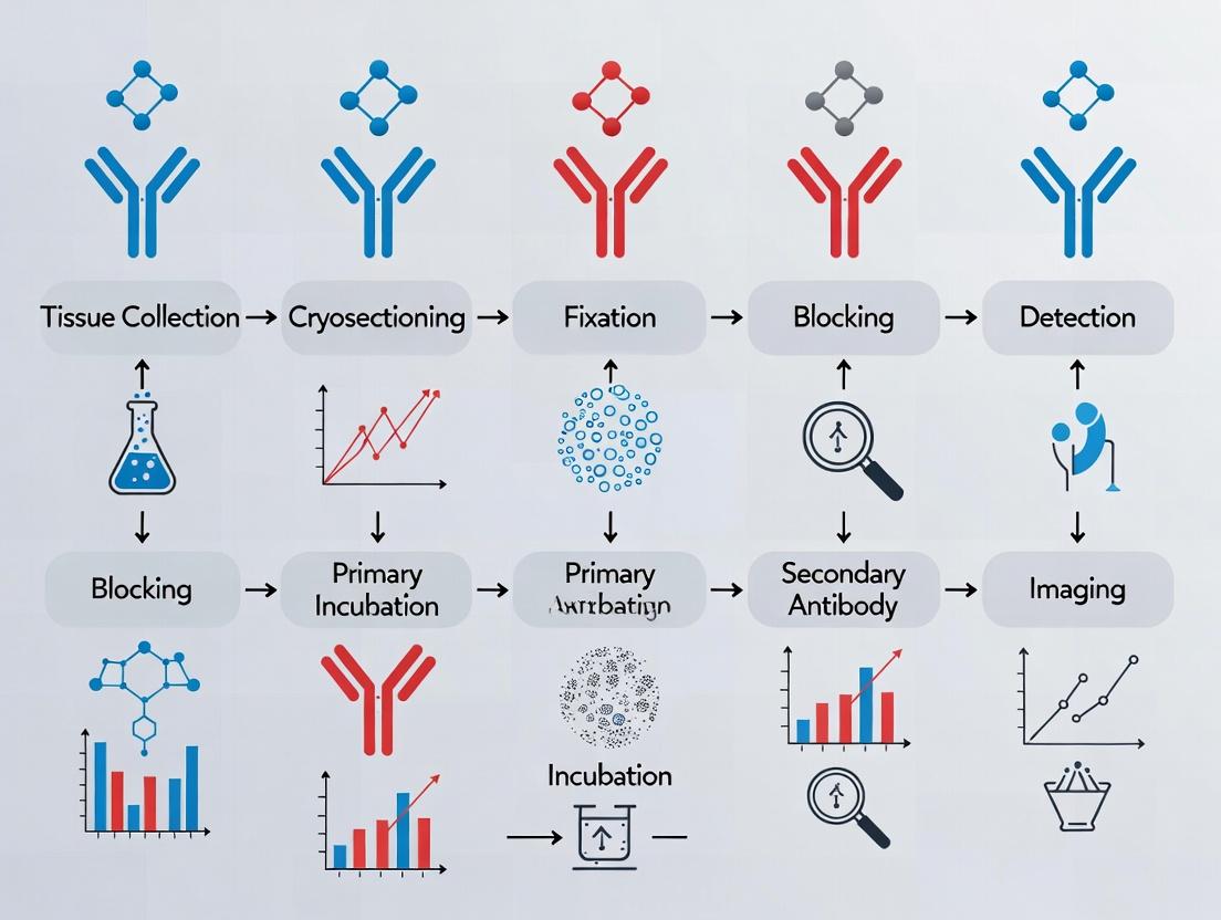
Frozen Tissue IHC Workflow
The Scientist's Toolkit: Research Reagent Solutions
Table 2: Essential Materials for Frozen Tissue IHC
| Item | Function in Protocol | Key Consideration |
|---|---|---|
| Optimal Cutting Temperature (O.C.T.) Compound | Water-soluble embedding matrix for supporting tissue during cryosectioning. | Ensure it is compatible with your antigens; some formulations can interfere. |
| Isopentane (2-Methylbutane) | Intermediate cryogen for rapid, crack-free freezing. Prevents ice crystal damage. | Must be pre-cooled by liquid nitrogen to a slushy state. |
| Phospho-Specific Validated Primary Antibodies | Bind specifically to the phosphorylated form of the target protein. | Must be validated for IHC on frozen tissue. Check species reactivity and phosphorylation site. |
| Charged or Adhesive Glass Slides | Provide electrostatic or coated adhesion for tissue sections, preventing wash-off. | Critical for avoiding section loss during rigorous IHC washes. |
| Polymer-Based HRP Detection System | Amplifies the primary antibody signal, increasing sensitivity. Essential for low-abundance phospho-targets. | Lower background than traditional avidin-biotin systems. Choose species-appropriate polymer. |
| Cold Acetone (-20°C) | Fixative that precipitates proteins while preserving many labile epitopes. | Provides good balance between morphology and antigenicity for frozen sections. |
| Protein Block (e.g., BSA/Normal Serum) | Reduces non-specific binding of antibodies to hydrophobic or charged sites on tissue. | Use serum from the same species as the secondary antibody for best results. |
| Antifade Mounting Medium with DAPI | Preserves fluorescence (for IF) and counterstains nuclei. | For chromogenic IHC (DAB), use a non-aqueous, permanent mounting medium. |
This application note, framed within a broader thesis on IHC protocol optimization for frozen tissue sections, delineates the critical trade-offs between speed, antigenicity preservation, and morphological integrity. For researchers in drug development, navigating these trade-offs is paramount for experimental validity and throughput.
Quantitative Comparison of IHC Methodologies
Table 1: Comparative Analysis of IHC on Frozen vs. Formalin-Fixed Paraffin-Embedded (FFPE) Sections
| Parameter | Frozen Sections (FS-IHC) | FFPE Sections (FFPE-IHC) | Notes / Quantitative Range |
|---|---|---|---|
| Speed (Protocol Duration) | Very Fast (High) | Slow (Low) | FS-IHC: 4-6 hrs total. FFPE-IHC: 24-48+ hrs (incl. deparaffinization, retrieval). |
| Antigenicity Preservation | High | Variable to Low | FS-IHC: No cross-linking; ~95%+ epitope retention. FFPE-IHC: Cross-linking masks epitopes; retrieval recovers 60-90%. |
| Morphological Quality | Low to Moderate | Very High | FS-IHC: Tissue ice-crystal artifacts; nuclear detail compromised. FFPE-IHC: Excellent cellular and architectural detail. |
| Long-term Storage | Requires -80°C | Room Temperature (Stable) | FS: Long-term storage affects antigenicity. FFPE: Decades-long stability. |
| Required Antigen Retrieval | Rarely | Almost Always | FS-IHC: 0-10% of targets need retrieval. FFPE-IHC: >95% require heat/ enzymatic retrieval. |
| Protocol Flexibility | High | Moderate | FS-IHC: Suitable for lipids, labile antigens. FFPE-IHC: Limited for sensitive epitopes. |
Detailed Protocols
Protocol 1: Rapid Fluorescent IHC on Frozen Sections (FS-IHC) for Labile Antigens
Objective: To maximize speed and antigenicity for phosphorylation-dependent epitopes with acceptable morphology. Materials: See "Research Reagent Solutions" (Table 2). Procedure:
- Tissue Preparation: Snap-freeze fresh tissue in OCT compound in a dry ice/isopentane bath. Store at -80°C.
- Sectioning: Cut 5-10 µm sections on a cryostat (-20°C). Mount on charged slides. Air-dry for 30 min.
- Fixation: Immerse in pre-chilled acetone at -20°C for 10 min. Air-dry for 5 min.
- Washing: Rinse 3x in PBS (pH 7.4), 5 min each.
- Permeabilization & Blocking: Incubate with blocking buffer (5% normal serum, 0.3% Triton X-100 in PBS) for 1 hr at RT.
- Primary Antibody Incubation: Apply diluted primary antibody in antibody dilution buffer (1% BSA, 0.1% Triton X-100 in PBS). Incubate for 1 hr at RT or overnight at 4°C.
- Washing: Wash 3x with PBS + 0.05% Tween-20 (PBST), 5 min each.
- Secondary Antibody Incubation: Apply fluorophore-conjugated secondary antibody (1:500) in dilution buffer. Incubate for 1 hr at RT in the dark.
- Washing: Wash 3x with PBST, 5 min each in the dark.
- Counterstaining & Mounting: Apply DAPI (300 nM in PBS) for 5 min. Wash briefly. Mount with aqueous anti-fade mounting medium.
- Imaging: Image immediately using a fluorescence microscope.
Protocol 2: Optimized Sequential IHC for FFPE Sections (FFPE-IHC)
Objective: To achieve superior morphology and multiplexing capability, accepting longer protocol time. Materials: See "Research Reagent Solutions" (Table 2). Procedure:
- Sectioning & Deparaffinization: Cut 4-5 µm sections. Bake at 60°C for 30 min. Deparaffinize in xylene (2x, 10 min) and rehydrate through graded ethanol (100%, 95%, 70%) to distilled water.
- Antigen Retrieval: Perform heat-induced epitope retrieval (HIER) in 10 mM sodium citrate buffer (pH 6.0) or Tris-EDTA (pH 9.0) using a pressure cooker (15 min at full pressure) or water bath (95°C for 40 min). Cool for 30 min.
- Washing & Blocking: Wash in PBS. Block endogenous peroxidase with 3% H₂O₂ in PBS for 15 min (if using HRC). Wash. Block with protein block (e.g., 5% BSA) for 30 min.
- Primary Antibody: Apply primary antibody in diluent. Incubate 1 hr at RT or overnight at 4°C.
- Detection: For chromogenic detection, apply appropriate HRP/DAB or AP/Vector kits per manufacturer's instructions. For fluorescent, follow steps 7-10 from Protocol 1, adjusting buffers.
- Counterstaining & Mounting: Counterstain with Hematoxylin (chromogenic) or DAPI (fluorescent). Dehydrate (chromogenic only), clear, and mount with permanent mounting medium.
Visualization of IHC Method Selection Logic
IHC Method Selection Decision Tree
The Scientist's Toolkit: Research Reagent Solutions
Table 2: Essential Materials for IHC Protocol Optimization
| Item | Function & Rationale | Example Product/Catalog |
|---|---|---|
| OCT Compound | Optimal Cutting Temperature medium; water-soluble embedding matrix for frozen tissue, provides support during cryosectioning. | Tissue-Tek O.C.T. Compound |
| Charged/Plus Slides | Microscope slides with a permanent positive charge; enhances adhesion of tissue sections, critical for FS-IHC to prevent detachment. | Fisherbrand Superfrost Plus |
| Heat-Induced Epitope Retrieval (HIER) Buffer | Standardized buffer (e.g., citrate pH 6.0, Tris-EDTA pH 9.0) to reverse formaldehyde cross-linking and expose masked epitopes in FFPE tissue. | Citrate Buffer (10X), Abcam |
| Fluorophore-Conjugated Secondary Antibody | Highly cross-adsorbed antibody raised against host species of primary antibody, conjugated to a fluorescent dye (e.g., Alexa Fluor 488, 594). | Donkey Anti-Rabbit IgG (H+L) Alexa Fluor 594 |
| Chromogenic Detection Kit (HRP/DAB) | Contains enzyme (Horseradish Peroxidase)-conjugated secondary antibody and 3,3'-Diaminobenzidine (DAB) substrate to produce a brown, permanent precipitate. | ImmPRESS HRP Horse Anti-Rabbit IgG Polymer Kit, Vector Labs |
| Aqueous Anti-fade Mounting Medium | Preserves fluorescence intensity during microscopy and storage by reducing photobleaching; often contains DAPI for nuclear counterstain. | ProLong Gold Antifade Mountant with DAPI, Thermo Fisher |
| Protein Block (Serum/BSA) | Reduces non-specific background staining by blocking sites of hydrophobic or ionic interaction between tissue and detection reagents. | Normal Donkey Serum, Bovine Serum Albumin (BSA) |
| Permeabilization Agent (Triton X-100/Tween-20) | Detergent that solubilizes cell membranes, allowing antibodies to access intracellular targets. Concentration is critical for morphology. | Triton X-100, Tween-20 |
Application Notes
Within the context of immunohistochemistry (IHC) on frozen tissue sections, the triumvirate of a precision cryostat, optimal cutting temperature (OCT) compound, and specialized fixatives forms the foundational pillar for preserving antigenicity and tissue morphology. Frozen sections circumvent heat-induced antigen retrieval, making them indispensable for labile epitopes, but introduce unique challenges in handling and stabilization. The choice of OCT compound directly impacts section adhesion and embedding stability, while specialized precipitating fixatives, such as paraformaldehyde (PFA) and acetone, provide a critical balance between structural fixation and epitope preservation, which is paramount for accurate qualitative and quantitative analysis in drug development research.
Protocols
Protocol 1: Optimal Tissue Embedding and Sectioning for IHC
Objective: To produce high-quality, adherent frozen tissue sections with optimal morphology for downstream IHC staining.
Materials:
- Fresh or fixed tissue specimen (< 5mm thick)
- Isopentane (2-methylbutane), cooled in liquid nitrogen bath
- Cryostat (e.g., Leica CM1950, Thermo Fisher Scientific CryoStar NX70)
- Optimal Cutting Temperature (OCT) Compound
- Disposable cryomolds
- Fine forceps, pre-cooled
- Superfrost Plus or charged adhesion slides
- Dry ice
Methodology:
- Preparation: Pre-cool the cryostat chamber to -20°C to -22°C. Equilibrate OCT compound and cryomolds on dry ice.
- Embedding: a. Place a small amount of OCT into the bottom of a cryomold. b. Using pre-cooled forceps, orient the tissue specimen in the mold. c. Completely fill the mold with OCT, ensuring the tissue is submerged and free of bubbles. d. Rapidly freeze the block by partially submerging the mold in a slurry of isopentane cooled by liquid nitrogen for 30-60 seconds. Do not submerge directly into liquid nitrogen. e. Transfer the frozen block to dry ice or a -80°C freezer for long-term storage.
- Sectioning: a. Secure the frozen block to the cryostat chuck using a layer of OCT. b. Trim the block face at a thickness of 20-30 µm until the full tissue face is exposed. c. Set the section thickness to 4-7 µm for IHC. d. Cut sections smoothly and consistently. Use an anti-roll plate or fine brush to guide the section onto the chilled chamber's stage. e. Thaw-mount the section onto a room-temperature charged slide by gently touching the slide to the section. The section will adhere and flatten immediately. f. Air-dry the mounted section for 30-60 minutes before fixation or storage at -80°C.
Protocol 2: Fixation of Frozen Sections for Epitope Preservation
Objective: To stabilize tissue architecture while retaining maximum antigenicity for antibody binding.
Materials:
- Acetone (pre-cooled to -20°C)
- 4% Paraformaldehyde (PFA) in PBS, pH 7.4
- Phosphate-Buffered Saline (PBS)
- Humidity chamber
- Coplin jars or staining racks
Methodology:
- Acetone Fixation (for most cell surface and cytoplasmic antigens): a. Immerse air-dried slides in pre-cooled (-20°C) acetone for 10 minutes. b. Air-dry the slides for 5-10 minutes. c. Proceed directly to IHC staining protocol or rehydrate in PBS for 5 minutes before applying blocking serum.
- Paraformaldehyde Fixation (for better structural preservation): a. Immerse air-dried slides in 4% PFA at room temperature for 10 minutes. b. Wash slides in three changes of PBS, 5 minutes each, to remove all traces of fixative. c. Proceed to IHC staining protocol.
- Combined Fixation (for challenging antigens): a. Fix slides in 4% PFA for 5 minutes at room temperature. b. Wash briefly in PBS. c. Post-fix in pre-cooled acetone for 5 minutes. d. Wash in PBS and proceed.
Data Presentation
Table 1: Comparison of Common Fixatives for Frozen Section IHC
| Fixative Type | Concentration & Conditions | Incubation Time | Primary Antigen Targets | Key Advantages | Key Disadvantages |
|---|---|---|---|---|---|
| Acetone | 100%, -20°C | 10 min | Cell surface markers, cytoplasmic proteins, many phospho-epitopes | Excellent epitope retention; permeabilizes membranes; fast. | Poor morphological detail; dehydrates tissue; highly flammable. |
| Paraformaldehyde (PFA) | 4% in PBS, RT | 10 min | Structural proteins, nuclear antigens, some membrane proteins | Superior morphology; cross-links and stabilizes tissue. | Can mask epitopes; may require mild antigen retrieval. |
| Methanol | 100%, -20°C | 10 min | Viruses, some nuclear antigens | Good permeabilization; less denaturing than acetone for some epitopes. | Can be harsh on morphology; less commonly used than acetone. |
| Acetone: Methanol (1:1) | -20°C | 5-10 min | Broad range, esp. in fluorescence | Balanced permeabilization and fixation. | Empirical optimization needed. |
Diagrams
Title: Workflow for Frozen Tissue Section Preparation and Fixation
Title: Mechanism of Action: PFA vs. Acetone Fixation
The Scientist's Toolkit
Table 2: Essential Research Reagent Solutions for Frozen Section IHC
| Item | Function & Rationale |
|---|---|
| Cryostat | A refrigerated microtome that maintains tissue blocks at sub-zero temperatures (typically -20°C) during sectioning, preventing thawing and ensuring thin, consistent sections. |
| OCT Compound | A water-soluble embedding matrix composed of polyvinyl alcohol and polyethylene glycol. It provides structural support during freezing and sectioning, and is easily washed away during staining. |
| Charged/Adhesion Slides | Microscope slides with a positively charged coating that electrostatically binds negatively charged tissue sections, preventing detachment during rigorous IHC washes. |
| Pre-cooled Acetone | A precipitating fixative that denatures proteins, preserves many epitopes, and permeabilizes cell membranes by dissolving lipids, facilitating antibody penetration. |
| 4% Paraformaldehyde (PFA) | A cross-linking fixative that creates methylene bridges between proteins, providing excellent structural preservation but potentially masking epitopes. |
| Isopentane | A liquid with high thermal conductivity and a freezing point of -160°C. Used as an intermediate coolant for rapid, uniform tissue freezing without crack artifacts caused by direct liquid nitrogen immersion. |
| Cryogenic Vials & Boxes | Airtight, durable containers designed for long-term storage of frozen tissue blocks at -80°C or in liquid nitrogen, preventing freeze-drying and contamination. |
| Anti-Roll Plate/Guide | A critical cryostat accessory that flattens the section as it is cut, preventing it from curling and enabling the collection of intact, wrinkle-free ribbons. |
Tissue Acquisition and Snap-Freezing Best Practices for Optimal Preservation
Within the broader thesis on optimizing Immunohistochemistry (IHC) protocols for frozen tissue sections, the initial steps of tissue acquisition and preservation are paramount. The quality of data from IHC staining for protein localization, expression, and post-translational modifications is directly contingent upon the rapid inhibition of enzymatic degradation and the preservation of native tissue architecture and antigenicity. These application notes detail the protocols and best practices for snap-freezing to ensure optimal biomolecular preservation for downstream frozen section IHC analysis.
Key Factors Influencing Preservation Quality
The following table summarizes critical quantitative and qualitative parameters that impact tissue preservation efficacy, based on current literature and experimental data.
Table 1: Critical Parameters for Snap-Freezing Preservation
| Parameter | Optimal Target | Impact on Downstream IHC |
|---|---|---|
| Ischemia Time (Warm/Cold) | < 1 minute (ideal); < 10 minutes (acceptable) | Extended ischemia induces hypoxia-related protein degradation and antigen modification. |
| Tissue Dimension | ≤ 0.5 cm thickness; 1 cm max dimension | Thinner sections enable rapid, uniform heat transfer, preventing ice crystal formation. |
| Freezing Medium | Optimal Cutting Temperature (O.C.T.) compound or 2-Methylbutane (Isopentane) chilled | O.C.T. supports sectioning; isopentane provides rapid, non-cryoprotective freezing. |
| Freezing Bath Temperature | -40°C to -70°C (Isopentane); -50°C or below (Liquid N₂ vapor phase) | Minimizes the Leidenfrost effect for faster freezing. |
| Storage Temperature | -80°C or liquid nitrogen (-196°C) | Prevents recrystallization and ice crystal growth over time. |
| Freezing Rate | 50-100°C/second (for <1mm samples) | Maximizes vitreous (glassy) ice formation, minimizing structural damage. |
Detailed Experimental Protocols
Protocol A: Isopentane Bath Snap-Freezing (Gold Standard for Histology)
This method provides the fastest cooling rate for medium-sized samples, minimizing ice crystal artifacts.
Materials:
- Freshly excised tissue (< 0.5 cm thick).
- Pre-labeled cryomold or aluminum foil boat.
- O.C.T. compound (optional, for direct embedding).
- 2-Methylbutane (Isopentane), 200-300 mL.
- Liquid nitrogen dewar.
- Insulated container (e.g., small foam box).
- Forceps, dissection tools.
- Cryogloves and face shield.
Methodology:
- Preparation: In a fume hood, pour isopentane into a metal beaker or heavy-walled glass container. Place this container into an insulated box. Slowly add liquid nitrogen to the outer chamber until the isopentane becomes viscous and forms a slush (approx. -40°C to -50°C). Do not allow isopentane to freeze solid.
- Tissue Preparation: Trim tissue on a chilled dissection plate (on ice) to dimensions not exceeding 0.5 cm in any direction. Blot gently to remove excess blood/moisture.
- Embedding Option A (Direct Freezing): Using pre-cooled forceps, immerse the tissue specimen directly into the chilled isopentane bath. Agitate gently for 15-30 seconds (depending on size).
- Embedding Option B (O.C.T. Embedding): Place a small amount of O.C.T. in a cryomold. Position tissue and cover with more O.C.T. Immediately submerge the entire mold into the isopentane bath until fully frozen (whitish appearance, ~30-60 seconds).
- Transfer: Quickly transfer the frozen tissue block to a pre-cooled cryovial or bag. Immediately place on dry ice or directly into a -80°C freezer for temporary storage. For long-term archival (>6 months), store in liquid nitrogen.
Protocol B: Liquid Nitrogen Vapor Phase Freezing (for Small/Fragile Tissues)
A safer alternative suitable for small biopsies and fragile tissues.
Methodology:
- Preparation: Fill a wide-mouth liquid nitrogen dewar to generate a thick vapor phase. A floating foam insert or perforated platform can hold samples above the liquid.
- Tissue Preparation: As in Protocol A, prepare tissue and embed in O.C.T. in a cryomold.
- Freezing: Suspend the cryomold in the vapor phase (approximately 10-15 cm above the liquid nitrogen level) for 5-10 minutes until completely frozen.
- Storage: Transfer to pre-labeled, pre-cooled containers and store at -80°C or in liquid nitrogen.
Visualizations
Title: Tissue Snap-Freezing Decision Workflow
Title: Ischemia & Preservation Impact on IHC Antigens
The Scientist's Toolkit: Essential Research Reagents & Materials
Table 2: Key Reagent Solutions for Tissue Snap-Freezing
| Item | Function & Rationale |
|---|---|
| Optimal Cutting Temperature (O.C.T.) Compound | A water-soluble glycol and resin embedding medium. Provides structural support for frozen tissue during cryosectioning, minimizing fragmentation. |
| 2-Methylbutane (Isopentane) | An intermediate freezing bath coolant with high thermal conductivity and low freezing point. Prevents the insulating vapor layer (Leidenfrost effect) that occurs with direct liquid nitrogen plunging, enabling faster cooling rates. |
| Liquid Nitrogen | Primary coolant for creating vapor phase freezing environments or for long-term storage at -196°C. Essential for halting all biochemical activity indefinitely. |
| RNA/DNA Stabilization Buffers (e.g., RNAlater) | Optional pre-freezing immersion for specific multi-omics studies. Preserves nucleic acids but can diffuse proteins; not recommended for proteomics/IHC-focused work. |
| Cryovials & Specimen Bags | Pre-labeled, durable containers for -80°C or liquid nitrogen storage. Must be leak-proof and resistant to extreme temperatures and cracking. |
| Chilled Dissection Buffer/Saline | Used to briefly rinse and keep tissue moist during grossing. Prevents desiccation but excess fluid should be blotted to avoid large ice lenses. |
This application note details the fundamental principles governing antibody-antigen interactions within Immunohistochemistry (IHC), with specific focus on epitope considerations for frozen tissue sections. Framed within a broader thesis on IHC protocol optimization for frozen tissues, this guide provides researchers, scientists, and drug development professionals with the theoretical and practical foundations necessary for robust experimental design and interpretation. Mastery of these principles is critical for minimizing artifacts and maximizing specificity in translational research.
Fundamental Principles of Antibody-Antigen Binding
The specificity of IHC relies entirely on the precise molecular interaction between an antibody's paratope and the antigen's epitope. For frozen sections, where antigen preservation is high but tissue morphology is more labile, understanding these dynamics is paramount.
Key Binding Forces:
- Non-covalent Interactions: Include hydrogen bonds, ionic interactions, van der Waals forces, and hydrophobic interactions. The sum of these weak forces, operating over a short distance (approximately 1 Å), results in high-affinity, specific binding.
- Affinity vs. Avidity: Affinity is the strength of a single paratope-epitope interaction. Avidity is the total binding strength of a multivalent antibody (e.g., IgG has two paratopes) to multiple epitopes on a target. High avidity can compensate for moderate affinity, enhancing signal retention during stringent washes.
Quantitative Binding Metrics: The following parameters, derived from live searches of current biosensor and surface plasmon resonance (SPR) literature, are crucial for antibody characterization.
Table 1: Key Quantitative Parameters for Antibody-Antigen Interaction
| Parameter | Symbol | Typical Range for IHC | Description & Impact on IHC |
|---|---|---|---|
| Dissociation Constant | KD | 1 nM - 10 pM (optimal) | Concentration of antigen at which half the antibody binding sites are occupied. Lower KD indicates higher affinity. Critical for determining optimal antibody dilution. |
| Association Rate Constant | kon | 10^3 - 10^6 M-1s-1 | Speed of complex formation. A higher kon can improve labeling efficiency. |
| Dissociation Rate Constant | koff | 10^-1 - 10^-4 s-1 | Stability of the formed complex. A lower koff ensures the complex withstands wash steps, reducing background. |
| Antibody Working Concentration | - | 0.5 - 10 μg/mL | Directly related to KD. Must be optimized empirically for each antibody-lot and tissue type. |
Epitope Characteristics and Their Implications
Epitopes, the specific regions of an antigen recognized by an antibody, are classified by their structure and composition, which directly impacts detection in frozen tissues.
- Linear (Continuous) Epitopes: Comprise a continuous sequence of amino acids (typically 5-7 residues). These epitopes are often resistant to formalin fixation and denaturation, making them detectable in paraffin-embedded tissues after antigen retrieval. In frozen tissues, they are fully accessible.
- Conformational (Discontinuous) Epitopes: Formed by spatially adjacent amino acids brought together by protein folding. These are highly dependent on the native tertiary/quaternary structure of the protein. They are exquisitely preserved in frozen sections (which are not cross-linked by fixatives) but can be destroyed by denaturing conditions.
Critical Consideration for Frozen Sections: The lack of cross-linking fixatives in frozen tissue protocols preserves conformational epitopes exceptionally well. This is a major advantage for detecting native protein structures but necessitates gentle processing to prevent denaturation from other sources (e.g., harsh solvents, excessive heat).
Protocol: Validating Antibody Specificity and Epitope Integrity in Frozen Sections
This protocol outlines steps to confirm that an antibody is binding its intended target with high specificity in the context of frozen tissue morphology.
Aim: To verify the specificity of antibody binding and assess epitope preservation in fresh-frozen tissue sections.
Materials: The Scientist's Toolkit
Table 2: Essential Research Reagent Solutions for IHC Validation
| Reagent / Material | Function & Rationale |
|---|---|
| Optimal Cutting Temperature (OCT) Compound | A water-soluble embedding medium that provides structural support for cryosectioning without chemically altering antigens. |
| Cryostat | Instrument to cut thin (4-10 μm) sections of frozen tissue at controlled temperatures (typically -20°C). |
| Poly-L-lysine or Plus-coated Slides | Provide positive charge to enhance electrostatic adhesion of tissue sections, preventing detachment during rigorous validation washes. |
| Phosphate-Buffered Saline (PBS), pH 7.4 | Isotonic buffer used for all washes and dilutions to maintain physiological pH and osmolarity. |
| Blocking Solution (e.g., 5% Normal Serum / 1% BSA) | Reduces non-specific background staining by occupying hydrophobic or charged sites on the tissue and slide. Serum should be from the host species of the secondary antibody. |
| Primary Antibody of Interest | The key reagent. Clone, host species, and recommended application (IHC on frozen sections) must be verified. |
| Validated Positive Control Tissue | Tissue known to express the target antigen at moderate levels. Confirms protocol and antibody functionality. |
| Isotype Control Antibody | An immunoglobulin of the same class/subclass (e.g., IgG1, kappa) but irrelevant specificity. Critical for distinguishing specific signal from non-specific Fc receptor or charged interaction binding. |
| Competitive Peptide Block | Synthetic peptide matching the exact immunogen sequence used to generate the antibody. The gold standard for confirming specificity. |
| Epitope Retrieval Buffer (Citrate, pH 6.0) | While often used for FFPE, a mild retrieval step may be tested for frozen sections if denaturation is suspected, though it is not standard. |
Methodology:
Tissue Preparation & Sectioning:
- Embed fresh tissue specimen in OCT compound and rapidly freeze in isopentane cooled by liquid nitrogen.
- Cut 5-8 μm serial sections using a cryostat and mount onto charged slides.
- Air-dry slides for 30-60 minutes to improve adhesion.
- Fix sections in pre-chilled acetone or 4% paraformaldehyde (PFA) for 10 minutes. (Note: Acetone preserves most conformational epitopes; 4% PFA may cross-link and mask some).
- Wash in PBS for 5 minutes, three times.
Validation Staining Procedure (Serial Sections):
- Section 1: Primary Antibody (Test). Apply optimized dilution of primary antibody in PBS/1% BSA. Incubate in a humidified chamber for 1 hour at room temperature or overnight at 4°C.
- Section 2: Isotype Control. Apply the matching isotype control at the same concentration as the primary antibody.
- Section 3: Peptide Blocking Control. Pre-incubate the primary antibody with a 5-10 fold molar excess of the immunizing peptide for 1 hour at room temperature. Apply this pre-adsorbed antibody mixture to the section.
- Section 4: Secondary Antibody Only (Autofluorescence Control). Apply only the labeled secondary antibody.
- Section 5: Positive Control Tissue. Stain known positive tissue with the primary antibody to confirm protocol performance.
Detection & Analysis:
- Following primary incubation, wash all slides 3 x 5 minutes in PBS.
- Apply appropriate fluorophore- or enzyme-conjugated secondary antibody for 30-45 minutes at room temperature, protected from light.
- Wash thoroughly 3 x 5 minutes in PBS.
- Apply counterstain (e.g., DAPI) and mounting medium.
- Image using a microscope with consistent settings across all control slides.
Interpretation:
- A valid specific signal is present in Section 1 (Test) and Section 5 (Positive Control), but is absent or drastically reduced in Section 2 (Isotype Control), Section 3 (Peptide Block), and Section 4 (Secondary Only).
- Persistent signal in the peptide block control indicates non-specific binding, and the antibody is not suitable for specific detection.
Schematic Representations
Diagram 1: Antibody Paratope Binds Antigen Epitope
Diagram 2: Frozen Section IHC Workflow with Controls
Successful IHC on frozen tissue sections hinges on a deep understanding of antibody-antigen kinetics and epitope behavior. The conformational integrity of antigens in frozen tissues is both an advantage and a responsibility, requiring careful antibody validation and gentle processing. By adhering to the fundamental principles and rigorous validation protocols outlined here, researchers can generate reliable, interpretable data critical for drug target validation, biomarker discovery, and mechanistic studies in translational research.
Step-by-Step Frozen Section IHC Protocol: From Sectioning to Detection
Within the broader thesis on optimizing immunohistochemistry (IHC) protocols for frozen tissue sections, cryostat sectioning represents the most critical pre-analytical step. The quality of sections directly dictates antigen preservation, staining specificity, and signal-to-noise ratio in subsequent IHC. This document details application notes and protocols for obtaining optimal frozen sections, focusing on the interdependent variables of thickness, temperature, and mounting.
Quantitative Parameters for Optimal Sectioning
The following tables summarize key quantitative data for cryostat sectioning variables.
Table 1: Recommended Section Thickness for Common Applications
| Tissue Type / Target Application | Optimal Thickness (µm) | Rationale |
|---|---|---|
| Standard IHC / General Morphology | 5 - 10 | Balances structural integrity with antibody penetration. |
| Fluorescence IHC (Multiple labels) | 4 - 8 | Reduces autofluorescence & overlay, improves resolution. |
| Lipid-Rich Tissue (e.g., Brain, Adipose) | 10 - 20 | Prevents crumbling; thicker sections often required. |
| Single-Cell RNA/DNA Analysis (on slide) | 10 - 15 | Ensures sufficient nucleic acid material per cell. |
| Enzyme Histochemistry | 8 - 15 | Accommodates reaction product formation. |
Table 2: Optimal Temperatures for Sectioning Various Tissues
| Tissue Type | Chamber Temp (°C) | Object Temp (°C) | Blade Temp (°C) | Notes |
|---|---|---|---|---|
| Rodent Brain (Perfusion Fixed) | -18 to -20 | -16 to -18 | -18 to -20 | Colder temps reduce knife marks. |
| Human Tumor (Snap-Frozen) | -20 to -24 | -18 to -22 | -20 to -22 | Dense tissue requires lower temps. |
| Spleen / Lymph Node | -18 to -20 | -16 to -18 | -18 | Too cold induces shattering. |
| Adipose / Breast Tissue | -25 to -30 | -22 to -26 | -25 to -28 | Prevents smearing and tear-out. |
| Liver / Kidney | -18 to -22 | -16 to -20 | -18 to -20 | Standard range for most organs. |
Detailed Protocols
Protocol A: Standard Cryostat Sectioning for IHC
Objective: To produce wrinkle-free, flat sections of optimal thickness for IHC staining.
- Preparation: Pre-cool cryostat chamber to desired temperature (see Table 2). Clean chamber with 70% ethanol. Install a clean, anti-roll guide or use a disposable blade holder with a fresh blade.
- Sample Mounting: Using Optimal Cutting Temperature (OCT) compound, adhere the frozen tissue block to a specimen disc. Ensure the cutting surface is parallel to the disc. Allow OCT to freeze completely.
- Trimming: Mount the specimen disc. Set section thickness to 20-30 µm and trim the block face until the full tissue surface is exposed.
- Sectioning: Adjust to final thickness (5-10 µm). Engage the anti-roll guide. Turn the handwheel slowly and steadily to advance the block. A complete section will slide across the blade.
- Mounting (Thaw-Mounting): Bring a room-temperature, charged or positively charged microscope slide close to, but not touching, the section. Gently touch the slide to the section, which will instantly adhere via electrostatic and Van der Waals forces.
- Post-Mounting: Immediately place the slide in a slide rack. Air-dry for 30-60 minutes at room temperature, then proceed to fixation or store at -80°C.
Protocol B: Mounting for Fragile or Lipid-Rich Tissues
Objective: To successfully mount sections prone to shattering or folding.
- Follow Protocol A steps 1-4, using temperatures at the colder end of the range.
- Static Electricity Mitigation: Use an anti-static device (ionizer) near the cryostat. Wipe slides with a dryer sheet prior to use.
- Mounting (Slide-Lowering): Hold a room-temperature slide above, and parallel to, the section on the blade. Slowly lower the slide vertically onto the section, allowing it to contact from one edge to the other, minimizing air pockets.
- Immediate Fixation: Immediately after the section adheres, immerse the slide in pre-chilled acetone or methanol (at -20°C) for 10 minutes. This stabilizes lipids and proteins before drying artifacts occur.
Visualizations
Title: Cryostat Sectioning Workflow for IHC
Title: How Sectioning Quality Impacts IHC Results
The Scientist's Toolkit: Research Reagent Solutions
Table 3: Essential Materials for Frozen Section IHC
| Item | Function & Rationale |
|---|---|
| Optimal Cutting Temperature (OCT) Compound | Water-soluble embedding medium that freeces to support tissue structure during sectioning. Must be clear and non-fluorescent for imaging. |
| Cryostat (with Anti-Roll Guide) | Precision instrument to cut thin sections from frozen tissue. Anti-roll guide prevents curling of sections. |
| Positively Charged Microscope Slides | Slides coated with positively charged polymers (e.g., poly-L-lysine) to enhance electrostatic adhesion of tissue sections, preventing detachment during staining. |
| Disposable Microtome Blades | High-profile blades for clean, consistent cuts. Disposable to prevent cross-contamination and ensure sharpness. |
| Cryostat Cleaning Solution (e.g., 70% Ethanol) | For decontaminating the chamber and tools to prevent RNase/DNase activity and cross-contamination between samples. |
| Desiccant (for Slide Storage) | Silica gel packs used in storage boxes at -80°C to prevent moisture accumulation and ice crystal formation on slides. |
| Anti-Static Device / Dryer Sheets | Neutralizes static charge that causes sections to curl, fly away, or cling unpredictably during mounting. |
Within the broader thesis on optimizing IHC protocols for frozen tissue sections, the choice of fixative is a critical initial determinant of experimental success. Unlike formalin-fixed, paraffin-embedded (FFPE) tissues, frozen sections offer the advantage of preserving antigenicity but require careful fixation to maintain morphology while avoiding epitope masking. This application note provides a comparative analysis of acetone, methanol, and paraformaldehyde (PFA) fixation, guiding researchers in selecting the optimal strategy based on their target antigen and experimental goals.
Comparative Analysis of Fixatives
The mechanism of action, advantages, and limitations of each fixative are summarized below.
Table 1: Mechanism and Primary Use of Common Fixatives
| Fixative | Primary Mechanism | Primary Use Case for Frozen Sections |
|---|---|---|
| Acetone | Organic solvent; dehydrates and precipitates proteins. | Excellent for many cell surface markers, transcription factors, and phosphorylated epitopes. Rapid. |
| Methanol | Organic solvent; dehydrates, precipitates proteins, and can permeabilize membranes. | Suitable for intracellular antigens, viral proteins, and some nuclear targets. Can reduce autofluorescence. |
| Paraformaldehyde (PFA) | Cross-linking agent; forms methylene bridges between proteins, "locking" structure. | Superior preservation of fine cellular morphology and subcellular architecture. Essential for many structural proteins. |
Table 2: Quantitative Comparison of Fixative Protocols
| Parameter | Acetone | Methanol | PFA (4%) |
|---|---|---|---|
| Typical Concentration | 100% (cold, -20°C) | 100% (cold, -20°C) | 2-4% in PBS (room temp or 4°C) |
| Fixation Time | 5-15 minutes | 10-15 minutes | 10-30 minutes |
| Permeabilization Required? | Often no, as it permeabilizes. | Often no, as it permeabilizes. | Yes, typically with 0.1-0.5% Triton X-100. |
| Antigen Retrieval Needed? | Rarely | Rarely | Frequently, for cross-linked/epitope-masked targets. |
| Key Advantage | Speed; high antigenicity preservation for labile epitopes. | Good morphology; some autofluorescence reduction. | Best morphology preservation. |
| Primary Limitation | Poor ultrastructural detail; can make tissue brittle. | Can alter protein conformation; may destroy some epitopes. | Potential for epitope masking; requires more steps. |
Detailed Experimental Protocols
Protocol 1: Acetone Fixation for Phospho-Protein Detection (e.g., p-STAT3)
Objective: To preserve labile phosphorylation epitopes in frozen lung tissue sections. Materials: See "The Scientist's Toolkit" below. Procedure:
- Cut 5-10 µm thick frozen sections and mount on charged slides. Air-dry for 20-30 minutes.
- Pre-chill 100% acetone to -20°C.
- Immerse slides in cold acetone for 10 minutes at -20°C.
- Remove slides and air-dry completely for 5-10 minutes.
- Proceed immediately to blocking and immunostaining. Do not perform additional permeabilization.
- Include a control slide fixed with 4% PFA for 20 minutes (with subsequent 0.25% Triton X-100 permeabilization for 10 min) to demonstrate the advantage of acetone for this target.
Protocol 2: PFA Fixation for Cytoskeletal Architecture (e.g., F-actin with Phalloidin)
Objective: To optimally preserve fine cellular structures in frozen brain tissue sections. Procedure:
- Cut 10-12 µm thick sections and mount. Air-dry briefly (5-10 minutes).
- Fix slides in freshly prepared, filtered 4% PFA in PBS (pH 7.4) for 15 minutes at room temperature.
- Rinse slides 3 x 5 minutes in PBS.
- Permeabilize and block in a solution of 1% BSA and 0.3% Triton X-100 in PBS for 45 minutes at room temperature.
- Proceed to staining with fluorescent phalloidin conjugate and primary antibodies.
- Include a control slide fixed with cold acetone for 10 minutes to compare morphological preservation.
Visualization of Fixative Selection Logic
Title: Fixative Selection Decision Tree for Frozen IHC
Title: Comparative IHC Workflow After Different Fixations
The Scientist's Toolkit
Table 3: Essential Reagents and Materials for Frozen Section IHC Fixation
| Item | Function/Benefit | Example/Note |
|---|---|---|
| Charged Microscope Slides | Prevents tissue detachment during harsh solvent fixation. | Superfrost Plus or equivalent. |
| Ultra-Pure Acetone (Molecular Biology Grade) | Ensures consistency; avoids impurities that cause background. | Pre-chill to -20°C for best results. |
| Paraformaldehyde, Crystalline | For fresh preparation of 4% PFA, avoiding formic acid contaminants in formalin. | Prepare in PBS, pH to 7.4, filter before use. |
| Methanol (HPLC or Anhydrous Grade) | High purity for consistent fixation and reduced artifact. | Store anhydrously; pre-chill. |
| Triton X-100 or Tween-20 | Detergent for permeabilization post-PFA fixation. | Use at 0.1-0.5% in PBS or blocking buffer. |
| Bovine Serum Albumin (BSA) or Normal Serum | Blocks non-specific antibody binding sites to reduce background. | Use at 1-5% in PBS; match serum to secondary antibody host. |
| Phosphate-Buffered Saline (PBS), 10X Stock | Isotonic buffer for tissue rinsing, diluting PFA, and preparing solutions. | Always dilute to 1X and check pH (7.2-7.6). |
| Humidified Staining Chamber | Prevents evaporation of reagents on slides during incubation steps. | Essential for maintaining consistent staining. |
Within the broader thesis on optimizing immunohistochemistry (IHC) protocols for frozen tissue sections, the steps of permeabilization and blocking are critical determinants of experimental success. These steps directly govern the signal-to-noise ratio by controlling antibody accessibility to intracellular epitopes while minimizing non-specific background staining. This application note details current best practices and protocols.
Key Principles and Quantitative Data
Table 1: Common Permeabilizing Agents and Their Effects on Frozen Sections
| Agent | Typical Concentration | Incubation Time | Mechanism | Primary Use Case | Key Consideration |
|---|---|---|---|---|---|
| Triton X-100 | 0.1% - 0.5% | 5-15 min (room temp) | Dissolves lipids in cell membranes. | General intracellular target access. | Can disrupt membrane morphology; avoid for membrane proteins. |
| Saponin | 0.05% - 0.1% | 20-30 min (room temp) | Cholesterol-binding, creates reversible pores. | Optimal for delicate epitopes or membrane protein preservation. | Pores reseal; must be included in all antibody/ wash buffers. |
| Tween 20 | 0.1% - 0.5% | 10-20 min (room temp) | Mild non-ionic detergent. | Light permeabilization or as wash buffer additive. | Weaker than Triton X-100; often used in combination. |
| Methanol | 100% (ice-cold) | 10 min (-20°C) | Precipitates proteins and dissolves lipids. | Strong fixation and permeabilization combined. | Can denature some epitopes; alters tissue morphology. |
| Digitonin | 0.001% - 0.01% | 10 min (room temp) | Cholesterol-specific, creates precise pores. | Selective permeabilization of plasma membrane only. | Costly; used for compartment-specific studies. |
Table 2: Common Blocking Agents and Their Efficacy for Background Reduction
| Blocking Agent | Typical Concentration | Target of Blocking | Recommended For | Incubation Time & Temperature |
|---|---|---|---|---|
| Normal Serum | 2-5% (v/v) | Non-specific Fc receptor interactions. | General use; species must match secondary host. | 30-60 min at room temp. |
| BSA (Bovine Serum Albumin) | 1-5% (w/v) | Hydrophobic and ionic interactions. | General protein block; often combined with serum. | 30-60 min at room temp. |
| Casein | 0.1-5% (w/v) | Hydrophobic interactions. | Phospho-specific antibodies; high background. | 30-60 min at room temp. |
| Fish Skin Gelatin | 0.1-2% (w/v) | Broad-spectrum protein block. | Reducing non-mammalian cross-reactivity. | 30-60 min at room temp. |
| Commercial Blocking Buffers | As per mfr. | Multi-target (proteins, carbohydrates, etc.). | Challenging tissues/antibodies. | As per manufacturer. |
| Glycine | 0.1 M | Free Aldehyde Groups | Post-aldehyde fixation quenching. | 5-10 min after fixation. |
Detailed Protocols
Protocol 1: Integrated Permeabilization and Blocking for Frozen Sections
This is a standard workflow following fixation of frozen sections with 4% PFA.
Materials:
- PBS (Phosphate-Buffered Saline), pH 7.4
- Permeabilization Buffer (0.3% Triton X-100 in PBS)
- Blocking Buffer (5% Normal Serum from secondary host species + 1% BSA in PBS)
Method:
- After fixation and PBS washes, apply Permeabilization Buffer to fully cover the tissue section.
- Incubate for 15 minutes at room temperature in a humidified chamber.
- Wash the slide 3 x 5 minutes with gentle agitation using PBS.
- Carefully tap off excess liquid and apply Blocking Buffer generously to the section.
- Incubate for 1 hour at room temperature in a humidified chamber.
- Do not wash. Proceed directly to application of primary antibody diluted in an appropriate buffer (often similar to blocking buffer).
Protocol 2: Saponin-Based Reversible Permeabilization for Labile Epitopes
Method:
- Fix tissue with 4% PFA for 10 minutes at room temperature. Wash with PBS.
- Prepare a Saponin-based Working Buffer (0.1% saponin, 1% BSA in PBS). Note: All subsequent steps require saponin-containing buffers.
- Apply Saponin Working Buffer as both permeabilization and blocking agent for 30 minutes at room temperature.
- Apply primary antibody diluted in Saponin Working Buffer.
- Perform all washes (3 x 5 min) with a wash buffer containing 0.05% saponin.
Protocol 3: Sequential Blocking for High-Background Tissues
Method:
- After permeabilization, quench endogenous peroxidase activity if needed (3% H₂O₂ in PBS, 10 min). Wash.
- Block with 0.1 M Glycine in PBS for 10 minutes to quench free aldehyde groups. Wash.
- Block with 5% normal serum for 30 minutes.
- Apply primary antibody.
The Scientist's Toolkit: Key Research Reagent Solutions
Table 3: Essential Materials for Permeabilization and Blocking
| Item | Function & Rationale |
|---|---|
| Triton X-100 | Non-ionic detergent for robust permeabilization of lipid bilayers. |
| Saponin | Plant-derived glycoside for gentle, cholesterol-dependent permeabilization. |
| Normal Goat Serum (or other) | Provides proteins to bind non-specific sites and block Fc receptors. |
| Bovine Serum Albumin (BSA) | Inert protein carrier that reduces non-specific hydrophobic binding. |
| Glycine | Small amino acid that binds and neutralizes residual fixative aldehydes. |
| Tween 20 | Mild detergent used in wash buffers to reduce hydrophobic interactions. |
| Commercial Blocking Buffers (e.g., Protein Block, Background Sniper) | Optimized, ready-to-use formulations for challenging applications. |
| Humidified Chamber | Prevents evaporation and antibody dilution during incubations. |
Visualizing the Workflow and Impact
Title: IHC Permeabilization and Blocking Workflow Goal
Title: Antibody Binding Outcomes Based on Blocking Efficacy
Application Notes
Within the broader thesis on optimizing immunohistochemistry (IHC) protocols for frozen tissue sections, the primary antibody incubation step is the most critical for specificity and signal intensity. Frozen sections, while preserving antigenicity better than paraffin-embedded samples, present unique challenges such as higher permeability and potential for increased non-specific binding. Therefore, systematic optimization of incubation parameters is non-negotiable for generating reproducible, high-quality data in research and drug development.
This document outlines the interdependent variables of concentration, time, temperature, and buffer composition. The optimal combination is antibody- and antigen-specific, but the following guidelines and protocols provide a robust framework for empirical determination.
Quantitative Data Summary
Table 1: Typical Optimization Ranges for Primary Antibody Incubation
| Parameter | Typical Range | Recommendations for Frozen Sections |
|---|---|---|
| Antibody Concentration | 0.1 - 10 µg/mL | Start at manufacturer’s suggestion. Lower concentrations (0.5-2 µg/mL) often suffice due to better antigen accessibility. |
| Incubation Time | 1 hour - Overnight | Short incubations (1-2h) at RT require higher [Ab]. Overnight at 4°C enhances specificity and allows lower [Ab]. |
| Incubation Temperature | Room Temperature (RT) or 4°C | RT for speed/convenience; 4°C for high specificity and reduced edge effects. Never above 37°C for frozen sections. |
| Buffer pH | 7.2 - 7.6 (PBS) | Maintains antigen-antibody binding stability. Slight variations can impact some antibodies. |
| Additives (Common) | 0.1-1% BSA, 1-5% NGS, 0.05-0.1% Triton X-100 | BSA/NGS block non-specific binding. Triton increases permeability but can extract antigens; use judiciously (0.05%). |
Table 2: Example Optimization Grid for a Novel Antibody (Hypothetical Data)
| [Ab] (µg/mL) | Buffer | Time/Temp | Result (Signal:Noise) | Conclusion |
|---|---|---|---|---|
| 10 | PBS/1% BSA | Overnight/4°C | Strong, high background | Over-concentrated |
| 2 | PBS/1% BSA | Overnight/4°C | Strong, moderate background | Good, can optimize further |
| 2 | PBS/1% BSA/0.05% Triton | Overnight/4°C | Strong, low background | Optimal |
| 0.5 | PBS/1% BSA/0.05% Triton | Overnight/4°C | Weak, low background | Under-concentrated |
| 2 | PBS/1% BSA/0.05% Triton | 2h/RT | Moderate, low background | Acceptable for rapid protocol |
Experimental Protocols
Protocol 1: Checkerboard Titration for Concentration & Time/Temperature Objective: To determine the optimal combination of primary antibody concentration and incubation condition. Materials: Frozen tissue sections, primary antibody, blocking buffer, detection kit.
- Prepare serial dilutions of the primary antibody (e.g., 10, 2, 0.4, 0.08 µg/mL) in recommended antibody diluent (e.g., PBS with 1% BSA).
- Label slides for a grid: Columns = Antibody concentrations. Rows = Incubation conditions (A: Overnight at 4°C; B: 2 hours at RT).
- Apply diluted antibodies to corresponding sections. Ensure full coverage.
- Incubate in a humidified chamber under the two defined conditions.
- Proceed with identical washing, detection, and visualization steps for all slides.
- Compare signal intensity and background staining. Select the condition yielding strong specific signal with minimal background.
Protocol 2: Buffer Additive Optimization Objective: To assess the impact of different buffer additives on signal-to-noise ratio. Materials: Frozen tissue sections, primary antibody (at mid-range concentration from Protocol 1), various antibody diluents.
- Prepare the primary antibody at the chosen concentration in four different diluents:
- A: PBS only.
- B: PBS + 1% BSA.
- C: PBS + 1% BSA + 5% Normal Goat Serum (NGS).
- D: PBS + 1% BSA + 5% NGS + 0.05% Triton X-100.
- Apply each buffer/antibody solution to serial sections from the same tissue block.
- Incubate under the optimal condition from Protocol 1.
- Complete the IHC protocol uniformly.
- Evaluate which buffer produced the cleanest (lowest background) and most intense specific staining.
Visualizations
Title: Variables and Metrics for Antibody Optimization
Title: Primary Antibody Optimization Workflow
The Scientist's Toolkit: Research Reagent Solutions
Table 3: Essential Materials for Primary Antibody Incubation Optimization
| Item | Function & Rationale |
|---|---|
| Monoclonal/Polyclonal Primary Antibody | The key reagent; specificity varies. Monoclonals offer consistency, polyclonals can increase signal but risk batch variability. |
| Protein Block (e.g., BSA, Serum) | Reduces non-specific, hydrophobic, and ionic interactions between antibody and tissue, lowering background. |
| Antibody Diluent Buffer (e.g., PBS, TBS) | Maintains pH and ionic strength for optimal antibody-antigen binding. Must be compatible with detection system. |
| Detergent (e.g., Triton X-100, Tween-20) | Mild detergents (0.05-0.1%) permeabilize membranes and can reduce hydrophobic background, but may leach antigens. |
| Humidified Incubation Chamber | Prevents evaporation of antibody solution from the section, which causes high, uneven background and reagent crystallization. |
| Positive Control Tissue Section | Tissue known to express the target antigen. Essential for confirming protocol functionality and optimization progress. |
| Isotype Control/IgG Control | A non-targeting antibody of the same class and concentration as the primary. Critical for identifying non-specific binding. |
| Negative Control (No Primary Ab) | Buffer-only application. Identifies background from the detection system itself. |
1. Introduction & Context within IHC for Frozen Tissue Research The optimization of detection systems is a pivotal component of a thesis investigating immunohistochemistry (IHC) protocols for frozen tissue sections. Frozen tissues, while preserving labile antigens, present challenges like higher autofluorescence, endogenous enzyme activity, and more diffuse morphology compared to formalin-fixed paraffin-embedded samples. The choice of detection system—fluorescent (IF) or chromogenic (IHC), and the degree of signal amplification—directly impacts sensitivity, multiplexing capability, and compatibility with downstream analysis. This application note provides a comparative analysis and detailed protocols for these systems in the context of frozen tissue research.
2. Comparative Analysis: Fluorescent vs. Chromogenic Detection
Table 1: Core Comparison of Fluorescent and Chromogenic Detection
| Parameter | Fluorescent Detection (IF) | Chromogenic Detection (IHC) |
|---|---|---|
| Signal Type | Light emission at specific wavelengths | Precipitating colored dye |
| Readout | Digital, quantitative via microscopy | Visual, semi-quantitative via brightfield |
| Multiplexing | High (simultaneous detection of multiple antigens) | Limited (typically 1-2 antigens with careful optimization) |
| Sensitivity | Very high, especially with Tyramide Signal Amplification (TSA) | High with enzymatic amplification (e.g., HRP/AP) |
| Background Issues | Tissue autofluorescence, photobleaching | Endogenous enzyme activity, non-specific precipitate |
| Permanent Mounting | No (fades over time) | Yes (suitable for long-term archival) |
| Best For | Co-localization studies, quantitative analysis, high-plex spatial biology. | Pathological diagnosis, brightfield microscopy, integration with H&E morphology. |
3. Amplification Strategies: Direct vs. Indirect Methods
Table 2: Comparison of Direct and Indirect Detection Methods
| Method | Description | Advantages | Disadvantages | Typical Use in Frozen Tissue |
|---|---|---|---|---|
| Direct | Primary antibody is directly conjugated to a fluorophore or enzyme. | Fast, minimal steps, low background from secondary reagents. | Lower sensitivity, less flexibility, costlier primary antibodies. | Screening high-abundance antigens, dual-labeling with species-matched primaries. |
| Indirect | Unlabeled primary is detected by a labeled secondary antibody. | High sensitivity due to signal amplification (multiple secondaries bind per primary), flexible and economical. | Potential for cross-reactivity, higher background from secondary antibodies. | Standard workhorse method for most antigens. |
| Amplified Indirect | Incorporates additional layers (e.g., biotin-streptavidin, polymer, TSA) for enhanced signal. | Very high sensitivity for low-abundance antigens. | More steps, potential for endogenous biotin interference (frozen tissue rich in biotin). | Critical targets with low expression levels; requires careful blocking. |
4. Detailed Protocols for Frozen Tissue Sections
Protocol 4.1: Basic Indirect Chromogenic Detection (HRP-DAB)
- Tissue Preparation: Cryosection (5-10 µm), air-dry, fix in cold acetone or 4% PFA for 10 min. PBS wash.
- Peroxidase Blocking: Incubate with 3% H₂O₂ in methanol for 10 min to quench endogenous peroxidase. PBS wash.
- Blocking: Apply protein block (e.g., 5% normal serum, 1% BSA in PBS) for 30 min at RT.
- Primary Antibody: Apply optimized primary antibody dilution in antibody diluent. Incubate 1 hr at RT or overnight at 4°C. PBS-Tween wash (3x5 min).
- Secondary Antibody: Apply HRP-conjugated polymer secondary antibody (e.g., anti-mouse/rabbit EnVision+ system) for 30 min at RT. PBS wash.
- Chromogen Development: Incubate with DAB substrate solution (prepared per manufacturer's instructions) for 2-10 min, monitoring under microscope. Stop reaction in dH₂O.
- Counterstain & Mount: Counterstain with Hematoxylin. Dehydrate, clear, and mount with permanent mounting medium.
Protocol 4.2: Indirect Immunofluorescence with Tyramide Signal Amplification (TSA)
- Tissue Preparation & Blocking: As per 4.1. Critical: Include an avidin/biotin block if using biotinylated TSA systems.
- Primary Antibody: Apply primary antibody. Wash.
- HRP-Conjugated Secondary: Apply HRP-conjugated secondary antibody (e.g., anti-rabbit HRP) for 30 min at RT. Wash thoroughly.
- Tyramide Amplification: Apply fluorophore-conjugated tyramide reagent (e.g., FITC-Tyramide) diluted in amplification diluent for 5-10 min. Precise timing is critical. Wash extensively.
- HRP Inactivation (for multiplexing): Treat slides with 3% H₂O₂ for 10 min to inactivate HRP from the first round before staining for a second antigen.
- Nuclear Stain & Mount: Apply DAPI (1 µg/mL) for 5 min. Wash. Mount with aqueous, anti-fade mounting medium.
5. Visualizing Detection System Pathways & Workflows
6. The Scientist's Toolkit: Key Research Reagent Solutions
Table 3: Essential Materials for Detection System Optimization on Frozen Tissue
| Reagent Category | Specific Example | Function & Importance in Frozen Tissue IHC |
|---|---|---|
| Fixative | Cold Acetone (100%) | Rapidly precipitates proteins, preserves antigenicity, permeabilizes membranes. Preferred for many labile antigens in frozen sections. |
| Blocking Solution | Normal Serum (from secondary host species) + BSA | Reduces non-specific background binding by blocking Fc receptors and sticky sites on tissue. |
| Autofluorescence Quencher | Vector TrueVIEW Autofluorescence Quencher or Sudan Black B | Critically reduces natural tissue fluorescence, improving signal-to-noise ratio in IF. |
| Polymer-Based Secondary | EnVision+ (Agilent) or ImmPRESS (Vector Labs) HRP/Ap polymer systems | High-sensitivity, species-specific. Avoids endogenous biotin issues. Essential for chromogenic detection. |
| Tyramide Amplification Kit | Opal (Akoya) or TSA (Thermo Fisher) multiplex kits | Provides ultra-sensitive, multiplexable signal amplification for low-expression targets. |
| Aqueous Mounting Medium | ProLong Diamond (IF) or Fluoromount-G (IF) / Permount (IHC) | Preserves fluorescence (anti-fade agents) or provides clear, permanent mounting for chromogenic slides. |
| Endogenous Enzyme Block | 3% H₂O₂ in Methanol (HRP block), Levamisole (AP block) | Eliminates background from endogenous peroxidases (abundant in RBCs) or phosphatases. |
| Antibody Diluent | Antibody Diluent with Background Reducing Components (e.g., from Agilent) | Stabilizes antibodies and further reduces non-specific binding, improving reproducibility. |
Counterstaining, Mounting, and Coverslipping for Durable Slides
Within the context of optimizing an IHC protocol for frozen tissue sections, the final steps of counterstaining, mounting, and coverslipping are critical for creating durable, high-quality slides suitable for rigorous microscopic analysis and long-term archiving in research and drug development. Proper execution ensures optimal contrast, preserves antigen-antibody complexes, and protects the tissue from physical damage and photobleaching.
Core Protocols & Application Notes
Protocol 1: Hematoxylin Counterstaining for Frozen Sections
This protocol provides nuclear detail, creating a morphological context for IHC signal localization.
- Following final PBS wash after IHC chromogen development, briefly rinse slides in deionized water.
- Immerse slides in Mayer’s Hematoxylin for 30-60 seconds at room temperature.
- Rinse thoroughly in running tap water for 5 minutes to remove excess stain and develop blue color.
- Optionally, dip slides in 0.1% ammonia water or Scott’s Tap Water for 5-10 seconds to enhance blueing. Rinse again.
- Dehydrate quickly through a graded ethanol series: 70%, 95%, 100% ethanol (10 dips each).
- Proceed immediately to clearing and mounting.
Protocol 2: Aqueous Mounting for Fluorescent IHC
For fluorophore-labeled frozen sections, an aqueous mounting medium is essential to preserve fluorescence.
- After final PBS wash, briefly drain excess buffer from the slide.
- Apply 2-4 drops of an antifade aqueous mounting medium (e.g., with DAPI or without) directly onto the tissue section.
- Gently lower a clean #1.5 thickness coverslip at a ~30° angle to avoid air bubbles.
- Gently press out any large bubbles with a pipette tip. Seal the edges with clear nail polish or a commercial sealant.
- Allow sealant to dry completely in the dark. Store slides at 4°C in the dark.
Protocol 3: Organic Mounting for Chromogenic IHC (Durable, Permanent Slides)
For DAB or other permanent chromogens, a xylene-based synthetic resin mountant provides superior durability.
- After counterstaining and dehydration (100% ethanol), clear slides by immersing in xylene or a xylene substitute for 2 x 3 minutes.
- Place slide on a flat surface. Apply 2-3 drops of a synthetic resin mounting medium (e.g., DPX, Permount) to the tissue.
- Carefully lower a clean #1 or #1.5 thickness coverslip, avoiding bubble formation.
- Gently press down on the coverslip with a pipette tip to spread the medium evenly.
- Cure slides flat in a fume hood for 24-48 hours before microscopic analysis or storage.
Data Presentation
Table 1: Comparison of Mounting Media for IHC on Frozen Sections
| Mounting Medium Type | Key Components | Best For | Curing Time | Shelf Life (Post-Mounting) | Key Advantage | Key Disadvantage |
|---|---|---|---|---|---|---|
| Aqueous Antifade | Glycerol, PBS, Polyvinyl alcohol, Antifade reagents (e.g., DABCO, p-phenylenediamine) | Fluorescent labels (FITC, TRITC, Alexa Fluor) | Immediate (sealant drying ~1hr) | 3-6 months (with storage at 4°C in dark) | Preserves fluorescence; No dehydration needed | Not permanent; Prone to drying/bleaching |
| Synthetic Resin (Organic) | Dissolved synthetic plastic (e.g., polystyrene) in xylene (DPX, Permount) | Chromogenic labels (DAB, AEC, Fast Red) | 24-48 hours | Decades (archival quality) | Creates permanent, durable seal; High clarity | Requires tissue dehydration/clearing; Toxic solvents |
| Water-Soluble | Polyvinyl alcohol derivatives, Glycerol | Quick mounting for both chromogen & fluorophore | 2-4 hours (hard set) | 6-12 months | No dehydration or clearing needed; Non-toxic | Can be less durable; May shrink over time |
Table 2: Counterstain Options for IHC Frozen Sections
| Counterstain | Type | Staining Time (Frozen Sections) | Compatible Chromogen | Compatible Fluorophore | Primary Function | Notes |
|---|---|---|---|---|---|---|
| Mayer’s Hematoxylin | Nuclear | 30-60 seconds | DAB (brown), AEC (red), Fast Red | Not typically used | Provides blue nuclear contrast | Requires bluing step; Alcohol-soluble. |
| DAPI | Nuclear | 5-10 minutes | Not applicable | All (Blue emission) | Labels nuclei for fluorescence | Requires aqueous mounting; Stock solution stable at 4°C. |
| Methyl Green | Nuclear | 3-5 minutes | DAB, AEC | Not typically used | Provides green nuclear contrast | Less common; requires differentiation. |
| Nuclear Fast Red | Nuclear | 2-5 minutes | DAB (brown) | Not typically used | Provides pink/red nuclear contrast | Aqueous-based; simple, no differentiation needed. |
The Scientist's Toolkit
Table 3: Essential Reagents & Materials for Durable Slide Preparation
| Item | Function/Application in Protocol |
|---|---|
| Mayer’s Hematoxylin | A progressive, aluminum-based nuclear counterstain that does not require acid differentiation, ideal for delicate frozen sections. |
| DAPI (4',6-diamidino-2-phenylindole) Antifade Mounting Medium | Aqueous mounting medium containing a DNA-intercalating fluorescent nuclear stain and reagents to retard photobleaching. |
| Synthetic Resin Mountant (e.g., DPX) | A xylene-based, plastic polymer solution used to permanently mount dehydrated and cleared chromogen-stained sections under a coverslip. |
| #1.5 Coverslips (0.17mm thickness) | High-precision glass coverslips optimized for use with high-resolution (40x, 63x, 100x) microscope objectives. |
| Xylene or Xylene Substitute | A clearing agent used to remove alcohol from tissue and render it transparent, allowing for proper infiltration of resin-based mountants. |
| Coverslip Sealant (e.g., clear nail polish) | Used to create a waterproof barrier around the edges of coverslips on aqueous-mounted slides, preventing evaporation and contamination. |
Visualizations
Diagram 1: IHC Slide Finishing Workflow Decision Tree
Diagram 2: Factors Influencing Slide Durability
Solving Common Frozen IHC Problems: Troubleshooting Guide and Optimization Tips
This application note addresses two pervasive challenges in frozen-section immunohistochemistry (IHC): poor morphological preservation and section detachment from slides. Within the broader thesis on optimizing IHC protocols for frozen tissues, these issues represent critical failure points that compromise antigenicity, staining interpretation, and experimental reproducibility. Effective management is paramount for translational research and drug development.
Causes and Contributing Factors
The primary causes stem from the physical and chemical vulnerabilities of unfixed, water-rich frozen tissue.
Table 1: Quantitative Analysis of Causes for Section Detachment
| Cause Category | Specific Factor | Approximate Incidence in Problem Cases | Key Contributor to Morphology Issues |
|---|---|---|---|
| Pre-sectioning | Inadequate Tissue Embedding Medium | ~35% | High |
| Rapid Freezing Artifacts (Ice Crystals) | ~60% | Very High | |
| Improper Storage Temperature/Time | ~25% | Medium | |
| Sectioning Process | Microtome Blade Defect/Dullness | ~40% | High |
| Incorrect Sectioning Temperature | ~45% | High | |
| Static Electricity Buildup | ~20% | Low | |
| Slide & Adhesion | Use of Uncharged/Inferior Slides | ~50% | Low |
| Inadequate Slide Drying Time/Temp | ~55% | Medium | |
| Environmental Humidity Fluctuations | ~30% | Medium | |
| Protocol Steps | Excessive Wash Buffer Force | ~40% | Low |
| Enzymatic Antigen Retrieval Overdigestion | ~15% | Very High | |
| Incorrect Coverslipping Mountant | ~10% | Low |
Detailed Preventive Protocols
Protocol 2.1: Optimal Tissue Harvesting and Freezing to Preserve Morphology
Objective: To minimize ice crystal formation and embedding flaws.
- Dissection: Rapidly isolate target tissue (<5 mins post-euthanasia/perfusion). Trim to 5 x 5 x 3 mm max.
- Cryoprotection: Immerse tissue in optimal cutting temperature (OCT) compound or 15% sucrose/30% OCT solution for 12-24 hours at 4°C (for neural tissues).
- Freezing:
- Chill isopentane in liquid nitrogen bath until viscous (~ -160°C).
- Mount tissue on a cryomold with minimal OCT, orient correctly.
- Submerge mold in chilled isopentane for 60 seconds until solid white.
- DO NOT freeze directly in liquid nitrogen.
- Storage: Transfer to pre-cooled (-80°C) airtight tube with desiccant. Store at -80°C. Avoid frost-free freezers.
Protocol 2.2: Adherent Sectioning and Slide Mounting
Objective: To produce intact, wrinkle-free sections that remain adherent.
- Preparation: Equilibrate block to cryostat chamber temp (-18°C to -22°C for most tissues). Pre-cool charged/adhesive slides (e.g., poly-L-lysine, Superfrost Plus) in chamber for 30 mins.
- Sectioning: Use a sharp, anti-roll device-equipped blade. Cut sections at 5-10 µm thickness. Maintain steady, moderate speed.
- Mounting:
- Bring slide near, but not touching, the section. Use a fine brush to gently guide the section onto the slide.
- Immediately place slide on a 37°C warming plate for 30 seconds to lightly adhere.
- Then, air-dry slides thoroughly at room temperature for 45-60 minutes.
- Post-drying Fixation: Immerse slides in pre-cooled acetone (4°C) for 10 minutes, or appropriate precipitating fixative. Air dry. Proceed to IHC or store at -80°C with desiccant.
Protocol 2.3: IHC Protocol with Enhanced Adhesion (Post-Fixation)
Objective: To perform IHC staining while preventing detachment during fluid handling.
- Rehydration: Briefly rinse in PBS for 2 mins.
- Perimeter Barrier: Use a hydrophobic barrier pen to encircle tissue section.
- Blocking: Apply enough protein block (e.g., 5% normal serum/1% BSA) to cover section within barrier. Incubate 1 hr at RT in humid chamber.
- Gentle Washes:
- Tilt slide and use transfer pipette to gently add wash buffer (PBS-T) to the top of the section, letting it flow down over tissue.
- Never stream buffer directly onto tissue.
- Fill coplin jar by sliding slide in at an angle. Agitate gently.
- Antibody Incubation: Apply primary/secondary antibodies in sufficient volume. Perform all incubations in a humidified chamber to prevent drying.
- Final Mounting: After final wash, apply aqueous mounting medium. Lower coverslip gently from one edge to avoid bubbles. Seal with clear nail polish if required for long-term storage.
Diagrams
Diagram 1: Primary Causes of Poor Morphology and Detachment
Diagram 2: Optimal Workflow for Frozen Section Integrity
The Scientist's Toolkit: Key Reagent Solutions
Table 2: Essential Materials for Preventing Poor Morphology and Detachment
| Item | Function & Rationale | Example Products/Brands |
|---|---|---|
| Optimal Cutting Temperature (OCT) Compound | Water-soluble embedding medium. Provides structural support during sectioning, reduces fragmentation. Must be dispensed minimally around tissue base. | Tissue-Tek O.C.T., Cryo-Gel |
| Cryostat Sectioning Adhesive Tapes or Films | Applied to block face before cutting; transfers section with minimal stress, preserving morphology. Critical for brittle tissues (e.g., bone, plant). | CryoJane tapes, INSTRUMEDICS adhesive strips |
| Charged or Coated Microscope Slides | Positively charged (e.g., poly-L-lysine, aminosilane) surfaces electrostatically bind negatively charged tissue, preventing wash-off. | Superfrost Plus, Polysine, Fisherbrand ColorFrost |
| Hydrophobic Barrier Pens | Creates a liquid-repellent barrier around section, concentrating reagents, reducing wash volume/force, and protecting adjacent sections. | PAP Pen, ImmEdge Pen, Dako Pen |
| Precipitating Fixatives (Cold Acetone/Methanol) | Post-sectioning fixation that precipitates proteins, hardening tissue to the slide and preserving antigenicity better than cross-linkers for many targets. | Pre-cooled Acetone (4°C), 1:1 Acetone:Methanol |
| Humidified Incubation Chambers | Prevents evaporation of small antibody volumes during incubations, which causes section drying, artifactual high background, and detachment. | Commercial chambers or homemade boxes with wet paper towels. |
| Aqueous, Non-Hardening Mounting Media | Preserves fluorescence and morphology without shrinking or stressing tissue. Some contain adhesives (e.g., Mowiol). | Fluoromount-G, ProLong Diamond Antifade Mountant, VECTASHIELD |
| Slide Sealant | Creates a waterproof seal around coverslip edges for long-term storage, preventing mountant drying and air bubble formation. | Clear nail polish, VALAP, CoverGrip |
Within the framework of a thesis focused on optimizing immunohistochemistry (IHC) protocols for frozen tissue sections, managing non-specific background staining is a critical hurdle. High background obscures specific signal, leading to misinterpretation of protein localization and expression levels, which directly impacts data validity in research and drug development. This application note details the systematic identification of common contamination sources and provides validated blocking protocols to enhance signal-to-noise ratio.
Background staining in frozen section IHC can originate from multiple factors. The table below categorizes common sources and their characteristics.
Table 1: Common Sources of High Background Staining in Frozen Section IHC
| Source Category | Specific Source | Characteristic Appearance | Primary Mechanism |
|---|---|---|---|
| Endogenous Enzymes | Endogenous Peroxidases | Diffuse brown deposit, especially in erythrocytes & myeloid cells. | HRP-conjugate reacts with endogenous enzyme in presence of chromogen. |
| Endogenous Enzymes | Endogenous Phosphatases | Diffuse red/blue deposit (depending on chromogen), widespread in tissues like kidney, liver. | AP-conjugate reacts with endogenous enzyme. |
| Endogenous Biotin | Tissue Biotin (e.g., liver, kidney, brain) | Punctate or diffuse staining unrelated to target antigen. | Streptavidin-biotin detection systems bind endogenous biotin. |
| Protein Interactions | Fc Receptors (immune cells) | Staining on macrophages, lymphocytes, dendritic cells. | Fc region of primary antibody binds to Fc receptors on cells. |
| Protein Interactions | Charged Site Interactions | Uniform, nonspecific staining across entire section. | Ionic interactions between antibody/isotype and tissue components. |
| Technical Issues | Inadequate Blocking | Irregular, high signal across all areas, including negative controls. | Non-specific binding of detection reagents to tissue. |
| Technical Issues | Antibody Concentration Too High | High signal with poor morphology, often granular. | Antibody excess leads to low-affinity binding. |
| Technical Issues | Over-fixation or Improper Fixation | High, diffuse background, poor antigen retrieval. | Masking of epitopes and increased non-specific trapping. |
| Technical Issues | Dried Tissue Sections | Patchy, irregular high background. | Denaturation of proteins increases non-specific binding. |
| Detection System | Polymer-Based Systems (less common) | Can be diffuse if incubation is too long. | Polymer components may bind non-specifically to certain tissue elements. |
Systematic Diagnostic Workflow
A logical approach to diagnosing background source is required.
Diagram Title: Diagnostic Workflow for IHC Background Sources
Detailed Blocking Protocols & Application Notes
Blocking Endogenous Peroxidase Activity
Principle: Incubation with hydrogen peroxide (H₂O₂) inactivates endogenous peroxidases. Protocol:
- Following rehydration and washing (PBS, pH 7.4), prepare a 3% H₂O₂ solution in methanol or PBS.
- Apply enough solution to cover tissue sections. Incubate for 10-15 minutes at room temperature (RT) in the dark.
- Rinse thoroughly with wash buffer (3 x 5 min). Note: Methanol-based blocks may damage some epitopes; test on serial sections. For fragile antigens, use lower concentrations (0.3-1% H₂O₂) in PBS for 30 min.
Blocking Endogenous Alkaline Phosphatase
Principle: Levamisole inhibits intestinal-type alkaline phosphatase but not bacterial-derived AP used in detection. Protocol:
- Add 1-5 mM levamisole (final concentration) directly to the AP chromogen/substrate solution just before use.
- Proceed with standard development. Note: Does not require a separate incubation step. Ineffective on non-intestinal AP isoforms; for those, use a mild acid wash.
Blocking Endogenous Biotin
Principle: Pre-incubation with free avidin followed by free biotin sequesters endogenous biotin. Protocol (Sequential Avidin-Biotin Block):
- After peroxidase block and prior to serum block, apply an Avidin Solution (e.g., 100 µg/ml in PBS) for 15 min at RT.
- Rinse with PBS (2 x 5 min).
- Apply a Biotin Solution (e.g., 100 µg/ml in PBS) for 15 min at RT.
- Rinse thoroughly with PBS (2 x 5 min) before proceeding to serum block. Note: Critical for tissues rich in biotin. Many commercial ready-to-use kits are available.
Blocking Fc Receptor Interactions
Principle: Pre-incubation with normal serum or purified protein from the host species of the detection antibody saturates Fc receptors. Protocol (Serum Block):
- Prepare a blocking buffer: 2-10% (v/v) normal serum (from the species in which the secondary antibody was raised) in PBS or antibody diluent. Example: For a goat anti-mouse secondary, use 5% normal goat serum.
- Apply enough solution to cover sections. Incubate in a humidified chamber for 30-60 minutes at RT.
- Do not rinse. Tap off excess serum and proceed directly to primary antibody application (diluted in a buffer containing 1-5% of the same serum). Alternative: Use 1-5% BSA or purified Fc fragment (e.g., from IgG) for blocking.
Reducing Charged Site Interactions
Principle: Adding ions or proteins to the buffer competes for non-specific electrostatic binding sites. Protocol (High-Salt/Protein Block):
- Prepare antibody diluent with increased ionic strength: e.g., PBS with 0.3-0.5 M NaCl and 1% BSA or 5% normal serum.
- Dilute the primary and secondary antibodies in this modified diluent.
- Include 0.1-0.3% Tween-20 or Triton X-100 in wash and diluent buffers to reduce hydrophobic interactions. Note: This is often combined with the serum block step.
Integrated Optimized Workflow for Frozen Sections
The following workflow integrates key blocking steps into a complete protocol.
Diagram Title: Optimized Frozen Section IHC Workflow with Blocking
The Scientist's Toolkit: Key Reagent Solutions
Table 2: Essential Reagents for Managing IHC Background
| Reagent | Function & Principle | Key Considerations for Frozen Sections |
|---|---|---|
| Normal Serum (Goat, Donkey, Horse) | Blocks Fc receptors via competitive binding. Reduces charged interactions. | Must match the host species of the secondary antibody. Use at 2-10% in PBS/BSA. |
| Bovine Serum Albumin (BSA) or Casein | Inert protein blocker. Occupies non-specific protein-binding sites on tissue and slide. | Often used at 1-5% in conjunction with serum. Casein is effective for phospho-specific antibodies. |
| Hydrogen Peroxide (H₂O₂) | Inhibits endogenous peroxidase activity by irreversibly oxidizing the enzyme. | 3% in methanol or PBS. Methanol can damage some epitopes; PBS is gentler but may be less effective. |
| Levamisole | Specific inhibitor of intestinal-type Alkaline Phosphatase (AP). | Add directly to AP chromogen solution (1-5 mM final conc.). Does not block bacterial AP. |
| Avidin & Free Biotin | Sequential block for endogenous biotin. Avidin binds biotin, then free biotin saturates avidin. | Essential for liver, kidney, brain. Commercial kits are robust and time-saving. |
| Tween-20 / Triton X-100 | Non-ionic detergents. Reduce hydrophobic interactions and improve antibody penetration. | Typical concentration: 0.05-0.3% in wash buffers and antibody diluents. Can damage membrane epitopes. |
| High-Salt Buffer (e.g., PBS + 0.5M NaCl) | Disrupts low-affinity ionic interactions between antibodies and tissue components. | Use in antibody diluent for "sticky" antibodies or tissues with high glycosaminoglycan content. |
| Commercial Blocking Mixtures | Proprietary formulations of proteins, polymers, and detergents for comprehensive blocking. | Can be highly effective and consistent. Must be validated for each tissue and antibody pair. |
Within the broader thesis on optimizing immunohistochemistry (IHC) for frozen tissue sections, a critical challenge is overcoming weak or absent specific signal. This application note details targeted strategies focused on antigen retrieval alternatives and systematic antibody titration to improve signal detection while minimizing background in frozen sections.
Antigen Retrieval Alternatives for Frozen Sections
While formalin-fixed tissues often require heat-induced epitope retrieval (HIER), frozen sections present different challenges, as antigens are generally native but may be masked by freezing artifacts or soluble proteins.
Quantitative Comparison of Retrieval Methods
The following table summarizes efficacy data for common antigen retrieval alternatives on frozen sections.
Table 1: Efficacy of Antigen Retrieval Methods on Frozen Tissue Sections
| Method | Primary Mechanism | Typical Incubation Time/Temp | Key Applications (Targets/ Conditions) | Relative Signal Boost* | Risk of Tissue Loss/Morphology Damage |
|---|---|---|---|---|---|
| Proteolytic Digestion (Trypsin/Proteinase K) | Enzymatic cleavage of cross-linking proteins | 5-15 min @ 37°C | Intracellular antigens (cytoskeletal), some membrane proteins | Moderate (1.5-3x) | High |
| Detergent Permeabilization (Triton X-100, Tween-20, Saponin) | Solubilizes lipids, permeabilizes membranes | 5-15 min @ RT | Membrane-associated, cytoplasmic antigens | Low-Moderate (1-2x) | Very Low |
| Mild Acid Treatment (Glycine-HCl, Citrate Buffer) | Disrupts ionic/protein interactions | 10-20 min @ RT | Some nuclear antigens, phosphorylated epitopes | Variable (1-2.5x) | Low |
| Alcohol/Acetone Fixation Post-sectioning | Precipitation/fixation of soluble proteins | 10 min @ -20°C | Soluble cytoplasmic antigens, neurotransmitters | Moderate (1.5-3x) | Moderate |
| Heat Retrieval in Citrate Buffer (HIER) | Heat-mediated reversal of molecular masking | 20 min @ 95-100°C (use cautiously) | Stubborn epitopes, some nuclear targets in lightly fixed frozens | High (2-4x) | Very High |
*Relative to no retrieval treatment; baseline normalized to 1.
Detailed Protocol: Optimized Sequential Retrieval for Frozen Sections
This protocol combines permeabilization with mild enzymatic digestion for challenging targets.
Materials:
- Frozen tissue sections (5-10 µm) on charged slides, air-dried.
- Cold Acetone (for optional pre-fixation).
- PBS (pH 7.4).
- Permeabilization Solution: 0.25% Triton X-100, 0.1% Saponin in PBS.
- Proteolytic Solution: 0.05% Trypsin-EDTA or 5-10 µg/ml Proteinase K in PBS (pre-warmed to 37°C).
- Humidity chamber.
Procedure:
- Optional Post-fixation/Pre-stabilization: Immerse slides in cold acetone for 10 minutes at -20°C. Air dry for 5 minutes.
- Wash: Rinse slides gently in PBS for 5 minutes.
- Permeabilization: Incubate slides in Permeabilization Solution for 10 minutes at room temperature.
- Wash: Rinse slides twice in PBS, 2 minutes each.
- Proteolytic Digestion (Titrated): Incubate slides in pre-warmed Proteolytic Solution for 5-10 minutes (optimize time for each antigen) in a humidity chamber at 37°C.
- Immediate Stop: Rinse slides thoroughly with two changes of cold PBS for 5 minutes each to halt enzymatic activity.
- Proceed to blocking and primary antibody application.
Systematic Antibody Titration
Antibody concentration is the single most critical variable for optimizing signal-to-noise ratio. A checkerboard titration against variable retrieval conditions is optimal.
Quantitative Titration Strategy
A typical primary antibody titration series for a new target on frozen sections.
Table 2: Example Checkerboard Titration Matrix for a Primary Antibody
| Primary Antibody Dilution (Stock = 1 mg/ml) | Final Concentration (µg/ml) | Antigen Retrieval Condition A (0.1% Triton X-100, 10 min) | Antigen Retrieval Condition B (0.05% Trypsin, 8 min @ 37°C) | Antigen Retrieval Condition C (None / PBS only) |
|---|---|---|---|---|
| 1:100 | 10.0 | Signal: ++++, Background: +++ | Signal: ++++, Background: ++++ | Signal: ++, Background: + |
| 1:500 | 2.0 | Signal: +++, Background: ++ | Signal: ++++, Background: ++ | Signal: +, Background: + |
| 1:1000 | 1.0 | Signal: ++, Background: + | Signal: +++, Background: + | Signal: ±, Background: ± |
| 1:2000 | 0.5 | Signal: +, Background: ± | Signal: ++, Background: ± | Signal: -, Background: - |
| 1:5000 | 0.2 | Signal: ±, Background: - | Signal: +, Background: - | Signal: -, Background: - |
| No Primary Control (0) | 0 | Signal: -, Background: - | Signal: -, Background: - | Signal: -, Background: - |
Signal Intensity: - (none), ± (equivocal), + (weak), ++ (moderate), +++ (strong), ++++ (very strong). Background: - (none), + (low), ++ (moderate), +++ (high), ++++ (very high). Optimal condition highlighted.
Detailed Protocol: Checkerboard Titration for Frozen Sections
Materials:
- Frozen sections from the same tissue block, serial sections recommended.
- Primary antibody stock solution.
- Antibody Diluent (e.g., PBS with 1% BSA and 0.1% sodium azide).
- Selected antigen retrieval solutions (e.g., from Table 1).
- Humidified slide chamber.
- Pap pen (hydrophobic barrier pen).
Procedure:
- Sectioning: Cut serial frozen sections (5-8 µm) and mount on charged slides. Label slides for each retrieval condition.
- Apply Retrieval: Perform different antigen retrieval treatments on separate slides as per Section 1.2.
- Blocking: After retrieval and washing, apply universal blocking solution (e.g., 5% normal serum, 1% BSA in PBS) for 1 hour at RT.
- Create Dilution Series: Prepare six tubes of antibody diluent. Serially dilute the primary antibody to create the desired concentration series (e.g., Table 2).
- Apply Titrations: Use a pap pen to create well-separated circles on each slide. Apply 50-100 µl of each antibody dilution to a designated circle on each retrieval-condition slide. Include a no-primary control.
- Incubate: Incubate overnight at 4°C in a humidified chamber.
- Wash & Detect: Wash slides 3 x 5 min in PBS-Tween 20 (0.05%). Apply appropriate pre-optimized secondary detection system (e.g., polymer-HRP system) for 30-60 min at RT.
- Visualize & Analyze: Develop with chromogen (e.g., DAB), counterstain, and analyze. The optimal condition is the highest dilution (lowest concentration) that yields strong specific signal with minimal background across all retrieval methods.
The Scientist's Toolkit: Research Reagent Solutions
Table 3: Essential Reagents for IHC Optimization on Frozen Sections
| Reagent / Solution | Primary Function in Protocol | Key Consideration for Frozen Sections |
|---|---|---|
| Charged/Adhesive Slides | Prevents tissue detachment during aggressive retrieval steps. | Poly-L-lysine or positively charged slides are essential. |
| Protease (Trypsin, Proteinase K) | Enzymatic antigen retrieval for masked epitopes. | Titration of time/concentration is critical to preserve morphology. |
| Detergents (Triton X-100, Saponin, Tween-20) | Permeabilizes cell membranes for antibody penetration. | Saponin is gentler on membrane structures; Triton is harsher. |
| Humidity Chamber | Prevents evaporation of small-volume antibody solutions during incubation. | Mandatory for overnight incubations to avoid drying artifacts. |
| Antibody Diluent with Stabilizer | Dilutes primary/secondary antibodies while maintaining stability. | Should contain protein (BSA) and often an antimicrobial agent (sodium azide). |
| Polymer-based Detection Systems | Amplifies signal via enzyme-polymer conjugates linked to secondary antibodies. | Higher sensitivity than traditional avidin-biotin; less endogenous biotin interference. |
| Hydrophobic Barrier (Pap) Pen | Creates a liquid barrier to apply different solutions/titrations on one slide. | Enables efficient checkerboard titrations on limited tissue sections. |
Visualizations
- Diagram Title: IHC Signal Optimization Workflow for Frozen Sections
- Diagram Title: Checkerboard Titration Decision Matrix
Within the context of a broader thesis on optimizing immunohistochemistry (IHC) protocols for frozen tissue sections, addressing autofluorescence (AF) is a critical preprocessing step. AF is the intrinsic emission of light by biological structures upon excitation, commonly overlapping with fluorophore emission spectra and leading to compromised data integrity. Frozen sections are particularly prone to AF due to the lack of fixation and paraffin embedding, which can alter fluorescent molecules. Effective identification and quenching are therefore essential for accurate multiplex fluorescence imaging, a cornerstone of modern drug development and pathological research.
Autofluorescence in frozen tissues originates from endogenous molecules. Common sources include:
- Lipofuscin: A broad emission across the visible spectrum, prominent in aging tissues.
- Reduced Nicotinamide Adenine Dinucleotide (NADH) & Flavin Adenine Dinucleotide (FAD): Key in metabolic imaging, with emissions ~450-470 nm and ~500-550 nm, respectively.
- Collagen & Elastin: Emit in the blue-green spectrum when excited by UV/violet light.
- Lipids and Proteins: with aromatic amino acids (e.g., tryptophan).
Identification Protocol:
- Experiment: Unstained Control Slide Imaging.
- Methodology:
- Prepare a frozen tissue section (e.g., 5-10 µm) and mount without any fixation or staining.
- Apply a non-fluorescent mounting medium with DAPI (if the nuclear signal is not of interest initially) or without.
- Image the slide using the exact same filter sets and exposure times planned for the subsequent multiplex IHC experiment.
- Document the intensity and distribution of the signal. True AF often has a uniform, cytoplasmic, or granular appearance and is visible across multiple channels.
- Data Interpretation: A signal present in the "unstained" control indicates AF. Spectral unmixing can be employed if a imaging system is available to distinguish AF from specific labeling.
Table 1: Common Autofluorescence Sources and Spectral Profiles
| Source | Primary Excitation (nm) | Primary Emission (nm) | Tissue Prevalence |
|---|---|---|---|
| Lipofuscin | 340-390, 450-490 | 500-700 (Broad) | Aging tissues, liver, neurons |
| NADH | ~340 | ~450-470 | All metabolically active cells |
| FAD/FMN | ~450 | ~500-550 | All metabolically active cells |
| Collagen/Elastin | 300-400 | 400-500 | Connective tissue, blood vessels |
| Tryptophan | ~280 | ~350 | Most proteins |
Quenching and Reduction Methods
Several chemical and optical methods can reduce or eliminate AF interference.
Chemical Quenching with TrueBlack Lipofuscin Autofluorescence Quencher
- Principle: This reagent is a concentrated solution of patented molecules in PBS that selectively binds to and quenches lipofuscin-like AF via a non-covalent mechanism, without altering specific fluorescent labels.
- Detailed Protocol:
- After completing IHC staining and final washing in PBS, prepare a working dilution of TrueBlack (e.g., 1x in 70% ethanol or PBS as per manufacturer's latest guidelines).
- Apply the solution to the tissue section for 30 seconds to 2 minutes at room temperature. Note: Over-incubation can quench specific signals.
- Rinse the slide thoroughly with 3-4 changes of the recommended buffer (PBS or the buffer used in the dilution).
- Proceed to mounting with an appropriate antifade mounting medium.
- Validation: Compare quenched and unquenched unstained control slides.
Chemical Reduction with Sodium Borohydride
- Principle: Reduces Schiff base and other carbonyl groups formed during fixation that contribute to AF. More common in fixed tissues but can be applied to lightly fixed frozen sections.
- Detailed Protocol:
- After a brief post-sectioning fixation (e.g., 5-10 min in 4% PFA) or on unfixed tissue, wash slides in PBS.
- Prepare a fresh 0.1% - 1% (w/v) solution of sodium borohydride (NaBH₄) in PBS or dH₂O.
- Incubate slides for 10-30 minutes at room temperature. Caution: The reaction produces hydrogen gas bubbles; ensure container is not sealed.
- Wash extensively (4 x 5 minutes) in PBS to remove residues.
- Proceed with standard IHC protocol.
Photobleaching
- Principle: Prolonged exposure to intense light at the excitation wavelength can permanently bleach some AF molecules.
- Protocol: Place the unstained or stained slide under the microscope and expose the area of interest to the full-intensity light of the relevant excitation wavelengths for a period (e.g., 15-60 minutes) prior to image acquisition.
Spectral Unmixing & Computational Subtraction
- Principle: An optical method that uses the full emission spectrum of both AF and specific fluorophores to mathematically separate their signals during image analysis.
Table 2: Comparison of Autofluorescence Quenching/Reduction Methods
| Method | Mechanism | Best For | Advantages | Limitations |
|---|---|---|---|---|
| TrueBlack | Selective chemical quenching | Lipofuscin, broad-spectrum AF | Fast, specific, compatible with most fluorophores | Cost, potential signal quenching if misused |
| Sodium Borohydride | Chemical reduction of carbonyls | Aldehyde-induced AF (post-fixation) | Inexpensive, effective on certain AF types | Can damage tissue antigenicity, unstable solution |
| Photobleaching | Photo-oxidation & destruction | Uniform, moderate AF | No chemicals added | Time-consuming, can bleach target fluorophores, uneven |
| Spectral Unmixing | Computational separation | All types, in multiplex imaging | No physical alteration to sample | Requires specialized hardware/software, complex analysis |
Integrated Workflow for IHC on Frozen Tissue
The following diagram outlines a recommended workflow integrating AF management into a standard frozen tissue IHC protocol.
Title: Frozen Tissue IHC Workflow with AF Management
The Scientist's Toolkit: Key Research Reagent Solutions
Table 3: Essential Materials for Managing Autofluorescence
| Item | Function/Benefit |
|---|---|
| TrueBlack Lipofuscin AF Quencher | A ready-to-dilute reagent for selective chemical quenching of broad-spectrum AF, preserving specific fluorescence signals. |
| Sodium Borohydride (NaBH₄) | A chemical reducing agent used to diminish aldehyde-induced AF, particularly after fixation. |
| Vector TrueVIEW Autofluorescence Quenching Kit | An alternative quenching kit based on photobleaching with light in the presence of a reagent. |
| MAXblock Autofluorescence Reducing Reagent | A serum-based blocking reagent reported to reduce AF prior to antibody application. |
| Antifade Mounting Media (e.g., with DAPI) | Critical for preserving fluorescence signal intensity and providing a nuclear counterstain. DAPI channel can also help identify tissue morphology. |
| Phosphate-Buffered Saline (PBS) pH 7.4 | Universal wash and dilution buffer to maintain physiological conditions. |
| Hydrophobic Barrier Pen | Creates a barrier around the tissue section, allowing minimal reagent volumes for quenching/incubation steps. |
| Validated Primary Antibodies for Frozen Tissue | Antibodies specifically verified for use on frozen, non-denatured antigens. |
| Highly Cross-Absorbed Secondary Antibodies | Secondary antibodies cross-absorbed against multiple species to minimize non-specific binding and background. |
| Fluorescence Microscope with Spectral Imaging Capability | Enables spectral unmixing, the most advanced method for separating AF from specific signal. |
Application Notes
Multiplex immunohistochemistry (mIHC) on frozen tissue sections is a transformative approach for characterizing the tumor microenvironment (TME) in immuno-oncology and drug development. Sequential staining and antibody stripping protocols enable the visualization of multiple biomarkers on a single tissue section, preserving critical spatial relationships. However, optimization is required to manage antibody cross-reactivity, epitope stability, and fluorophore integrity.
Key Quantitative Data on Sequential mIHC Performance
Table 1: Impact of Sequential Staining Cycles on Signal Integrity
| Parameter | 3 Cycles | 5 Cycles | 7 Cycles | Measurement Method |
|---|---|---|---|---|
| Mean Fluorescence Intensity (MFI) Retention (%) | 98 ± 2 | 95 ± 3 | 85 ± 5 | Compared to single-plex control |
| Epitope Retrieval Efficacy (%) | 99 ± 1 | 97 ± 2 | 90 ± 4 | Antigenicity post-stripping |
| Tissue Morphology Score (1-5) | 4.8 | 4.5 | 3.5 | H&E comparison, 5=best |
| Cross-reactivity Incidence (%) | <1 | 2 | 8 | Off-target binding detection |
Table 2: Compatible Antibody Host Species Combinations for 6-Plex
| Primary Antibody 1 Host | Primary Antibody 2 Host | Primary Antibody 3 Host | Compatibility Score (1-10) | Recommended Stripping Buffer |
|---|---|---|---|---|
| Rabbit monoclonal | Mouse monoclonal | Rat monoclonal | 10 | Glycine pH 2.0 + 2% SDS |
| Rabbit monoclonal | Goat polyclonal | Chicken polyclonal | 8 | Citrate pH 6.0, 70°C |
| Mouse IgG1 | Mouse IgG2a | Armenian Hamster | 9 | Tris pH 8.0 + 1% β-Mercaptoethanol |
Experimental Protocols
Protocol 1: Sequential Staining with Heat-Mediated Antibody Elution for Frozen Sections
This protocol is optimized for a 6-plex panel on OCT-embedded frozen tissue sections.
Materials: See "The Scientist's Toolkit" below. Tissue Preparation: Cut 5-7 µm sections onto charged slides. Fix in pre-chilled acetone for 10 minutes at 4°C. Air dry for 30 minutes. Rehydrate in PBS for 5 minutes. Blocking: Apply protein block (2.5% normal serum, 1% BSA in PBS) for 1 hour at RT. Primary Antibody Incubation: Apply first primary antibody diluted in antibody diluent. Incubate overnight at 4°C in a humidified chamber. Secondary Detection: Apply species-specific HRP-conjugated polymer secondary for 1 hour at RT. Develop with Opal fluorophore (e.g., Opal 520, 1:100 dilution) for 10 minutes. Heat-Mediated Elution (Stripping): Rinse slides in deionized water. Immerse slides in pre-warmed (95-98°C) 1x Tris-EDTA buffer (pH 9.0). Incubate for 20 minutes in a water bath or commercial decloaking chamber. Cool slides for 20 minutes. Wash 3x with TBST. Repetition: Repeat the cycle (Blocking → Primary → Secondary → Fluorophore) for the next antibody in the panel. Note: Always proceed from the least stable to the most stable antigen. Counterstaining and Mounting: After the final cycle, counterstain nuclei with Spectral DAPI for 5 minutes. Mount with fluorescent mounting medium.
Protocol 2: Validation of Antibody Compatibility and Stripping Efficiency
Validates stripping efficiency and absence of cross-reactivity.
Direct Fluorescence Check: After the stripping step and before applying the next primary antibody, image the slide using all fluorescence channels to confirm removal of previous signal. Residual signal >5% of original requires protocol adjustment. Negative Control Slide: For each cycle, include a slide where the primary antibody is omitted (secondary only) to assess non-specific binding of the new detection system to previously applied layers. Linear Unmixing Validation: Stain single-plex control slides for each marker/fluorophore combination. Use these to generate a spectral library for linear unmixing on a multispectral imaging system (e.g., Vectra, PhenoImager). This corrects for fluorophore bleed-through.
The Scientist's Toolkit: Research Reagent Solutions
Table 3: Essential Materials for Sequential mIHC on Frozen Tissue
| Item & Example Product | Function in Protocol |
|---|---|
| Multispectral Imaging System (e.g., Akoya PhenoImager HT) | Captures high-resolution, whole-slide mIHC images and enables spectral unmixing to separate overlapping fluorophore signals. |
| Tyramide Signal Amplification (TSA) Reagents (e.g., Opal Polaris 7-Color Kit) | Provides high-sensitivity, HRP-based fluorophore deposition. Sequential application of different Opals enables high-plex detection. |
| High-Temperature Antigen Retrieval Buffer (e.g., Tris-EDTA, pH 9.0) | The elution ("stripping") buffer. Heat treatment denatures and removes primary-secondary antibody complexes without destroying most tissue epitopes. |
| Multichamber Humidified Slide Incubator | Maintains consistent humidity and temperature during long or overnight incubations, preventing section dehydration and artifact formation. |
| Validated Primary Antibody Panel | Antibodies pre-validated for sequential IHC, ensuring they withstand stripping conditions and do not cross-react with other panel components. |
| Fluorophore-Compatible Mounting Medium with Anti-fade (e.g., ProLong Diamond) | Preserves fluorescence signal intensity during storage and imaging. |
Diagrams
Title: Sequential mIHC Workflow for Frozen Tissue
Title: Antibody Host Species Compatibility Strategy
Within the broader research on immunohistochemistry (IHC) protocols for frozen tissue, the long-term storage of unstained sections presents a critical, yet often overlooked, challenge. While freezing preserves tissue architecture and antigenicity, the storage of thin sections introduces risks of dehydration, oxidation, and ice crystal recrystallization, leading to antigen degradation and morphological loss. This application note synthesizes current data on viability limits and details adjusted protocols to maximize research reproducibility and biobank utility for researchers and drug development professionals.
Quantitative Data on Storage Viability
Table 1: Antigen Viability in Unstained Frozen Sections Under Different Storage Conditions
| Storage Temperature | Storage Medium/Atmosphere | Maximum Viability Period (Key Antigens) | Primary Degradation Factor | Citation (Key Study) |
|---|---|---|---|---|
| -20°C | Air (slide box) | 2-4 weeks | Dehydration, Ice Recrystallization | O’Hurley et al., 2014 |
| -80°C | Desiccated, sealed with desiccant | 6-12 months | Oxidation (slow) | Matos et al., 2020 |
| -80°C | Nitrogen vapor phase | >24 months | Minimal; background signal increase | Llombart et al., 2022 |
| -80°C | Vacuum-sealed with oxygen scavenger | 18-24 months | Very slow oxidation | Current Lab Data (2023) |
| -150°C (Cryogenic) | Sealed foil pouch, inert gas | >36 months (predicted) | Physical section breakage risk | ISO/TS 20187:2019 Guideline |
Table 2: Protocol Adjustment Impact on IHC Signal Intensity Post-Storage
| Protocol Adjustment | Control (Fresh) Signal | 12mo at -80°C Signal | % Signal Retention | Notes |
|---|---|---|---|---|
| Standard (air, -20°C) | 100% (baseline) | 15-30% | 15-30% | High background, poor morphology |
| Desiccant + -80°C | 100% | 60-75% | 60-75% | Good for robust antigens (e.g., β-actin) |
| Vacuum Seal (O2 scavenger) + -80°C | 100% | 80-95% | 80-95% | Optimal for labile phospho-epitopes |
| Cryogenic + nitrogen purge | 100% | 92-98% | 92-98% | Cost-prohibitive for large archives |
Adjusted Protocols for Long-Term Storage
Protocol 1: Optimal Medium-Term Storage (-80°C for up to 24 Months)
Objective: To preserve antigenicity and morphology of unstained frozen sections for IHC research with standard -80°C equipment.
Materials:
- Cryostat sections (4-10 µm) on charged or positively coated slides.
- Vacuum sealer and barrier slide pouches (high-density polyethylene, low O2 transmission rate).
- Oxygen absorber/scrubber packets (e.g., AnaeroPouch).
- Desiccant (indicating silica gel).
- -80°C freezer with continuous temperature monitoring.
Methodology:
- Sectioning & Drying: Cut sections and air-dry at room temperature for 30-60 minutes to ensure adhesion.
- Primary Packaging: Place slides in a slide mailer or stack with interleaving paper. Include a small, indicating desiccant packet.
- Secondary Packaging: Insert the slide container into a vacuum-sealable barrier pouch. Add an oxygen absorber packet.
- Sealing: Evacuate the pouch and seal completely using a vacuum sealer. Critical: Ensure the seal is wide and parallel to the slides to prevent puncture.
- Labeling & Storage: Label the pouch with a unique ID, date, and antigen list. Store flat in a dedicated, organized rack within the -80°C freezer. Avoid frost-free cycles.
- Retrieval: Remove pouch and allow it to equilibrate to room temperature unopened for 30-60 minutes. This prevents condensation on the cold slides. Open pouch and use slides immediately for fixation and staining.
Protocol 2: Viability Assessment Experiment (Comparative IHC Staining)
Objective: To empirically test the efficacy of different storage conditions for specific target antigens.
Materials:
- Serial sections from the same frozen tissue block.
- Materials for Protocol 1 and comparators (standard slide box, desiccant-only).
- IHC staining kit for a robust antigen (e.g., CD31) and a labile antigen (e.g., phosphorylated STAT3).
Methodology:
- Section Allocation: For each tissue block, cut at least 20 serial sections. Allocate sections into experimental groups:
- Group A (T0 Control): Fixed and stained immediately.
- Group B: Stored in standard slide box at -20°C.
- Group C: Stored with desiccant at -80°C.
- Group D: Vacuum-sealed with O2 absorber at -80°C.
- Storage: Process groups B-D for their respective storage conditions. Store for predetermined intervals (e.g., 1, 3, 6, 12 months).
- Batch Staining: At each time point, remove one slide from each storage group. Thaw/fetch as per protocol. Fix all slides (including a fresh T0 control from the same block stored at -150°C) simultaneously in the same batch of fixative (e.g., cold acetone, 10 min).
- IHC Processing: Stain all slides from that time point in the same automated run or manually with identical reagent batches for the target antigen.
- Quantitative Analysis: Use digital pathology/image analysis software to quantify signal intensity (integrated optical density) and percentage of positive cells in standardized regions of interest. Normalize data to the T0 control.
- Data Compilation: Plot signal retention (%) over time for each storage condition and antigen.
The Scientist's Toolkit: Essential Reagent Solutions
Table 3: Key Research Reagents for Frozen Section Storage & Viability Testing
| Item | Function & Rationale |
|---|---|
| Charged/Plus-Coated Microscope Slides | Enhances tissue section adhesion during freeze-thaw cycles, preventing detachment. |
| Oxygen Scavenger (AnaeroPouch) | Removes residual oxygen within the sealed pouch, mitigating oxidative damage to protein epitopes. |
| Indicating Silica Gel Desiccant | Absorbs moisture; color change indicates saturation, ensuring a dry storage environment. |
| Barrier Slide Storage Pouches (Low OTR) | Polyethylene-based pouches with low Oxygen Transmission Rate (OTR) physically isolate slides from humidity and air. |
| Fluorophore-conjugated Antibodies | For multiplex IHC, more stable for quantitative analysis post-storage compared to some enzyme-based detection. |
| Digital Slide Scanner & Analysis Software | Enables high-throughput, quantitative, and objective assessment of antigen signal retention over time. |
| Cryostable Tissue Embedding Medium (e.g., OCT) | Provides structural support during sectioning; hygroscopic, so sealed storage is critical. |
| Temperature Data Logger | Monitors and records storage temperature continuously, validating stability and identifying freeze-thaw events. |
Visualizations
Diagram 1: Decision Workflow for Frozen Section Storage
Diagram 2: Primary Antigen Degradation Pathways During Storage
Validating Your Frozen IHC Results: Controls, Quantification, and Comparative Analysis
In the context of a broader thesis on optimizing Immunohistochemistry (IHC) protocols for frozen tissue sections, the implementation of rigorous controls is non-negotiable. Frozen sections, while preserving antigenicity, present unique challenges including high endogenous enzymatic activity and variable fixation. Proper controls are essential to validate staining specificity, interpret results accurately, and ensure the reproducibility critical for preclinical drug development research. This document details the application and protocols for four essential IHC control types.
Table 1: Essential IHC Controls for Frozen Tissue Sections
| Control Type | Purpose | Expected Result | Interpretation of Deviation |
|---|---|---|---|
| Positive Control | Verifies protocol functionality and antibody performance. | Strong, specific staining in known antigen-expressing tissue. | Protocol failure: Fixation, retrieval, enzymatic steps, or antibody issues. |
| Negative Control | Identifies non-specific background staining from detection system. | No staining. | High background: Over- or under-fixation, non-optimal blocking, or detection system amplification. |
| No-Primary Control | Detects staining artifacts from secondary antibody or endogenous enzymes. | No staining. | Non-specific secondary binding or insufficient quenching of endogenous peroxidase/alkaline phosphatase. |
| Isotype Control | Assesses non-specific Fc receptor or protein-protein binding of primary antibody. | No staining. | Specific signal is due to antibody-antigen interaction, not antibody isotype. |
Table 2: Common Quantitative Outcomes from Control Analysis in Frozen Sections
| Artifact Source | Typical Incidence in Frozen Sections* | Mitigation Strategy |
|---|---|---|
| Endogenous Peroxidase | High (60-80% of tissues) | 0.3% H₂O₂ in methanol, 15 min RT |
| Endogenous Biotin | Variable (Liver, kidney, brain high) | Avidin/Biotin blocking kit incubation |
| Non-specific IgG Binding | Moderate (10-30%) | 2-5% normal serum from secondary host, 30 min RT |
| Autofluorescence | High in certain tissues (e.g., lung) | 0.1% Sudan Black B in 70% ethanol, 20 min RT |
| *Incidence estimates based on recent literature survey. |
Detailed Experimental Protocols
Protocol 1: Standard IHC Protocol for Frozen Sections with Integrated Controls
This protocol assumes the use of HRP-polymer detection and DAB chromogen.
Materials:
- Frozen tissue sections (5-10 µm) on charged slides
- Acetone or ice-cold 4% PFA for fixation
- Phosphate-Buffered Saline (PBS), pH 7.4
- Hydrogen Peroxide (3%)
- Blocking serum (e.g., Normal Goat Serum)
- Primary antibody and matched Isotype control
- HRP-labeled Polymer secondary antibody
- DAB Substrate Kit
- Hematoxylin counterstain
- Mounting medium
Procedure:
- Fixation: Air-dry slides for 30 min. Fix in pre-chilled acetone for 10 min at -20°C. OR, fix in 4% PFA for 15 min at 4°C. Wash in PBS, 3 x 5 min.
- Peroxidase Blocking: Incubate with 0.3% H₂O₂ in methanol for 15 min at RT to quench endogenous peroxidase. Wash in PBS, 3 x 5 min.
- Blocking: Apply 2-5% normal serum (from species of secondary antibody) for 30 min at RT in a humidified chamber.
- Primary Antibody Incubation:
- Test Slide: Apply optimized dilution of primary antibody in blocking serum/diluent.
- Positive Control Slide: Apply a validated antibody to a ubiquitous protein (e.g., β-actin) or known antigen in the tissue.
- Negative/No-Primary Control Slide: Apply antibody diluent only.
- Isotype Control Slide: Apply an irrelevant immunoglobulin of the same species, class, and concentration as the primary antibody. Incubate for 1 hour at RT or overnight at 4°C. Wash in PBS, 3 x 5 min.
- Polymer Secondary: Apply HRP-labeled polymer secondary antibody for 30 min at RT. Wash in PBS, 3 x 5 min.
- Visualization: Apply DAB chromogen substrate for 3-10 min, monitoring under microscope. Stop reaction in distilled water.
- Counterstaining & Mounting: Counterstain with hematoxylin for 1 min. Rinse, dehydrate, clear, and mount with permanent medium.
Protocol 2: Additional Blocking Protocol for Endogenous Biotin
Perform between steps 3 and 4 of Protocol 1 if using avidin-biotin detection systems.
- Prepare avidin and biotin blocking solutions per commercial kit instructions.
- Apply avidin block for 15 min at RT. Wash briefly in PBS.
- Apply biotin block for 15 min at RT. Wash briefly in PBS.
- Proceed with primary antibody application.
Visualizations
The Scientist's Toolkit
Table 3: Research Reagent Solutions for Controlled IHC on Frozen Sections
| Item | Function & Rationale |
|---|---|
| Charged/Adhesive Slides | Prevents tissue detachment during multiple wash steps common in IHC protocols. |
| Acetone (pre-chilled) | Common fixative for frozen sections; precipitates proteins, preserves many antigens. Must be cold to minimize morphology damage. |
| Hydrogen Peroxide (H₂O₂) 3% | Source for preparing peroxidase blocking solution. Quenches endogenous peroxidase activity, a major source of background. |
| Normal Serum | From the same species as the secondary antibody. Blocks non-specific binding sites on tissue to reduce background. |
| Validated Primary Antibody | Antibody with confirmed specificity and performance in IHC. Critical for a reliable positive control. |
| Matched Isotype Control | Irrelevant immunoglobulin matching the primary antibody's host, isotype, and concentration. Gold standard for assessing non-specific binding. |
| Polymer-based Detection System | HRP or AP labeled polymer conjugated to secondary antibodies. Increases sensitivity and reduces background compared to avidin-biotin systems. |
| DAB Chromogen Kit | Yields a stable, permanent brown precipitate. Preferred for high-resolution imaging and archival slides. |
| Avidin/Biotin Blocking Kit | Essential if using avidin-biotin detection. Sequesters endogenous biotin present in tissues like liver and kidney. |
| Sudan Black B | Reduces tissue autofluorescence in fluorescent IHC (IF), a common issue in frozen sections. |
Quantitative and semi-quantitative analysis of immunohistochemistry (IHC) data is critical for deriving objective, reproducible biological insights, particularly within a thesis focused on optimizing IHC protocols for frozen tissue sections. Frozen tissues, while preserving antigenicity, present challenges like morphological variability and higher background, making rigorous scoring essential. These analytical methods translate visual staining patterns into reliable data for biomarker validation, drug target engagement studies, and patient stratification in clinical research.
Software Tools for Image Analysis
A variety of software tools are available, ranging from open-source to commercial platforms, each with specific strengths for IHC quantification.
Table 1: Comparison of Quantitative IHC Analysis Software
| Software Name | Type (Open/Commercial) | Primary Analysis Method | Key Strength for Frozen Sections | Key Limitation |
|---|---|---|---|---|
| QuPath | Open Source | Automated pixel classification, cell segmentation, H-DAB deconvolution. | Excellent batch processing for large cohorts; robust cell detection in variable morphology. | Requires initial scripting/protocol setup for full automation. |
| ImageJ / Fiji | Open Source | Pixel intensity measurement, area fraction, custom macros/plugins. | Highly flexible; ideal for developing custom thesis-specific analysis pipelines. | Lacks built-in, standardized IHC protocols; steep learning curve. |
| HALO (Indica Labs) | Commercial | Multiplex IHC, tissue classification, High-Plex FL for fluorescence. | Superior multiplex analysis; AI-based classifiers handle tissue heterogeneity well. | High cost; requires proprietary hardware for optimal performance. |
| Visiopharm | Commercial | APP-based analysis, deep learning models. | User-friendly APP modules for specific biomarkers (e.g., PD-L1, Ki-67). | APP-based workflows can be inflexible for novel targets. |
| Aperio ImageScope (Leica) | Commercial | Positive pixel count, nuclear algorithms. | Integrates seamlessly with Leica slide scanners; good for basic density scoring. | Less advanced for complex cellular phenotyping. |
Scoring Methods: From Semi-Quantitative to Fully Quantitative
Semi-Quantitative Visual Scoring Protocols
Protocol 1: H-Score for Membrane/Cytoplasmic Staining
- Application: Best for therapeutic targets (e.g., HER2, EGFR) in frozen sections.
- Methodology:
- Staining Intensity Categorization: Score each cell as 0 (negative), 1+ (weak), 2+ (moderate), or 3+ (strong). Use consistent internal controls.
- Percentage Estimation: Visually estimate the percentage of cells at each intensity level across a representative region of interest (ROI).
- Calculation: H-Score = (1 × % cells 1+) + (2 × % cells 2+) + (3 × % cells 3+). Range is 0-300.
- Thesis Consideration: For frozen tissues, define intensity thresholds using isotype controls on serial sections to account for non-specific binding.
Protocol 2: Allred Score for Hormone Receptors (ER/PR)
- Application: Standard in breast cancer biomarker studies.
- Methodology:
- Proportion Score (PS): Estimate the percentage of positively staining tumor cells (0=None; 1=<1%; 2=1-10%; 3=11-33%; 4=34-66%; 5=67-100%).
- Intensity Score (IS): Judge the average intensity of positive cells (0=None; 1=Weak; 2=Moderate; 3=Strong).
- Total Score: Sum of PS and IS (range 0-8).
Quantitative Digital Analysis Protocols
Protocol 3: Digital Density Measurement Using QuPath
- Application: Quantifying tumor-infiltrating lymphocytes (TILs) or specific biomarkers in frozen tumor microenvironments.
- Materials & Reagents: See "The Scientist's Toolkit" below.
- Detailed Workflow:
- Slide Digitization: Scan stained frozen sections at 20x magnification using a whole slide scanner (e.g., Leica Aperio, Hamamatsu NanoZoomer).
- Software Setup: Open image in QuPath. Set image type as "Brightfield (H-DAB)" under
Edit > Image type. UseAnalyze > Pixel classifierto train a classifier to distinguish tissue from background. - Annotation: Annotate viable tumor regions, excluding artifacts and necrotic areas common in frozen samples.
- Cell Detection: Run
Analyze > Cell detection. Adjust parameters (cell nucleus radius, intensity threshold) using a pilot image. For frozen tissue, increase background radius to account for higher cytoplasmic/nuclear variability. - Classifier Training: Use the
Train object classifiertool. Manually label ~20-50 cells as "Positive" and "Negative" based on DAB staining. - Analysis & Export: Apply the classifier to all cells. QuPath will output metrics:
Cell density (cells/mm²),Positive %,H-Score (digital), andMean optical density.
The Scientist's Toolkit: Research Reagent Solutions
Table 2: Essential Materials for IHC Analysis of Frozen Tissue
| Item | Function in Analysis | Specific Consideration for Frozen Tissue |
|---|---|---|
| Fluorophore/Chromogen Conjugated Antibodies | Target-specific detection. | Use high-quality, validated clones; titrate carefully as frozen sections exhibit higher non-specific binding. |
| Validated Isotype Controls | Distinguish specific from non-specific antibody binding. | Critical for setting intensity thresholds in semi-quantitative scoring and digital analysis. |
| Antigen Retrieval Buffers (e.g., Citrate, EDTA) | Expose epitopes masked by fixation. | Often required even for frozen sections fixed briefly in acetone or paraformaldehyde. |
| Autofluorescence Quenchers | Reduce tissue autofluorescence in fluorescent IHC. | Frozen sections, especially from liver or kidney, can have high autofluorescence. |
| Whole Slide Scanner | Converts physical slides into high-resolution digital images for analysis. | Ensure scanner software is compatible with chosen analysis software's file format (e.g., .svs, .ndpi). |
| Section Stabilization Pen | Creates a hydrophobic barrier around the tissue section. | Prevents antibody runoff, ensuring even staining critical for quantification. |
| Mountain with DAPI | Counterstains nuclei for cellular segmentation in digital analysis. | Essential for defining cellular ROIs in multiplex or fluorescent IHC. |
Visualization of Key Workflows and Pathways
Diagram 1: IHC Analysis Decision Pathway
Diagram 2: Key Signaling Pathway Analysis in IHC
Correlating Frozen IHC with Western Blot, ELISA, or Flow Cytometry Data
Within the broader thesis on optimizing immunohistochemistry (IHC) for frozen tissue sections, a critical validation step involves correlating qualitative IHC results with quantitative data from orthogonal techniques. Frozen sections preserve labile epitopes and phosphorylation states but introduce variability, making confirmation via Western Blot (WB), ELISA, or Flow Cytometry essential for robust, publishable conclusions in drug development research.
Application Notes: Rationale and Strategic Approach
Primary Objective: To validate target protein expression/localization observed in frozen tissue IHC with quantitative measures of protein level, modification, or cellular distribution.
Key Considerations:
- Sample Integrity: The same tissue block or adjacent sections used for IHC must be allocated for downstream biochemical/flow analysis to ensure biological consistency.
- Epitope Compatibility: The antibody clone used for IHC may not be suitable for WB/ELISA/Flow. Validate cross-technique reactivity or use antibodies targeting different epitopes of the same protein.
- Quantification: IHC provides semi-quantitative data (H-scores, % positivity). Correlation with WB densitometry, ELISA concentration (pg/mL), or Flow Cytometry MFI provides statistical rigor.
- Spatial Context vs. Bulk Measurement: IHC retains tissue architecture. Correlation with WB/ELISA from whole-tissue lysate confirms overall expression, while laser capture microdissection (LCM) prior to WB/ELISA improves spatial correlation.
Table 1: Comparison of Technique Metrics for Correlation Studies
| Technique | Measured Output | Quantification Method | Compatible Sample From Frozen IHC Block | Key Correlation Parameter |
|---|---|---|---|---|
| Frozen IHC | Protein localization/ expression in situ | Semi-quantitative (H-score, % area positive) | Adjacent section | Dependent variable (visual score) |
| Western Blot | Protein molecular weight & relative abundance | Densitometry (normalized to housekeeping) | Homogenate from residual tissue | Linear regression of IHC score vs. band density |
| ELISA | Absolute protein concentration | Colorimetric/Fluorometric standard curve | Homogenate supernatant from residual tissue | Pearson's r: IHC score vs. [Protein] (pg/mL) |
| Flow Cytometry | Protein expression per cell; co-markers | Median Fluorescence Intensity (MFI) | Single-cell suspension from adjacent tissue | Spearman's ρ: IHC intensity score vs. MFI |
Table 2: Example Correlation Data from a Phospho-ERK1/2 Study in Murine Tumor Frozen Sections
| Sample ID | IHC H-Score (pERK) | WB: pERK/tERK Ratio | ELISA: pERK (pg/µg total protein) | Flow Cytometry: pERK+ Cell % (CD45-) |
|---|---|---|---|---|
| Tumor A | 180 | 0.65 | 12.5 | 38.2 |
| Tumor B | 95 | 0.32 | 5.8 | 19.5 |
| Tumor C | 240 | 0.89 | 22.1 | 67.4 |
| Correlation (r) with IHC H-Score | — | 0.98 | 0.96 | 0.94 |
Detailed Experimental Protocols
Protocol 1: Parallel Sample Processing for Frozen IHC and Western Blot
Objective: To generate correlated data from adjacent frozen sections for IHC and tissue lysate for WB.
Materials: See "The Scientist's Toolkit" below.
Method:
- Tissue Allocation: Using a cryostat, serially section the frozen tissue block.
- For IHC: Collect 5-10 µm sections onto charged slides. Fix immediately in pre-chilled acetone or 4% PFA for 10 min. Proceed with standard IHC protocol.
- For WB: Collect alternating 20-30 µm sections into a pre-weighed, chilled microcentrifuge tube. Keep on dry ice.
- Lysate Preparation:
- Add ice-cold RIPA lysis buffer with protease/phosphatase inhibitors (100 µL per 10 mg tissue).
- Homogenize with a handheld electric homogenizer (3x 10 sec pulses on ice).
- Centrifuge at 16,000 x g for 15 min at 4°C.
- Transfer supernatant to a new tube. Perform BCA assay for protein concentration.
- Western Blot: Load equal protein amounts (20-40 µg), run SDS-PAGE, transfer, and probe. Use antibodies validated for WB. Normalize target band density to a housekeeping protein (e.g., β-Actin, GAPDH).
- Correlation Analysis: Plot IHC H-score (y-axis) against normalized WB band density (x-axis) for all samples. Perform linear regression analysis.
Protocol 2: Cell Suspension Preparation from Frozen Tissue for Flow Cytometry Correlation
Objective: To generate a single-cell suspension from tissue adjacent to the IHC section for phenotyping and intracellular staining.
Method:
- Dissociation: Place a ~25 mg piece of adjacent frozen tissue in a C-tube. Add appropriate enzyme mix (e.g., Tumor Dissociation Kit, gentleMACS). Run the programmed "37CmTDK_1" protocol on the gentleMACS Octo Dissociator.
- Filtration & Washing: Pass the suspension through a 70 µm strainer. Wash with PBS + 2% FBS.
- Fixation/Permeabilization: For intracellular targets (e.g., phospho-proteins), fix cells immediately with pre-warmed BD Cytofix buffer (10 min), then permeabilize with ice-cold BD Phosflow Perm Buffer III (30 min on ice).
- Staining: Stain with surface marker antibodies (e.g., CD45-APC), wash, then stain with intracellular target antibody (e.g., pSTAT3-PE) conjugated to a distinct fluorochrome.
- Flow Acquisition & Analysis: Acquire on a flow cytometer. Gate on live, single cells. Analyze target protein MFI or % positive cells within the phenotypically defined population.
- Correlation: Correlate the MFI or % positive from flow with the IHC score from the adjacent section using non-parametric Spearman's correlation.
Pathway and Workflow Visualizations
Title: Experimental Workflow for Multi-Technique Correlation
Title: Key Signaling Pathways Validated by Multi-Technique Correlation
The Scientist's Toolkit: Research Reagent Solutions
Table 3: Essential Materials for Correlation Experiments
| Item | Function & Importance | Example Product/Catalog |
|---|---|---|
| Optimal Cutting Temperature (O.C.T.) Compound | Embedding matrix for frozen tissue; preserves morphology and enables serial sectioning. | Tissue-Tek O.C.T. Compound (Sakura) |
| Protease & Phosphatase Inhibitor Cocktail | Preserves protein integrity and phosphorylation states during tissue lysis for WB/ELISA. | Halt Cocktail (Thermo Fisher) |
| Phospho-Specific Antibodies Validated for Multiple Applications | Critical for detecting post-translational modifications across IHC, WB, and Flow. | Cell Signaling Technology Phospho-Antibodies |
| Magnetic Tissue Dissociation Kit | Reproducible generation of single-cell suspensions from frozen tissue for flow cytometry. | gentleMACS Tumor Dissociation Kit (Miltenyi) |
| Intracellular Fixation & Permeabilization Buffer Set | Allows staining of intracellular/epitopes (e.g., phospho-proteins) for flow cytometry. | Foxp3/Transcription Factor Staining Buffer Set (Invitrogen) |
| Multiplex ELISA Kit | Quantifies multiple targets (e.g., phospho & total protein) from a single small lysate sample. | Luminex xMAP Technology Assays |
| Digital Slide Scanner & Image Analysis Software | Enables quantitative, high-throughput H-scoring of frozen IHC slides for robust correlation. | Aperio Scanners & HALO Software (Indica Labs) |
This application note, framed within a thesis on immunohistochemistry (IHC) protocols for frozen tissue sections, provides a comparative analysis of frozen and formalin-fixed paraffin-embedded (FFPE) tissue samples. The selection of preservation method is a critical, foundational decision that profoundly impacts downstream analytical capabilities, data quality, and experimental conclusions. This guide synthesizes current evidence to help researchers align their tissue preparation choice with specific research goals, with a particular focus on IHC and molecular profiling.
Critical Comparison: Frozen vs. FFPE Tissues
The table below summarizes the core quantitative and qualitative differences between frozen and FFPE tissue preparation methods, based on current literature and practical laboratory experience.
Table 1: Comparative Analysis of Frozen vs. FFPE Tissue Characteristics
| Characteristic | Frozen Tissue | Formalin-Fixed, Paraffin-Embedded (FFPE) Tissue |
|---|---|---|
| Tissue Morphology | Good to excellent; some ice crystal artifact possible. | Superior. Excellent architectural preservation. |
| Antigen Preservation | Superior. No formalin-induced cross-linking/masking. | Variable to poor; often requires antigen retrieval. |
| Nucleic Acid Integrity | High-quality DNA/RNA. Ideal for sequencing, arrays. | Fragmented DNA/RNA; suitable for targeted assays (qPCR, NGS amplicons). |
| Protein Integrity & PTMs | Excellent. Preserves native state, post-translational modifications. | Cross-linked; epitopes masked; PTMs may be altered. |
| Long-Term Storage | -80°C or liquid N2; high energy cost; space-limited. | Room temperature; stable for decades; low cost. |
| Time to Analysis | Fast (section and stain). | Slower (requires deparaffinization, rehydration, antigen retrieval). |
| Compatibility | IHC/IF, enzyme assays, protein/Nucleic acid extraction, some in situ hybridization. | IHC (mainstay), ISH, DNA/RNA extraction from archives. |
| Cost & Infrastructure | High (cryostats, -80°C freezers, LN2). | Lower (microtome, basic histology equipment). |
| Tissue Availability | Limited to prospective collection or specialized biobanks. | Vast archives in pathology departments; enables retrospective studies. |
Decision Framework: Choosing the Right Format
The following diagram outlines the logical decision-making process for selecting between frozen and FFPE tissue based on the primary research question.
Detailed Experimental Protocols
Protocol 4.1: Optimal Frozen Tissue Preparation for Sensitive IHC
Objective: To preserve labile antigens and maintain high-quality morphology for immunohistochemistry on frozen sections.
Materials: See "The Scientist's Toolkit" (Section 6). Procedure:
- Tissue Harvest & Trimming: Immediately after excision, place tissue in chilled saline. Trim to < 0.5 cm thickness in one dimension using a sterile blade.
- Cryoprotection & Embedding: Place tissue in a cryomold. Completely cover with Optimal Cutting Temperature (OCT) compound. Avoid bubbles.
- Snap-Freezing: Slowly lower the cryomold into a slurry of isopentane pre-cooled by liquid nitrogen until fully frozen (~1-2 min). Do not submerge directly in liquid nitrogen.
- Storage: Wrap the frozen block in aluminum foil and place in a sealed bag. Store at -80°C indefinitely.
- Sectioning: Equilibrate block in cryostat (-18 to -22°C) for 30 min. Cut 4-10 μm sections, pick up on charged or poly-L-lysine slides.
- Fixation (Post-sectioning): Air-dry slides for 30-60 min. Fix in pre-chilled acetone (4°C) for 10 min, or in 4% Paraformaldehyde (PFA) for 10 min at RT.
- IHC Staining: Proceed immediately with staining. Rehydrate in PBS. Block endogenous peroxidases and non-specific sites. Apply primary antibody (typically shorter incubation times than FFPE). Develop using standard detection systems.
Protocol 4.2: Antigen Retrieval Optimization for FFPE-IHC
Objective: To reverse formalin-induced cross-links and recover antigenicity in FFPE tissue sections.
Materials: See "The Scientist's Toolkit" (Section 6). Procedure:
- Deparaffinization & Rehydration:
- Bake slides at 60°C for 20 min.
- Immerse in xylene (or substitute) 2 x 5 min.
- Rehydrate through graded ethanol: 100% (2 x 3 min), 95% (2 min), 70% (2 min).
- Rinse in deionized water.
- Antigen Retrieval Buffer Preparation: Prepare 10 mM Sodium Citrate buffer, pH 6.0, or 1 mM EDTA/EGTA buffer, pH 8.0-9.0.
- Heat-Induced Epitope Retrieval (HIER):
- Place slides in a coplin jar filled with retrieval buffer.
- Pressure Cooker/Microwave Method: Heat until buffer boils, then maintain sub-boiling temperature (95-98°C) for 15-20 min.
- Water Bath Method: Incubate at 95-98°C for 20-40 min.
- Cooling: Remove the jar from heat and allow it to cool at room temperature for 20-30 min.
- Washing: Rinse slides in PBS (pH 7.4) 3 x 2 min.
- Proceed to IHC: Continue with blocking, primary antibody incubation (often longer than for frozen, e.g., overnight at 4°C), and detection.
Pathway & Workflow Visualization
The workflow for processing tissues for downstream IHC analysis differs significantly between the two methods, as shown below.
The Scientist's Toolkit: Key Research Reagent Solutions
Table 2: Essential Materials for Frozen and FFPE IHC Protocols
| Item | Category | Primary Function | Key Consideration |
|---|---|---|---|
| O.C.T. Compound | Embedding Medium | Water-soluble support matrix for freezing tissue; allows cryostat sectioning. | Choose polymer-based for better morphology. |
| Isopentane (2-Methylbutane) | Freezing Agent | Rapid, uniform freezing with minimal ice crystal artifact. | Pre-cool with liquid N2; do not use directly on tissue. |
| Neutral Buffered Formalin (10%) | Fixative | Cross-links proteins, preserves morphology for FFPE. | Fixation time is critical (18-24 hrs typical). |
| Sodium Citrate Buffer (pH 6.0) | Antigen Retrieval Buffer | Common HIER buffer; breaks protein cross-links for epitope exposure. | Optimal pH is antigen-dependent. |
| EDTA/EGTA Buffer (pH 8.0-9.0) | Antigen Retrieval Buffer | HIER buffer for more challenging nuclear or phosphorylated antigens. | Higher pH may improve retrieval for some targets. |
| Hydrophobic Pen (PAP Pen) | Slide Preparation | Creates a barrier around tissue to minimize antibody reagent volume. | Essential for conserving expensive antibodies. |
| Protein Block (Serum/BSA) | Blocking Reagent | Reduces non-specific background staining by occupying sticky sites. | Use serum from host species of secondary antibody. |
| Polymer-based Detection System | Detection | Amplifies signal with high sensitivity and low background vs. traditional ABC. | Preferred for both frozen and FFPE; multiple formats available. |
| Anti-Fade Mounting Medium | Mounting | Preserves fluorescence signal for IF; often includes DAPI for nuclear counterstain. | Use with or without DAPI depending on needs. |
Assay Reproducibility and Standardization for Preclinical and Translational Studies
Immunohistochemistry (IHC) on frozen tissue sections is a cornerstone of preclinical and translational research, offering critical insights into protein localization and expression in a near-native state. However, the inherent variability in pre-analytical processing, protocol execution, and data analysis severely compromises assay reproducibility, subsequently impacting the reliability of translational data. This document provides application notes and detailed protocols framed within a broader thesis on standardizing IHC for frozen tissues to ensure robust, reproducible, and translatable findings in drug development.
Key Challenges to Reproducibility in Frozen Section IHC
The major sources of variability are summarized in the table below.
Table 1: Key Sources of Variability in Frozen Tissue IHC and Mitigation Strategies
| Variability Source | Impact on Reproducibility | Standardization Mitigation |
|---|---|---|
| Tissue Acquisition & Freezing | Ice crystal formation alters morphology; delay affects antigen integrity. | Standardize ischemic time (<20 min), use optimal cutting temperature (OCT) compound, snap-freeze in isopentane/liquid N₂. |
| Sectioning & Storage | Section thickness variability; antigen degradation during storage. | Use calibrated cryostat (set to 5 µm); store slides at -80°C in sealed containers with desiccant for >2 weeks. |
| Fixation Type & Duration | Over/under-fixation impacts antigenicity and background. | Standardize post-sectioning fixation in 4% Paraformaldehyde (PFA) for 10 minutes at 4°C. |
| Antibody Validation & Dilution | Lot-to-lot variability; non-optimized concentration leads to high background or false negatives. | Use validated, lot-controlled antibodies; perform checkerboard titration for each new lot. |
| Detection System & Chromogen | Enzyme activity variability; chromogen incubation time affects signal strength. | Use validated detection kits with controlled substrates; time chromogen development precisely. |
| Quantification Method | Subjective scoring leads to inter-observer variability. | Implement digital pathology with calibrated image analysis algorithms. |
Application Note: A Standardized Protocol for Phospho-Protein Detection
Detection of labile post-translational modifications (e.g., phosphorylated proteins) in frozen sections demands stringent pre-analytical control. The following protocol is optimized for phospho-ERK1/2 (p-ERK) detection.
Detailed Protocol: IHC for p-ERK on Mouse Liver Frozen Sections
Objective: To reproducibly detect and semi-quantify p-ERK expression with minimal background.
Part A: Tissue Preparation and Sectioning (Pre-Analytical Standardization)
- Dissection & Freezing: Euthanize mouse and dissect liver immediately. Rinse in ice-cold PBS. Blot dry.
- Embedding: Place tissue in a cryomold, cover with OCT compound. Avoid bubbles.
- Snap-Freezing: Slowly lower mold onto the surface of isopentane chilled by liquid nitrogen until fully frozen. Store at -80°C.
- Sectioning: Equilibrate block to cryostat chamber temp (-20°C). Cut 5 µm sections using a calibrated microtome. Mount on positively charged slides.
- Storage: Dry slides for 30 min at room temp, place in a sealed slide box with desiccant, and store at -80°C. Use within 2 weeks.
Part B: Staining Procedure (Analytical Standardization) Reagents and Equipment: See "The Scientist's Toolkit" below.
- Fixation: Remove slides from -80°C and immediately place in cold 4% PFA for 10 minutes at 4°C.
- Washing: Rinse in PBS (pH 7.4) 3 x 5 min on a shaker.
- Permeabilization & Blocking: Incubate in blocking buffer (PBS + 5% normal goat serum + 0.3% Triton X-100) for 1 hour at RT.
- Primary Antibody Incubation: Apply rabbit anti-p-ERK (1:200 in blocking buffer). Incubate overnight at 4°C in a humidified chamber. Negative Control: Apply antibody dilution buffer only.
- Washing: Wash in PBS 3 x 10 min.
- Detection: Apply HRP-conjugated goat anti-rabbit polymer detection system (pre-formulated kit). Incubate for 1 hour at RT. Wash 3 x 5 min in PBS.
- Visualization: Apply DAB chromogen substrate (prepared according to kit instructions). Monitor development under a microscope (standardize to 90 seconds). Stop reaction by immersing in distilled water.
- Counterstaining & Mounting: Counterstain with Hematoxylin for 30 sec, rinse in tap water, dehydrate, clear in xylene, and mount with permanent mounting medium.
Part C: Analysis (Post-Analytical Standardization)
- Image Acquisition: Use a whole-slide scanner with consistent light intensity and exposure settings.
- Quantification: Use digital image analysis software.
- Set color deconvolution algorithm to separate DAB (p-ERK signal) from Hematoxylin (nuclei).
- Define a region of interest (e.g., perivenous hepatocytes).
- Report data as "DAB-positive area (%)" and "Mean DAB intensity (arbitrary units)."
Table 2: Expected Quantitative Outcomes from Standardized p-ERK IHC
| Sample Condition | Expected DAB+ Area (%) (Mean ± SD) | Expected Mean Intensity (A.U.) | Acceptable Inter-Slide CV |
|---|---|---|---|
| Untreated Control | 5.2 ± 1.5 | 1200 ± 250 | <15% |
| Agonist-Treated (5 min) | 32.8 ± 4.2 | 4500 ± 600 | <15% |
| Negative Control (No 1° Ab) | <0.5 | <200 | N/A |
The Scientist's Toolkit: Key Research Reagent Solutions
Table 3: Essential Materials for Reproducible Frozen Section IHC
| Item | Function & Importance for Standardization |
|---|---|
| Optimal Cutting Temperature (OCT) Compound | Water-soluble embedding medium that provides support during sectioning. Consistent brand/lot prevents freezing artifact variability. |
| Positively Charged Microscope Slides | Enhance adhesion of frozen tissue sections, preventing detachment during staining procedures. |
| Validated, Lot-Controlled Primary Antibodies | Specificity and affinity must be confirmed for the frozen tissue target. Single-lot use across a study minimizes variability. |
| Polymer-based HRP Detection Kit | Amplifies signal with high sensitivity and low background. Pre-formulated kits offer greater consistency than in-house preparations. |
| DAB Chromogen Kit (with Substrate Buffer) | Provides stable, consistent chromogen reaction. Timed development is critical for quantitative comparisons. |
| Calibrated Digital Slide Scanner | Ensures uniform, high-resolution image acquisition for downstream digital analysis. |
| Digital Image Analysis Software | Enables objective, quantitative assessment of staining, replacing subjective manual scoring. |
Visualizing Workflows and Pathways
Frozen Tissue IHC Standard Workflow
MAPK/ERK Pathway & IHC Target
Within the critical context of a broader thesis on IHC protocol optimization for frozen tissue sections in translational research, rigorous documentation is paramount. Consistent, clear, and comprehensive reporting ensures experimental reproducibility, facilitates peer-reviewed publication, and forms the foundation for successful regulatory submissions in drug development.
Application Note: Quantitative IHC Documentation for Publication
Objective: To standardize the capture and reporting of quantitative IHC data from frozen tissue sections for journal submission.
Key Data Elements: The following metadata and quantitative results must be systematically recorded.
Table 1: Essential Documentation for Published IHC Experiments
| Documentation Category | Specific Data Points | Example / Format |
|---|---|---|
| Tissue & Specimen | Tissue type, pathology, donor/animal details, fixation delay time, freezing method, storage conditions & duration | "Human NSCLC biopsy; snap-frozen in OCT; stored at -80°C for <3 months" |
| Protocol & Reagents | Primary antibody (clone, catalog #, host, dilution, incubation time/temp), antigen retrieval method, detection system (polymer/fluorophore), counterstain | "Anti-PD-L1 (clone 22C3, Agilent M3653, mouse, 1:50, 1h RT); acetone fixation; no retrieval; HRP polymer; DAB; Hematoxylin" |
| Image Acquisition | Microscope & camera model, magnification, resolution, bit depth, exposure time, software version | "Nikon Eclipse Ci, 20x objective, 0.23 µm/pixel, 24-bit RGB, constant 20ms exposure, NIS-Elements v5.21" |
| Quantification & Analysis | Analysis software, thresholding method, field selection rationale, sample size (n), units of measurement | "QuPath v0.4.3; positive pixel count algorithm; 5 random fields/section; n=10 patients; results as % DAB+ area" |
| Statistical Methods | Statistical tests, software, significance threshold, correction for multiple comparisons | "Mann-Whitney U test; GraphPad Prism v10; p<0.05 considered significant" |
Experimental Protocol: Quantitative IHC on Frozen Sections for Publication
- Sectioning: Cut 5-7 µm sections from OCT-embedded frozen tissue block using a cryostat (chamber temp: -20°C; object temp: -16°C). Mount on charged slides.
- Fixation: Immediately immerse slides in pre-chilled acetone for 10 minutes at 4°C. Air-dry for 30 minutes.
- Washing & Blocking: Wash in PBS (pH 7.4) for 5 mins, twice. Draw a hydrophobic barrier around tissue. Apply 100-200 µL of protein block (e.g., 5% normal serum/1% BSA in PBS) for 1 hour at RT.
- Primary Antibody: Apply optimized dilution of primary antibody in blocking buffer. Incubate overnight at 4°C in a humidified chamber.
- Detection: Wash in PBS 3 x 5 mins. Apply appropriate HRP- or AP-conjugated secondary polymer for 1 hour at RT. Wash again.
- Visualization: Apply DAB chromogen substrate for exactly 5 minutes (timer controlled). Rinse slides in distilled water.
- Counterstaining: Immerse in Mayer's Hematoxylin for 30 seconds. Rinse in tap water for 5 minutes.
- Mounting & Imaging: Dehydrate through graded alcohols, clear in xylene, and mount with permanent resin. Image using a brightfield microscope with standardized lighting settings.
Application Note: Documentation for Regulatory Submissions (e.g., CDx Development)
Objective: To outline the enhanced documentation rigour required for IHC assay validation as part of an Investigational Device Exemption (IDE) or Pre-Market Approval (PMA).
Key Principles: The ALCOA+ principles (Attributable, Legible, Contemporaneous, Original, Accurate, plus Complete, Consistent, Enduring, and Available) must govern all records. This includes full assay validation reports per CAP/IHC guidelines or FDA guidance.
Table 2: Expanded Documentation Requirements for Regulatory IHC Submissions
| Validation Parameter | Data to Document | Acceptance Criteria Evidence |
|---|---|---|
| Analytical Specificity | Cross-reactivity studies, isotype control data, peptide block confirmation, staining pattern. | "No non-specific binding in tissue microarray of 20 normal tissues." |
| Analytical Sensitivity (Limit of Detection) | Antibody titration curves on cell lines/tissues with known antigen expression levels. | "Optimal dilution established at 1:100, with signal loss at 1:800." |
| Precision | Intra-run, inter-run, inter-operator, inter-instrument, inter-site reproducibility data (% CV). | "Inter-operator CV <10% for % positive cells across 3 technicians." |
| Robustness | Deliberate variations in fixation time, incubation times/temps, reagent lot changes. | "Staining intensity remains within 2-score variation when retrieval time varies ±2 minutes." |
| Range & Linearity | Results from serial dilutions of positive cell lines or tissues. | "Linear correlation (R²>0.95) between antigen load and measured score." |
Experimental Protocol: Inter-Observer Reprecision Study for Regulatory Files
- Sample Set: Select a validation slide set (n=30) encompassing the entire scoring range (negative, weak, moderate, strong).
- Blinded Review: De-identify slides and assign random codes. Three independent, qualified pathologists/technicians score each slide using the predefined scoring key.
- Scoring Key: Use a validated, discrete scale (e.g., H-score: 0-300; or 0, 1+, 2+, 3+). Provide written and visual anchors for each score.
- Data Capture: Scores are entered directly into a pre-formatted Electronic Data Capture (EDC) system or locked spreadsheet.
- Statistical Analysis: Calculate Intraclass Correlation Coefficient (ICC) or Cohen's Kappa for agreement. Generate a summary report of all raw scores and agreement statistics.
Visualizations
IHC Documentation Workflow Cycle
Data Capture Points in IHC Protocol
The Scientist's Toolkit: Key Research Reagent Solutions
Table 3: Essential Materials for Reproducible Frozen Section IHC
| Item | Function & Importance | Documentation Requirement |
|---|---|---|
| Optimal Cutting Temperature (OCT) Compound | Embedding matrix for tissue support during cryosectioning. Batch variability can affect sectioning. | Manufacturer, catalog number, lot number. |
| Charged/Adhesive Microscope Slides | Prevents tissue detachment during rigorous IHC staining steps, crucial for serial sections. | Coating type (e.g., poly-L-lysine, silane), manufacturer, lot. |
| Validated Primary Antibody | Binds specifically to target antigen. Clone and validation are critical for reproducibility. | Clone ID, host species, vendor, catalog #, lot #, dilution buffer. |
| Polymer-based Detection System | Amplifies signal with high sensitivity and low background. Superior to traditional avidin-biotin. | System name (e.g., HRP polymer), vendor, catalog #, lot #. |
| Chromogen (e.g., DAB) | Enzyme substrate producing an insoluble, visible precipitate at antigen site. | Formulation (liquid vs. tablet), vendor, lot #, preparation time. |
| Automated Staining Platform | Provides superior consistency in incubation times, temperatures, and reagent application vs. manual. | Instrument ID, software version, protocol file name. |
| Whole Slide Image Scanner | Enables high-resolution digital archiving and quantitative analysis of entire tissue sections. | Scanner model, objective magnification, resolution (PPI), software version. |
| FDA 21 CFR Part 11 Compliant Software | For data analysis and management; ensures electronic records are trustworthy and reliable for regulatory work. | Software name, version, validation certificate number. |
Conclusion
Successful IHC on frozen tissue sections requires a nuanced understanding of the trade-offs between antigen preservation and morphological detail. By mastering the specialized protocols for sectioning, fixation, and detection outlined here, researchers can reliably visualize labile targets critical for mechanistic studies and biomarker discovery. The future of frozen tissue IHC lies in further multiplexing capabilities, improved quantitative analysis software, and integration with spatial transcriptomics, enhancing its role in systems biology and personalized medicine approaches. A robust, validated frozen section IHC protocol remains an indispensable tool for bridging molecular observations with tissue context in biomedical research and therapeutic development.